
Introduction
Vasoactive Intestinal Peptide (VIP) is a 28–amino acid neuropeptide widely distributed throughout the central nervous system, peripheral nerves, and immune tissues. Originally identified in the gastrointestinal tract, VIP is now recognized as a multifunctional signaling molecule involved in neuroimmune communication, circadian rhythm regulation, vascular tone modulation, and anti-inflammatory transcriptional control in research models.
Molecular Structure and Biosynthesis
VIP is synthesized as part of a larger prepropeptide and processed through proteolytic cleavage into its active 28–amino acid form. It belongs to the secretin/glucagon peptide family and adopts an alpha-helical conformation critical for receptor binding. This structural arrangement enables high-affinity interactions with class B G‑protein–coupled receptors.
VIP Receptor Biology
VIP exerts its effects primarily through VPAC1 and VPAC2 receptors, with additional interactions involving PAC1 under certain conditions. These receptors are class B GPCRs coupled mainly to Gs proteins, leading to adenylate cyclase activation and increased intracellular cAMP. Downstream signaling cascades include PKA activation, CREB phosphorylation, and transcriptional regulation of immune and metabolic genes.
Neuroimmune Modulation
VIP is extensively studied for its role in immune regulation. Research models demonstrate VIP-mediated shifts toward anti-inflammatory cytokine profiles, including modulation of IL‑10, TNF‑α, IL‑6, and interferon-related pathways. VIP signaling influences T‑cell differentiation, macrophage polarization, and dendritic-cell activity, positioning it as a key neuroimmune regulator.
Circadian Rhythm and Suprachiasmatic Nucleus Signaling
VIP plays a critical role in circadian biology through its activity in the suprachiasmatic nucleus (SCN), the brain’s central circadian pacemaker. VIP–VPAC2 signaling synchronizes neuronal firing within the SCN and regulates clock-gene transcription, including CLOCK, BMAL1, PER, and CRY families. Research links VIP signaling to stability of circadian rhythms and temporal coordination of peripheral tissues.
Vascular and Smooth Muscle Research
VIP is a potent vasodilatory peptide studied for its effects on smooth muscle relaxation and vascular tone. These effects are mediated through cAMP-dependent pathways and nitric oxide interactions in endothelial research models. VIP signaling is also examined in pulmonary, cerebral, and gastrointestinal vascular systems.
Metabolic and Gastrointestinal Pathways
In metabolic research, VIP influences gastrointestinal secretion, motility, and epithelial barrier regulation. Studies examine its role in nutrient absorption, mucosal immune balance, and enteric nervous system signaling. VIP also appears in research on pancreatic function and metabolic homeostasis.
Neuroprotection and Cellular Stress Response
VIP is evaluated in models of neuronal stress for its influence on cell-survival pathways, antioxidant gene expression, and mitochondrial integrity. Research explores VIP-mediated activation of CREB-dependent survival programs and suppression of stress-induced apoptosis markers.
Summary
VIP is a multifunctional neuropeptide studied for its roles in neuroimmune regulation, circadian rhythm synchronization, vascular signaling, gastrointestinal physiology, and cellular stress adaptation. Its broad receptor distribution and cAMP-mediated signaling make VIP a central molecule in integrative neuroendocrine and immune research.
Educational & Research Disclaimer
This article is for educational and scientific research purposes only. No therapeutic claims or usage recommendations are provided. Compounds referenced are not approved for human use and are intended solely for controlled laboratory experimentation.
PMID:
- PMID: 10983204 – VIP as a neuroimmune signaling peptide
- PMID: 17666521 – VIP receptor biology (VPAC1 / VPAC2)
- PMID: 20363920 – VIP in immune and inflammatory regulation
- PMID: 21983043 – Circadian and autonomic effects of VIP
- PMID: 31570126 – Systemic regulatory roles of VIP in research models
FAQ:
What is VIP in research models?
Vasoactive Intestinal Peptide (VIP) is a 28-amino-acid neuropeptide studied for its roles in neuroimmune communication, circadian rhythm regulation, and systemic signaling.
How does VIP influence immune signaling?
VIP modulates cytokine production and immune cell activity through VPAC receptors, contributing to immune balance in experimental systems.
Is VIP involved in circadian regulation?
Yes. VIP signaling in the suprachiasmatic nucleus plays a key role in synchronizing circadian rhythms and neuronal timing.
What receptors does VIP act on?
VIP primarily activates VPAC1 and VPAC2 receptors, which are expressed across nervous, immune, and peripheral tissues.
Why is VIP considered a systemic regulatory peptide?
Research shows VIP integrates neural, immune, and autonomic signaling pathways, influencing multiple organ systems simultaneously.
RELATED SEARCHES:
Thymosin Alpha-1 (Tα1): Immune Resilience and the Science of Thymic Restoration
KPV: The Anti-Inflammatory Tripeptide and Cellular Repair Mechanism
LL-37: The Antimicrobial Peptide and Innate Immunity Blueprint
Introduction
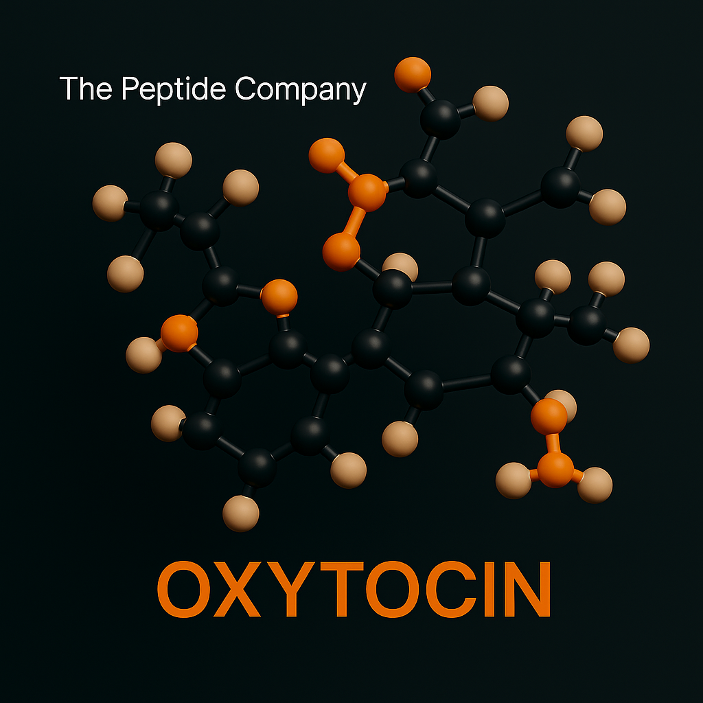
Oxytocin is a nonapeptide neurohormone synthesized primarily in the hypothalamus and released through the posterior pituitary. It functions as both a hormone and a neuromodulator, with research spanning social cognition, stress regulation, autonomic balance, immune signaling, and metabolic coordination. Due to its dual central and peripheral actions, oxytocin is a core molecule in neuroendocrine research.
Molecular Structure and Biosynthesis
Oxytocin is composed of nine amino acids with a characteristic disulfide bond that stabilizes its cyclic structure. It is synthesized as part of a larger precursor protein (prepro-oxytocin-neurophysin I) and processed through enzymatic cleavage. This structural configuration contributes to receptor specificity and signaling kinetics in research models.
Oxytocin Receptor Biology
The oxytocin receptor (OXTR) is a G-protein–coupled receptor expressed in the brain, heart, gastrointestinal tract, reproductive tissues, and immune cells. Activation of OXTR primarily couples to Gq/11 proteins, triggering phospholipase C signaling, intracellular calcium release, and downstream kinase cascades. Research also explores context-dependent coupling to Gi/o pathways.
Neural Circuitry and Social Cognition
Central oxytocin signaling is studied extensively in relation to social behavior, bonding, trust, emotional recognition, and affiliative processing. Research examines oxytocin’s influence on limbic structures including the amygdala, hippocampus, and prefrontal cortex, as well as its modulation of salience and reward networks.
Stress Response and Autonomic Regulation
Oxytocin interacts with stress-response systems by modulating hypothalamic–pituitary–adrenal (HPA) axis activity and autonomic nervous system balance. Research models show associations with reduced stress signaling, altered cortisol dynamics, and enhanced parasympathetic tone.
Immune and Inflammatory Pathways
Oxytocin receptors are expressed on various immune cells. Studies investigate oxytocin’s role in cytokine regulation, immune-cell migration, and neuroimmune communication. These pathways link oxytocin signaling to inflammatory balance and systemic stress responses.
Metabolic and Cardiovascular Research
Beyond neural effects, oxytocin is studied for its involvement in metabolic coordination and cardiovascular signaling. Research explores its influence on glucose regulation, lipid metabolism, vascular tone, and cardiac contractility, highlighting its role as a systemic regulatory peptide.
Developmental and Reproductive Signaling
Oxytocin is well known for its role in reproductive biology, including parturition and lactation, but research also extends to developmental neurobiology. Studies examine how oxytocin signaling influences early-life neural circuit formation and long-term behavioral phenotypes.
Summary
Oxytocin is a multifunctional neuroendocrine peptide studied for its roles in social cognition, stress modulation, immune signaling, metabolic regulation, and cardiovascular biology. Its widespread receptor distribution and context-dependent signaling make it a central molecule in integrative physiology and neuroscience research.
Educational & Research Disclaimer
This article is for educational and scientific research purposes only. No therapeutic claims or usage recommendations are provided. Compounds referenced are not approved for human use and are intended solely for controlled laboratory research.
PMID:
- PMID: 10983204 – Oxytocin as a central neuromodulator
- PMID: 16123301 – Oxytocin and social behavior signaling
- PMID: 17666521 – Oxytocin receptor distribution and function
- PMID: 23665558 – Oxytocin in stress and autonomic regulation
- PMID: 31289336 – Neuroendocrine and systemic effects of oxytocin
FAQs
What is oxytocin in research models?
Oxytocin is a nonapeptide neurohormone studied for its role in neuroendocrine signaling, social cognition, and systemic regulatory processes.
How does oxytocin function in the brain?
Oxytocin acts as a neuromodulator, influencing neuronal activity in regions associated with social behavior, stress processing, and emotional regulation.
What receptors does oxytocin act on?
Oxytocin binds to the oxytocin receptor (OXTR), a G-protein–coupled receptor expressed in both central and peripheral tissues.
Is oxytocin involved in stress and autonomic regulation?
Yes. Research indicates oxytocin modulates autonomic balance and interacts with stress-response pathways.
Is oxytocin studied outside of social behavior?
Yes. Research models examine oxytocin’s roles in immune signaling, metabolism, cardiovascular regulation, and neurodevelopment.
Related Searches:
Dihexa — Neurotrophic Peptide Research Article (Educational • Research Use Only)
Semax : ACTH(4–10)-Derived Heptapeptide and Neurotrophic Research Pathways

Introduction
Short peptide bioregulators—ultrashort amino acid sequences typically 2–4 residues long—are studied for their ability to influence transcriptional activity, chromatin structure, and intracellular signaling within specific tissues. ProstaMax is a prostate-targeting bioregulator examined for its regulatory interactions with prostate epithelial and stromal tissues, including androgen-associated gene networks, stromal–epithelial signaling, and nuclear regulatory pathways.
Overview of Prostate Tissue Biology
The prostate is composed of luminal epithelial cells, basal epithelial cells, stromal fibroblasts, smooth muscle cells, neuroendocrine cells, and resident immune cells. Regulatory behavior depends heavily on stromal–epithelial cross-talk mediated by growth factors, androgen signaling, cytokines, and extracellular matrix components.
Short Peptide Bioregulators
Bioregulators differ from classical peptides because they act intracellularly rather than through membrane-bound receptors. Their ultrashort size allows passive diffusion, nuclear penetration, and interactions with nuclear proteins, transcription factors, chromatin remodelers, and peptide-binding proteins.
Molecular Basis of ProstaMax
ProstaMax is derived from conserved amino acid motifs found in prostate-regulatory proteins. Its structure allows intracellular and nuclear access, potential affinity for chromatin-associated proteins, androgen receptor co-regulators, nuclear matrix proteins, and DNA-binding proteins.
Mechanistic Pathways
Research explores ProstaMax’s potential influence on transcriptional modulation, chromatin accessibility, androgen-regulated gene networks, MAPK and PI3K/AKT signaling intersections, stromal–epithelial communication pathways, and nuclear scaffold interactions.
Intracellular Transport and Nuclear Uptake
Ultrashort peptides can enter cells through diffusion or transporter-mediated uptake. Once inside, they may bind cytoplasmic proteins, diffuse toward the nucleus, or interact with nuclear import machinery. Due to their size, they may pass through nuclear pores and affect transcriptional protein complexes.
Gene Networks of Interest
Prostate research models investigate ProstaMax in relation to luminal cell markers (PSA/KLK3, TMPRSS2, NKX3-1), basal cell markers (KRT5, KRT14, p63), stromal genes (TGF-β–associated pathways, extracellular matrix remodeling), cytokine expression signatures, and androgen-responsive transcriptional circuits.
Tissue-Level Research Themes
ProstaMax appears in studies examining epithelial differentiation, stromal structural regulation, extracellular matrix turnover, luminal/basal identity markers, prostate-specific secretory genes, and transcriptional homeostasis in androgen-responsive tissues.
Summary
ProstaMax is a prostate-targeting short peptide bioregulator studied for its influence on transcriptional modulation, chromatin structure, stromal–epithelial regulatory pathways, and prostate-specific gene expression. Its ultrashort size and intracellular/nuclear accessibility position it as a unique research tool in prostate regulatory biology.
Educational & Research Disclaimer
This article is for educational and scientific research purposes only. No therapeutic claims or usage recommendations are made. Compounds referenced are not approved for human use and are intended solely for controlled laboratory research.
PMID:
- PMID: 11957224 — Tissue-specific regulatory peptides and gene expression control
- PMID: 15004429 — Short peptides as regulators of transcription and chromatin structure
- PMID: 15894536 — Peptide regulation of cell differentiation and tissue homeostasis
- PMID: 17606801 — Prostate-specific peptide signaling and stromal–epithelial interactions
FAQ:
What is ProstaMax studied for in research models?
ProstaMax is studied as a short peptide bioregulator associated with prostate tissue–specific gene expression, stromal–epithelial signaling, and transcriptional regulation in experimental research settings.
How do short peptide bioregulators function at the cellular level?
Short peptide bioregulators are investigated for their ability to influence chromatin structure, transcription factor activity, and intracellular signaling pathways due to their small size and nuclear accessibility.
Is ProstaMax intended for human or clinical use?
No. ProstaMax is referenced exclusively for educational and laboratory research purposes. It is not approved for human use, clinical treatment, or therapeutic applications.
RELATED SEARCHES:
Bronchogen: Short Peptide Bioregulator for Bronchial and Pulmonary Tissue Research
Cardiogen: Short Peptide Bioregulator for Cardiac and Myocardial Tissue Research
Introduction
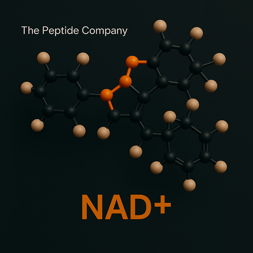
Nicotinamide adenine dinucleotide (NAD⁺) is a ubiquitous redox cofactor essential for cellular energy metabolism, mitochondrial function, and regulatory signaling. Beyond its classical role in oxidation–reduction reactions, NAD⁺ serves as a substrate for multiple enzyme families that govern DNA repair, chromatin remodeling, stress responses, and metabolic adaptation. As a result, NAD⁺ occupies a central position in modern cellular and mitochondrial research.
Chemical Structure and Redox Function
NAD⁺ consists of two nucleotides joined through their phosphate groups: one containing an adenine base and the other nicotinamide. The nicotinamide moiety undergoes reversible reduction to NADH, enabling electron transfer reactions. This NAD⁺/NADH redox couple is fundamental to glycolysis, the tricarboxylic acid cycle, and oxidative phosphorylation.
Role in Mitochondrial Energy Metabolism
Within mitochondria, NAD⁺ accepts electrons generated during the TCA cycle and delivers them to complex I of the electron transport chain via NADH. This process drives proton pumping, establishes the electrochemical gradient, and ultimately supports ATP synthesis. Research examines how compartmentalized NAD⁺ pools influence mitochondrial efficiency, redox balance, and adaptive responses to energetic stress.
NAD⁺-Consuming Enzymes
NAD⁺ functions not only as a redox cofactor but also as a consumable substrate for several enzyme families. Sirtuins (SIRT1–SIRT7) utilize NAD⁺ for deacetylation and ADP-ribosylation reactions that regulate gene expression, mitochondrial protein function, and stress resistance. Poly(ADP-ribose) polymerases (PARPs) consume NAD⁺ during DNA repair, linking NAD⁺ availability to genomic maintenance.
NAD⁺ and Chromatin Regulation
Through sirtuin activity, NAD⁺ levels influence chromatin structure and transcriptional programs. Research models show that NAD⁺-dependent deacetylation affects histones, transcription factors, and co-regulators, thereby coordinating metabolic state with gene expression. This positions NAD⁺ as a molecular bridge between metabolism and epigenetic control.
Cellular Stress, DNA Repair, and Redox Homeostasis
During oxidative or genotoxic stress, NAD⁺ consumption by PARPs increases to facilitate DNA repair. Excessive activation can deplete cellular NAD⁺ pools, disrupting energy metabolism. Research explores how cells balance NAD⁺ regeneration, redox homeostasis, and repair processes to maintain viability under stress.
NAD⁺ Salvage and Biosynthetic Pathways
Cells maintain NAD⁺ levels through de novo synthesis and salvage pathways. The salvage pathway recycles nicotinamide into NAD⁺ via intermediates such as NMN, coordinated by enzymes including NAMPT and NMNATs. Research focuses on how these pathways regulate intracellular NAD⁺ availability across nuclear, cytosolic, and mitochondrial compartments.
Systemic and Intercellular Signaling Roles
Beyond individual cells, NAD⁺ metabolism influences intercellular communication and systemic physiology. Studies examine extracellular NAD⁺ turnover, ectoenzyme activity, and the role of NAD⁺-derived metabolites in immune and inflammatory signaling. These findings expand the relevance of NAD⁺ beyond classical metabolism.
Summary
NAD⁺ is a central molecular hub integrating redox chemistry, mitochondrial energy production, DNA repair, chromatin regulation, and stress-response signaling. Its dual role as both a cofactor and a consumable substrate makes NAD⁺ a key determinant of cellular resilience and metabolic adaptation in advanced biological research.
Educational & Research Disclaimer
This article is for educational and scientific research purposes only. No therapeutic claims or usage recommendations are provided. Compounds referenced are not approved for human use and are intended solely for controlled laboratory experimentation.
PMID:
- PMID: 33353981 (NAD⁺ metabolism in ageing) PMC
- PMID: 24786309 (NAD⁺ + sirtuins in aging/disease) PubMed
- PMID: 29883761 (NAD metabolism in aging/longevity) PubMed
- PMID: 29482842 (CD38 biology; NAD⁺ consumption) PubMed
- PMID: 30355082 (Sirtuins/NAD⁺ in cardio-metabolic disease models)
FAQ:
What is NAD⁺ in research models?
NAD⁺ (nicotinamide adenine dinucleotide) is an essential redox cofactor that supports cellular energy metabolism and acts as a substrate for enzymes involved in DNA repair and gene regulation.
How does NAD⁺ support cellular energy production?
NAD⁺ shuttles electrons in glycolysis and the TCA cycle, enabling oxidative phosphorylation and ATP generation through mitochondrial respiration.
What’s the difference between NAD⁺ and NADH?
NAD⁺ is the oxidized form and NADH is the reduced form. The NAD⁺/NADH ratio is a core indicator of cellular redox state and metabolic flux.
Why is NAD⁺ linked to sirtuins and longevity pathways?
Sirtuins use NAD⁺ to regulate protein deacetylation, influencing mitochondrial biogenesis, stress responses, and metabolic adaptation in research systems.
How is NAD⁺ regulated inside cells?
Cells maintain NAD⁺ through biosynthesis and salvage pathways (notably from nicotinamide), while enzymes like CD38 and PARPs consume NAD⁺ during signaling and repair processes.
How is NAD⁺ typically measured in lab studies?
Common approaches include LC–MS/MS quantification, enzymatic cycling assays, and paired measurement of NAD⁺/NADH to assess redox balance.
Is NAD⁺ itself a peptide?
No. NAD⁺ is a nucleotide-derived coenzyme, not a peptide—though it’s often discussed alongside bioactive molecules studied for cellular optimization.
Related Searches:
MOTS-c: The Mitochondrial-Encoded Peptide for Metabolic Regulation and Cellular Resilience

NAD+ 1000mg
NAD+ 1000mg is a research compound studied for cellular energy metabolism, redox balance, mitochondrial function, and sirtuin-associated signaling pathways. For research use only.
Introduction
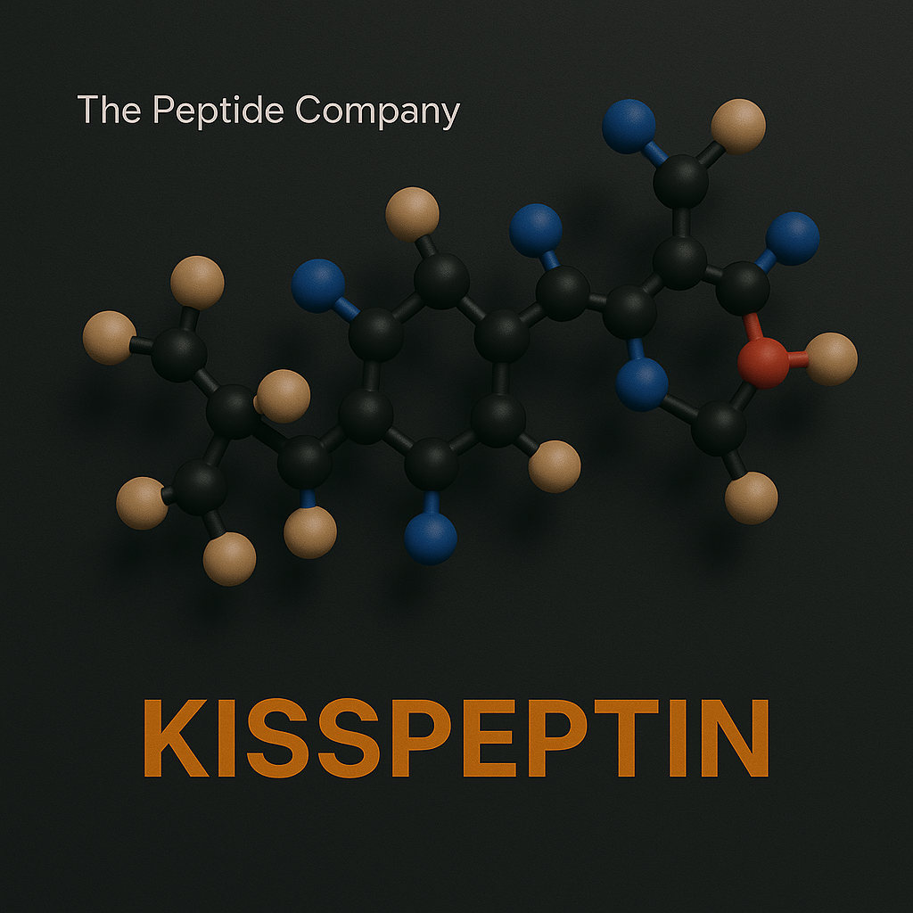
Kisspeptin refers to a family of neuropeptides encoded by the KISS1 gene that play a central role in regulating the hypothalamic–pituitary–gonadal (HPG) axis. Discovered through cancer-metastasis research and later identified as a master regulator of reproductive neuroendocrinology, kisspeptin signaling is now a foundational topic in neuroscience and endocrine research.
Molecular Structure and Peptide Variants
Kisspeptin peptides are derived from a common precursor and exist in multiple biologically active forms, including kisspeptin‑54, kisspeptin‑14, kisspeptin‑13, and kisspeptin‑10. All variants share a conserved C‑terminal sequence essential for receptor binding. Research focuses on how peptide length influences stability, diffusion, and signaling dynamics.
KISS1 Receptor Biology (GPR54)
Kisspeptin exerts its effects through the G‑protein–coupled receptor KISS1R (also known as GPR54). This receptor is highly expressed on gonadotropin‑releasing hormone (GnRH) neurons within the hypothalamus. Receptor activation couples primarily to Gq/11 proteins, leading to phospholipase C activation, intracellular calcium release, and downstream transcriptional signaling.
Control of GnRH Pulsatility
Kisspeptin signaling is a primary upstream driver of GnRH pulsatility. Research demonstrates that kisspeptin neurons integrate metabolic, circadian, and stress-related signals to regulate the timing and amplitude of GnRH release. This pulsatile control is essential for downstream secretion of luteinizing hormone (LH) and follicle‑stimulating hormone (FSH).
Integration with Metabolic and Energy Signals
Kisspeptin neurons receive input from metabolic regulators including leptin, insulin, and AMPK‑related pathways. Research explores how energy availability and nutritional status influence reproductive signaling through kisspeptin-mediated neuroendocrine integration.
Sex Steroid Feedback Mechanisms
Kisspeptin signaling mediates both positive and negative feedback effects of sex steroids such as estrogen and testosterone. Distinct populations of kisspeptin neurons within the hypothalamus are involved in feedback regulation, allowing precise control of reproductive hormone cycles in research models.
Developmental and Puberty-Related Research
Kisspeptin is a key factor in the initiation of puberty. Research investigates how developmental changes in kisspeptin expression and receptor sensitivity trigger activation of the HPG axis and long-term reproductive competence.
Extra-Reproductive Signaling Pathways
Beyond reproductive control, kisspeptin and KISS1R are expressed in other tissues including the pancreas, liver, and cardiovascular system. Studies explore potential roles in metabolic regulation, cell migration, and tissue-specific signaling outside the classical HPG axis.
Summary
Kisspeptin is a central neuropeptide regulator of the hypothalamic–pituitary–gonadal axis, integrating metabolic, circadian, and hormonal signals to control reproductive endocrine function. Its receptor-mediated signaling and developmental importance make it a cornerstone of neuroendocrine research.
Educational & Research Disclaimer
This article is for educational and scientific research purposes only. No therapeutic claims or usage recommendations are provided. Compounds referenced are not approved for human use and are intended solely for controlled laboratory experimentation.
PMID:
- PMID: 12124405 – Discovery of kisspeptin and GPR54
- PMID: 15126532 – Kisspeptin regulation of GnRH secretion
- PMID: 15998810 – Central control of reproductive axis
- PMID: 18332454 – Kisspeptin neurons and puberty onset
- PMID: 31117005 – Kisspeptin signaling in reproductive research models
FAQ:
What is kisspeptin in research models?
Kisspeptin is a neuropeptide that activates the GPR54 receptor and serves as a key upstream regulator of the hypothalamic–pituitary–gonadal (HPG) axis.
How does kisspeptin regulate reproductive signaling?
Kisspeptin stimulates gonadotropin-releasing hormone (GnRH) neurons, triggering downstream release of LH and FSH in experimental systems.
Is kisspeptin involved in puberty and fertility research?
Yes. Kisspeptin signaling is essential for puberty onset and reproductive function, making it a central target in neuroendocrine research.
Where is kisspeptin expressed?
Research shows kisspeptin expression in the hypothalamus, placenta, and peripheral tissues involved in reproductive regulation.
How is kisspeptin studied in laboratory research?
Studies examine receptor activation, GnRH neuron firing, hormone pulsatility, and developmental timing in controlled research models.
Related Searches:
Sermorelin: GHRH Fragment Research and Growth Hormone Pulsatility Models

Introduction
Humanin is a small mitochondrial-derived peptide (MDP) encoded within the mitochondrial genome and translated in the cytoplasm. It was originally identified in studies of neuronal survival and has since become a central molecule in research on mitochondrial stress signaling, apoptosis resistance, metabolic regulation, and neuroprotection. Humanin represents a class of peptides that function as retrograde signals, allowing mitochondria to communicate cellular stress states to the nucleus.
Mitochondrial Origin and Structure
Humanin consists of 24 amino acids and is encoded within the 16S rRNA region of mitochondrial DNA. Despite its small size, Humanin exhibits a conserved sequence across species, suggesting evolutionary importance. Structural studies indicate that Humanin adopts conformations compatible with receptor binding and intracellular protein–protein interactions.
Humanin Receptor and Signaling Complexes
Humanin interacts with both intracellular targets and cell-surface receptor complexes. One well-studied signaling route involves a trimeric receptor complex composed of CNTFRα, WSX‑1, and gp130, leading to activation of STAT3-dependent transcriptional pathways. Research also explores Humanin’s receptor-independent intracellular actions.
Anti-Apoptotic Mechanisms
Humanin is extensively studied for its ability to suppress apoptotic signaling. It directly interacts with pro-apoptotic BCL‑2 family members, including BAX, tBID, and BimEL, preventing mitochondrial outer membrane permeabilization and cytochrome c release. These mechanisms position Humanin as a key modulator of intrinsic apoptosis pathways in research models.
Mitochondrial Stress and Retrograde Signaling
As a mitochondrial-derived peptide, Humanin participates in retrograde signaling networks that link mitochondrial function to nuclear gene expression. Research investigates its role in modulating stress-response transcription, mitochondrial quality control, and cellular adaptation to oxidative and metabolic challenges.
Neuroprotective Research Pathways
Humanin is widely studied in neuronal models for its influence on cell survival, synaptic integrity, and resistance to neurotoxic stressors. Research examines its interactions with amyloid-associated stress, excitotoxic signaling, and mitochondrial dysfunction in neural tissues.
Metabolic and Endocrine Research
Beyond the nervous system, Humanin is explored for its effects on metabolic regulation. Studies examine its influence on insulin signaling pathways, glucose metabolism, lipid handling, and cellular energy balance. These findings position Humanin within a broader network of mitochondrial peptides involved in systemic metabolic communication.
Inflammatory and Immune Signaling
Humanin signaling intersects with inflammatory pathways through modulation of cytokine expression and stress-responsive immune transcription. Research explores its potential role in balancing pro‑ and anti‑inflammatory signaling during cellular stress conditions.
Summary
Humanin is a mitochondrial-derived peptide studied for its roles in apoptosis suppression, mitochondrial stress signaling, neuroprotection, metabolic regulation, and immune-modulatory pathways. Its ability to function as a retrograde signal highlights the mitochondrion as an active regulator of cellular fate and resilience in advanced biological research.
Educational & Research Disclaimer
This article is for educational and scientific research purposes only. No therapeutic claims or usage recommendations are provided. Compounds referenced are not approved for human use and are intended solely for controlled laboratory experimentation.
PMID:
- PMID: 11416100 – Discovery of humanin and neuroprotective effects
- PMID: 14593182 – Humanin and mitochondrial-mediated apoptosis
- PMID: 18755891 – Cytoprotective signaling pathways of humanin
- PMID: 23336290 – Humanin analogs and stress resistance
- PMID: 30559258 – Humanin as a mitochondrial-derived peptide in aging research
FAQ:
What is humanin in research models?
Humanin is a mitochondrial-derived peptide studied for its role in cellular protection, stress resistance, and survival signaling under pathological conditions.
How does humanin protect cells?
Humanin interferes with pro-apoptotic signaling pathways and supports mitochondrial integrity, reducing oxidative and metabolic stress–induced cell death.
Is humanin linked to mitochondrial function?
Yes. Humanin originates from mitochondrial DNA and is closely associated with mitochondrial communication and bioenergetic regulation.
What systems are studied with humanin?
Research models explore humanin in neuroprotection, metabolic regulation, aging, and cellular stress response pathways.
How is humanin investigated in laboratory settings?
Humanin is examined through receptor binding studies, apoptosis assays, mitochondrial stress models, and signaling pathway analyses.
Related Searches:
MOTS-c: The Mitochondrial-Encoded Peptide for Metabolic Regulation and Cellular Resilience
NMN: NAD⁺ Precursor Biology, Cellular Metabolism, and Mitochondrial Research

Introduction
Hexarelin is a synthetic hexapeptide belonging to the growth hormone secretagogue (GHS) class. It is widely studied for its interaction with the growth hormone secretagogue receptor (GHS‑R1a) and its downstream signaling effects on endocrine, metabolic, and tissue‑repair pathways. Unlike endogenous growth hormone–releasing hormone, Hexarelin engages ghrelin‑related signaling mechanisms, making it a distinct tool for growth‑axis research.
Molecular Structure and Peptide Class
Hexarelin is composed of six amino acids arranged to optimize binding affinity for the GHS‑R1a receptor. Its compact structure confers resistance to rapid enzymatic degradation compared to native peptides. Research examines how structural modifications influence receptor activation kinetics, signaling bias, and downstream transcriptional effects.
Growth Hormone Secretagogue Receptor Biology
The GHS‑R1a receptor is a G‑protein–coupled receptor expressed in the pituitary, hypothalamus, cardiac tissue, skeletal muscle, and other peripheral tissues. Hexarelin binding activates Gq/11‑mediated signaling, leading to intracellular calcium mobilization, phospholipase C activation, and growth hormone pulse initiation in experimental models.
Endocrine Signaling and Growth Axis Research
Hexarelin is studied for its effects on the hypothalamic–pituitary axis, particularly its role in modulating growth hormone release patterns. Research investigates pulse amplitude, feedback regulation, and interactions with somatostatin signaling. These studies help clarify growth hormone dynamics independent of direct GHRH stimulation.
Cardiovascular and Myocardial Research
Beyond endocrine effects, Hexarelin is examined in cardiovascular research for its influence on myocardial signaling pathways. Studies explore interactions with cardiomyocyte survival mechanisms, calcium handling, and gene expression related to cardiac stress adaptation. These properties distinguish Hexarelin from other GHS compounds in tissue‑specific research contexts.
Metabolic and Tissue-Level Effects
Hexarelin research extends into metabolic regulation, including effects on lipid metabolism, energy balance, and insulin‑related signaling pathways. Tissue‑level studies examine its influence on skeletal muscle protein turnover, connective tissue signaling, and regenerative transcriptional programs.
Receptor Desensitization and Signaling Dynamics
Continuous exposure to GHS compounds can lead to receptor desensitization. Research on Hexarelin investigates receptor internalization, resensitization kinetics, and biased signaling profiles that differentiate acute versus chronic receptor engagement.
Summary
Hexarelin is a growth hormone secretagogue studied for its interaction with the GHS‑R1a receptor, its modulation of growth hormone signaling, and its broader tissue‑level effects on cardiovascular, metabolic, and regenerative pathways. Its distinct receptor dynamics and signaling profile make it a valuable compound in growth‑axis and integrative physiology research.
Educational & Research Disclaimer
This article is for educational and scientific research purposes only. No therapeutic claims or usage recommendations are provided. Compounds referenced are not approved for human use and are intended solely for controlled laboratory experimentation.
PMID:
- PMID: 7510399 – Discovery and GH-releasing activity of hexarelin
- PMID: 10459801 – GHS-R binding and endocrine effects
- PMID: 14523018 – IGF-1 and GH axis modulation
- PMID: 16029934 – Cardiovascular and metabolic research effects
- PMID: 18265864 – Comparison with other GH secretagogues
FAQ:
What is hexarelin in research models?
Hexarelin is a synthetic growth hormone secretagogue that activates the growth hormone secretagogue receptor (GHS-R), stimulating pulsatile GH release in experimental systems.
How does hexarelin differ from GHRH analogs?
Unlike GHRH analogs that act directly on the pituitary, hexarelin works through GHS-R activation, influencing both hypothalamic and pituitary signaling.
Does hexarelin affect IGF-1 levels?
Research shows hexarelin-induced GH release can secondarily influence IGF-1 signaling, depending on study duration and model.
Is hexarelin studied outside of GH signaling?
Yes. Research explores cardiovascular, metabolic, and tissue-protective effects independent of GH release.
How is hexarelin used in laboratory research?
Hexarelin is examined for receptor binding, GH pulsatility, endocrine feedback loops, and comparative secretagogue potency.
Related Searches:
Sermorelin: GHRH Fragment Research and Growth Hormone Pulsatility Models
IGF-1 Analogues: LR3 and DES Structural Variations and Receptor Binding in Research Models
CJC-1295: GHRH Analog, DAC Conjugation, and Growth Hormone Pulsatility in Research

Introduction
Glutathione (GSH) is a tripeptide composed of glutamate, cysteine, and glycine and is one of the most abundant intracellular antioxidants. It plays a central role in maintaining cellular redox balance, detoxification processes, mitochondrial function, and regulation of cellular signaling pathways. Due to its broad involvement in oxidative stress management, glutathione is a foundational molecule in biochemical and cellular research.
Molecular Structure and Biosynthesis
Glutathione’s unique γ-glutamyl bond between glutamate and cysteine confers stability and functional specificity. Biosynthesis occurs in two ATP-dependent steps catalyzed by glutamate–cysteine ligase (GCL) and glutathione synthetase (GS). Cellular availability of cysteine is rate-limiting, linking glutathione synthesis to amino acid metabolism and redox demand.
Redox Cycling and Antioxidant Function
Glutathione exists in reduced (GSH) and oxidized (GSSG) forms, enabling dynamic redox cycling. Glutathione peroxidases (GPxs) utilize GSH to neutralize hydrogen peroxide and lipid peroxides, while glutathione reductase regenerates GSH from GSSG using NADPH. The GSH:GSSG ratio is a key indicator of cellular redox status in research models.
Detoxification and Conjugation Pathways
Glutathione participates in phase II detoxification through conjugation reactions catalyzed by glutathione S-transferases (GSTs). These reactions increase solubility of electrophilic compounds, facilitating their removal. Research explores glutathione’s role in xenobiotic metabolism, environmental toxin handling, and cellular defense against reactive intermediates.
Mitochondrial Function and Oxidative Stress
Mitochondrial glutathione pools are essential for maintaining oxidative phosphorylation efficiency and preventing oxidative damage to mitochondrial DNA, lipids, and proteins. Studies examine glutathione transport into mitochondria and its role in preserving mitochondrial membrane integrity and respiratory chain function under stress conditions.
Regulation of Cellular Signaling
Beyond antioxidant activity, glutathione modulates redox-sensitive signaling pathways. Protein S-glutathionylation serves as a reversible post-translational modification that influences enzyme activity, transcription factor binding, and signal transduction. This positions glutathione as a regulator of cellular communication in research settings.
Immune and Inflammatory Research
Glutathione status influences immune cell function, cytokine production, and inflammatory signaling. Research investigates how glutathione levels affect lymphocyte proliferation, macrophage activity, and the balance between pro- and anti-inflammatory transcriptional programs.
Summary
Glutathione is a central tripeptide antioxidant studied for its roles in redox homeostasis, detoxification, mitochondrial protection, and regulation of cellular signaling. Its integration into metabolic, immune, and stress-response pathways makes glutathione a cornerstone molecule in cellular and biochemical research.
Educational & Research Disclaimer
This article is for educational and scientific research purposes only. No therapeutic claims or usage recommendations are provided. Compounds referenced are not approved for human use and are intended solely for controlled laboratory experimentation.
- PMID: 16492803 – Meister & Anderson, glutathione metabolism overview
- PMID: 17021671 – Redox regulation and cellular signaling
- PMID: 18973918 – Role of glutathione in detoxification pathways
- PMID: 23766848 – Mitochondrial glutathione and oxidative stress
- PMID: 33208454 – Glutathione depletion and redox imbalance in disease models
FAQ:
What is glutathione and why is it important in research models?
Glutathione (GSH) is a tripeptide composed of glutamate, cysteine, and glycine that functions as the primary intracellular antioxidant, maintaining redox balance and protecting cells from oxidative damage.
How does glutathione regulate cellular redox homeostasis?
Glutathione cycles between reduced (GSH) and oxidized (GSSG) states, buffering reactive oxygen species and preserving redox-sensitive signaling pathways.
What role does glutathione play in detoxification pathways?
Glutathione conjugates xenobiotics and endogenous toxins via glutathione S-transferases, facilitating cellular detoxification and excretion.
Is glutathione involved in mitochondrial function?
Yes. Mitochondrial glutathione protects respiratory chain components from oxidative stress and supports ATP production and mitochondrial integrity.
How is glutathione studied in laboratory research?
Research models examine glutathione synthesis, depletion, redox ratios (GSH:GSSG), and enzyme activity to assess oxidative stress, detoxification capacity, and cellular resilience.
Related Searches:
MOTS-c: The Mitochondrial-Encoded Peptide for Metabolic Regulation and Cellular Resilience
NMN: NAD⁺ Precursor Biology, Cellular Metabolism, and Mitochondrial Research
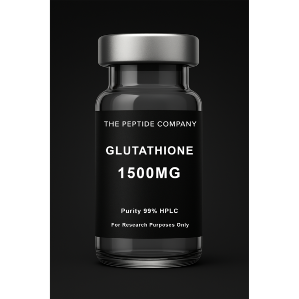
Glutathione 1500mg
Glutathione 1500mg is a research compound studied for cellular redox balance, oxidative stress modulation, and detoxification pathway mechanisms. For research use only.
Overview
AOD‑9604 is a 16‑amino‑acid fragment derived from the C‑terminus of growth hormone (residues 177–191 with an added N‑terminal tyrosine). It was developed to study lipid‑metabolism pathways attributed to this region while minimizing classical growth‑hormone effects. Published work spans in‑vitro systems, animal models, and early clinical exploration focused on fat metabolism and energy balance.
Mechanism of Action (Research Context)
Evidence from preclinical and early clinical literature indicates modulation of lipid handling via β‑adrenergic pathways, with signals consistent with increased lipolysis and reduced lipogenesis. Summaries report little to no change in IGF‑1, suggesting activity distinct from the canonical GH/IGF‑1 axis. Findings vary by model, dose, and study design.
Reported Findings in the Literature
The following points consolidate outcomes commonly described across studies and reviews. Exact magnitude and reproducibility depend on protocol, population, and duration:
• Stimulates fat breakdown (lipolysis): activation of pathways associated with mobilizing stored fatty acids for energy.
• Inhibits fat creation (anti‑lipogenic signals): down‑regulation of processes linked to new fat cell formation and triglyceride storage.
• Increases metabolic activity: observations consistent with higher energy expenditure in several models.
• Targets adipose depots: reports describe preferential effects on stubborn or abdominal fat in some trials.
• Muscle preservation signals: weight‑management studies emphasize fat‑focused effects with limited impact on lean mass.
• Glycemic/IGF‑1 profile: summaries frequently note minimal effect versus full‑length GH in short‑term observations.
Key Research‑Context Takeaways
• Weight‑management research focus centered on fat reduction rather than increases in lean tissue.
• Signals consistent with metabolic‑rate support observed in multiple models.
• Short‑term tolerability generally favorable; efficacy outcomes mixed across longer studies.
Chemical / Physical Information
• Sequence: Tyr‑Leu‑Arg‑Ile‑Val‑Gln‑Cys‑Arg‑Ser‑Val‑Glu‑Gly‑Ser‑Cys‑Gly‑Phe (16 aa) • Approx. molecular weight: ~1815 Da • Handling (general peptide guidance): store lyophilized material at −20 °C, protected from moisture/light; aliquot reconstituted solutions and avoid repeated freeze–thaw.
Notes on Formats Studied
Across the literature, AOD‑9604 has been evaluated in various experimental formats, including subcutaneous administration and oral formulations. Formulation, dosing, and endpoints differ by study and should be interpreted within each protocol’s design.
Regulatory & Compliance Notes
Referenced by anti‑doping organizations and not approved as an obesity therapy by major health authorities. Acquisition and use must follow applicable laws and institutional policies. Verify status before any regulated activity.
References (Selection)
• Endocrinology (2001): lipid metabolism and β‑adrenergic context. • Journal of Endocrinology & Metabolism (2013–2014): safety/tolerability summaries and IGF‑1 observations. • Obesity pharmacotherapy reviews (2013–2015): efficacy overview across trials. • Compound databases (e.g., PubChem) for identifiers and mass. • Anti‑doping organization statements/lists for status in sport.
Disclaimer
This is only intended for research purposes only. None of this is intended for consumption. This is only for educational purposes.
——————————–
Selected References
PMID: 18469936 — AOD-9604 mechanisms in fat metabolism and lipolysis
PMID: 20826578 — Growth-hormone–related peptides and metabolic regulation
PMID: 21753056 — Peptide modulation of visceral adiposity and body composition
PMID: 25826926 — Endocrine and metabolic outcomes of GH-axis analogs
Frontiers in Endocrinology — Peptide-based metabolic therapeutics
Journal of Clinical Endocrinology & Metabolism — GH-derivative peptides and metabolic effects
FAQ:
What is AOD-9604?
AOD-9604 is a modified fragment of human growth hormone (hGH 176-191) studied for its potential effects on lipid metabolism and fat-oxidation pathways in research settings.
How does AOD-9604 work in research models?
A2: AOD-9604 interacts with metabolic signaling related to fat breakdown and energy use without activating full GH receptor pathways, making it unique for studying selective metabolic effects.
Is AOD-9604 approved for human or medical use?
No. AOD-9604 discussed here is a research compound and is not approved for therapeutic or general consumer use.
What are researchers studying AOD-9604 for?
Research evaluates AOD-9604 in contexts involving fat metabolism, adipocyte signaling, and energy balance in controlled experimental models.
How is AOD-9604 different from full growth hormone?
AOD-9604 is a short peptide fragment that does not stimulate IGF-1 production or systemic GH activity, allowing researchers to isolate GH-independent metabolic pathways.
How is AOD-9604 evaluated in studies?
AOD-9604 is examined using in vitro metabolic assays and animal models that track fat oxidation, adipocyte response, and energy utilization.
Are there known effects or side effects in research settings?
Studies report variable responses depending on model and dosing, and long-term safety is not established for human use.
Related Research Compounds
Frag 176–191: Growth Hormone–Derived Fragment and Lipolytic Research Mechanisms
GLP-1 Pathway Peptides: Comparative Research on Semaglutide, Tirzepatide & Retatrutide

AOD-9604 5mg
AOD-9604 5mg is a research compound studied for lipolytic signaling pathways, adipocyte metabolism regulation, and growth hormone-derived fragment activity. For research use only.

Overview
Melanotan II (MT‑II) is a synthetic cyclic heptapeptide analog of α‑melanocyte‑stimulating hormone (α‑MSH). It activates melanocortin receptors, particularly MC1R on melanocytes, to increase eumelanin synthesis. Interest has centered on pigmentation enhancement, photobiology/photoprotection, neuromodulatory effects (including appetite and sexual arousal pathways), and broad melanocortin signaling.
Mechanism of Action (Research Context)
By agonizing MC1R on melanocytes, MT‑II up‑regulates eumelanin (brown/black melanin) production and can augment the tanning response; eumelanin is associated with improved photoprotection compared with pheomelanin. MT‑II and related analogs can interact with other melanocortin receptors (e.g., MC3/4/5) expressed in the CNS and peripheral tissues, which is consistent with reported effects on appetite and sexual function in experimental settings.
Potential Research Benefits (Reported in Literature)
• Pigmentation enhancement and tanning response
Multiple investigations describe increases in eumelanin content and an exaggerated tanning response to UV light. In controlled settings, α‑MSH analogs increased pigmentation without pathologic findings at exposed sites, and effects were synergistic with UV exposure in some protocols.
• Photoprotection / photobiology
Because eumelanin is photoprotective, increased MC1R signaling is mechanistically linked to reduced UV‑induced damage in skin models. Reviews emphasize the central role of the α‑MSH/MC1R axis in photoprotection and support the rationale for studying MT‑II in this context.
• Appetite modulation (research context)
Melanocortin pathways in the hypothalamus regulate energy balance. Reports and commentaries note decreased appetite in some subjects exposed to MT‑II analogs, consistent with central MC3/MC4 receptor activation; magnitude of effect varies by dose and protocol.
• Sexual arousal / libido effects (research context)
Early randomized, placebo‑controlled experiments reported increases in sexual desire and erectile response in male subjects following MT‑II exposure; subsequent pharmacologic development led to bremelanotide for HSDD in premenopausal populations. These observations support ongoing interest in melanocortin‑mediated sexual function pathways.
Potential Reported Side Effects / Adverse Events
Across trials, observational reports, and safety statements, subjects most commonly report transient flushing, nausea, decreased appetite, yawning/somnolence, headache, and GI upset. Erection‑related events have been described in males (including prolonged erections in rare cases). Dermatologic changes such as new or darkening nevi and freckling have been described in surveillance reports; any pigmentary change warrants clinical evaluation. Regulatory advisories also caution about unapproved products and potential serious risks with unsupervised use.
Reported Findings / Key Points
• Photobiology: increased eumelanin and enhanced tanning response in controlled settings.
• Sexual function signals: increased sexual desire metrics in early MT‑II studies; modern development of bremelanotide built on this pathway.
• Appetite: qualitative reports of reduced appetite are consistent with central melanocortin signaling.
• Tolerability: nausea and flushing are the most frequent short‑term effects; severity is dose‑dependent in early studies.
• Safety notes: pigmentary lesion changes have been reported; unregulated products raise additional risk concerns.
Chemical / Physical Information
• Sequence (example): Ac‑Nle‑c[Asp‑His‑D‑Phe‑Arg‑Trp‑Lys]‑NH₂ (cyclic heptapeptide) • Target receptors: primarily MC1R (pigmentation), with activity at MC3/4/5 (CNS/peripheral effects) • General handling (peptide guidance): store lyophilized material at −20 °C, protect from light/moisture; aliquot reconstituted solutions; avoid repeated freeze–thaw.
Notes on Formats Studied
Published protocols have investigated different experimental formats (e.g., subcutaneous administration). Dosing schedules and endpoints vary; interpret results within each study’s design and population.
Regulatory & Compliance Notes
Regulatory agencies have issued advisories regarding unapproved products marketed for tanning or other purposes. Status and legal frameworks differ by jurisdiction; confirm local requirements before any regulated activity.
References (Selection)
• Dorr RT, et al. Effects of a superpotent melanotropic peptide in volunteers. JAMA Dermatology, 2004. • Wessells H, et al. Melanocortin agonists and sexual function; early MT‑II data. • Kingsberg SA, et al. Bremelanotide for HSDD: phase 3 results (2019). • Böhm M, et al. Chronic MC1R signaling: benefits/risks overview (2024). • DermNet NZ: Melanotan II overview and side effects. • TGA advisory on melanotan products (2025). • Reviews on α‑MSH/MC1R and photoprotection (2024).
Disclaimer
This is only intended for research purposes only. None of this is intended for consumption. This is only for educational purposes.
————————————
Selected References
PMID: 16133878 — Melanotan peptides and melanocortin receptor activation
PMID: 12893986 — Melanocortin pathways in pigmentation and photoprotection
PMID: 18573760 — Melanotan II effects on energy balance and neuroendocrine signaling
PMID: 21717461 — Peptide-based modulation of skin pigmentation and UV defense
Frontiers in Endocrinology — Melanocortin system and neuroregulatory mechanisms
Journal of Peptide Science — Melanocortin-derived peptides and physiological activity
FAQ:
What is Melanotan II?
Melanotan II is a synthetic analog of α-MSH (alpha-melanocyte-stimulating hormone) studied for its effects on pigmentation pathways and melanocortin receptor signaling in research settings.
How does Melanotan II work in research models?
Melanotan II activates melanocortin receptors—primarily MC1R for pigmentation and MC3R/MC4R for autonomic and behavioral responses—allowing researchers to study a range of signaling pathways.
Is Melanotan II approved for human use?
No. Melanotan II discussed here is a research compound and is not approved for clinical or general consumer use.
What are researchers studying Melanotan II for?
Research explores Melanotan II in pigmentation models, photoprotection studies, melanocortin pathway mapping, and autonomic signaling.
Does Melanotan II affect tanning or pigmentation?
In research models, Melanotan II increases melanogenesis via MC1R activation, influencing melanin production.
How is Melanotan II typically evaluated in studies?
Studies use in vitro models, ex vivo skin systems, and experimental animals to examine pigmentation, receptor activity, and downstream signaling markers.
Are there known effects or side effects in research settings?
Research models sometimes report flushing, appetite changes, or autonomic responses, though findings vary by protocol and dosage.
Related Research Compounds
GLP-1 Pathway Peptides: Comparative Research on Semaglutide, Tirzepatide & Retatrutide
Sermorelin: GHRH Fragment Research and Growth Hormone Pulsatility Models
IGF-1 Analogues: LR3 and DES Structural Variations and Receptor Binding in Research Models
CJC-1295: GHRH Analog, DAC Conjugation, and Growth Hormone Pulsatility in Research