The Forgotten Immune Regulator
For decades, the thymus — the small gland behind the sternum — has been quietly training the immune system’s most specialized soldiers: T-cells. With age, chronic stress, or illness, this gland shrinks and loses efficiency. What follows is an immune system that’s reactive, fragmented, and often inflamed rather than balanced.
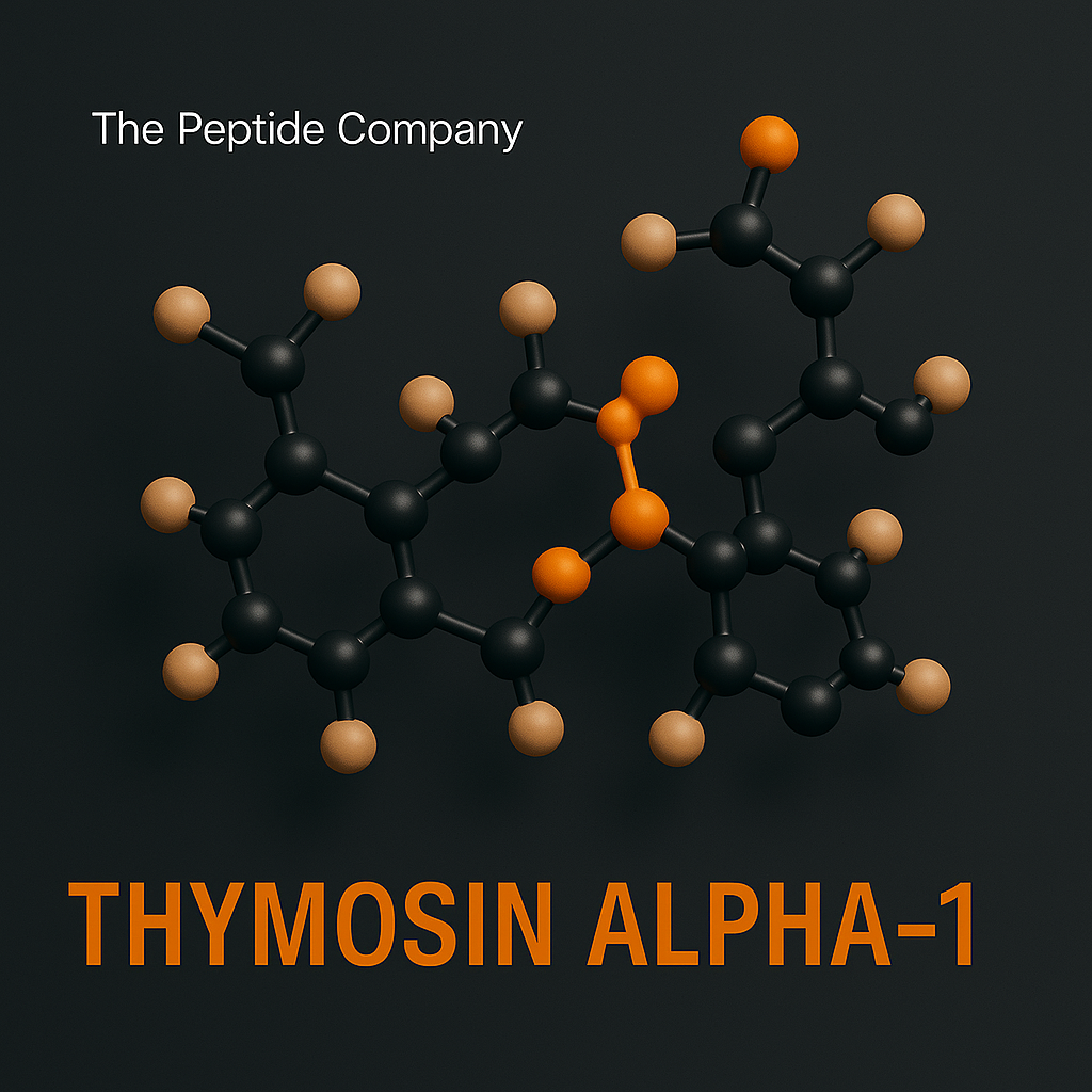
Among the most interesting molecules emerging from thymic research is Thymosin Alpha-1 (Tα1) — a naturally occurring 28-amino-acid peptide fragment first isolated in the 1970s. It has been studied for its ability to modulate immunity, rebalance cytokine signaling, and enhance the body’s intrinsic defense system rather than overstimulate it.
Today, Tα1 is recognized in research as a model for immune precision — restoring function where it’s weak and calming it where it’s overactive.
What Is Thymosin Alpha-1?
Tα1 originates from the thymus gland, a critical component of immune maturation. As an endogenous peptide, it’s part of the body’s natural regulatory framework — released to support T-cell differentiation, dendritic cell activation, and immune surveillance.
While discovered nearly half a century ago, its significance is now resurfacing with renewed attention from immunologists studying:
- Viral defense mechanisms
- Cancer immunotherapy adjuvants
- Age-related immune decline
- Immune “reset” models for chronic inflammation
Tα1 is not a stimulant. Instead, it appears to act as a modulator, tuning the immune system’s responsiveness — a concept known as immunorestoration.
Mechanism of Action: TLR Signaling and Immune Precision
Research suggests Thymosin Alpha-1 interacts with Toll-like Receptors (TLR-2 and TLR-9) located on dendritic cells and lymphocytes. This interaction sparks a cascade that reshapes cytokine balance:
- Upregulates Th1 cytokines → supports antiviral and antitumor defense
- Promotes interferon-α and interferon-γ → strengthens innate immunity
- Regulates IL-10 and TNF-α → reduces chronic, misdirected inflammation
- Enhances antigen presentation → improves immune “education” and tolerance
Rather than suppressing inflammation outright, Tα1 encourages the immune system to return to equilibrium — a key distinction from conventional immunosuppressants.
Research Highlights
1. Antiviral ActivityStudies have shown Tα1 to enhance host resistance in viral models, improving immune responsiveness without toxicity. It has been evaluated in hepatitis, influenza, and SARS-related contexts for its ability to optimize interferon signaling and T-cell activation.
2. Cancer ImmunomodulationTα1 has been explored as a co-adjuvant in several oncology studies. Its immune-modulating properties make it a candidate for enhancing checkpoint inhibitor efficacy and reducing treatment-induced immune fatigue.
3. Immune Aging (Immunosenescence)Aging leads to thymic involution — a gradual shrinking and functional decline. Research indicates that Tα1 may help re-establish naïve T-cell production and recalibrate immune responsiveness, positioning it as an intriguing molecule in longevity and immune rejuvenation research.
4. Cytokine Balance and Autoimmune ModulationBy fine-tuning IL-2, IL-6, and interferon pathways, Tα1 has demonstrated potential in restoring balance in hyperinflammatory or immunodeficient states. This is the foundation of its description as a bi-directional regulator.
Cellular Pathways Overview
At a cellular level, Thymosin Alpha-1 orchestrates immune recalibration through several mechanisms:
| Function | Molecular Target | Research Effect |
| Innate Immunity Activation | TLR-2 / TLR-9 | Enhances dendritic and NK cell activity |
| Cytokine Modulation | NF-κB pathway | Reduces chronic inflammation |
| T-Cell Maturation | Thymic microenvironment | Promotes balanced Th1/Th2 ratios |
| Antiviral Defense | Interferon induction | Strengthens host defense |
| Adaptive Tolerance | Antigen presentation | Supports immune precision |
Collectively, these effects represent a restoration of immune rhythm — not a blunt enhancement, but an optimization of communication between immune cells.
Synergistic Combinations (Research Context)
While each molecule has distinct properties, certain compounds show conceptual synergy with Tα1 in research models:
- KPV — complements Tα1’s immune regulation with localized anti-inflammatory effects (gut, skin, mucosa).
- LL-37 — enhances antimicrobial defense and innate immunity, pairing well in “programmable immunity” studies.
- GHK-Cu — may support tissue repair following inflammatory stress, aligning with the regenerative theme of Tα1.
- Glutathione — the antioxidant counterpart to Tα1’s immunomodulation, providing redox balance and cellular protection.
These associations help position Tα1 at the intersection of immune modulation, regeneration, and cellular protection — the three pillars of resilience biology.
Research Use and Safety
In research contexts, Tα1 has been explored across a wide range of concentrations.Typical study protocols involve 1.6–3.0 mg subcutaneous doses, though all data are drawn from controlled research environments.
Across published literature, reported tolerability is high, with mild injection-site reactions being the most common observation.It’s important to note that Thymosin Alpha-1 is not approved for consumer or clinical use in many regions, and is supplied strictly for in-vitro or laboratory research.
All references to Tα1 in this article are intended solely for educational and research purposes.
Summary: Restoring Balance, Not Forcing Response
Thymosin Alpha-1 stands out among bioactive molecules for one key reason — it doesn’t push, it calibrates.It represents a paradigm shift in immunology: moving from suppression and stimulation toward precision modulation.
As research continues, Tα1 may serve as a model for a new class of immune regulators — ones that restore homeostasis across inflammation, aging, and resilience.
Key Concepts
- Immune calibration over stimulation
- Thymic peptides and immune education
- Cytokine homeostasis and redox control
- Regeneration through modulation
References (Selection)
- Goldstein AL, et al. Proc Natl Acad Sci USA. (1977).
- Romani L, et al. Front Immunol. (2020).
- Garaci E, et al. Ann N Y Acad Sci. (2007).
- Di Cesare S, et al. Clin Exp Immunol. (2021).
- Khanna N, et al. Immunol Rev. (2018).
Educational & Research Disclaimer
This content is for educational and research purposes only.No medical advice or product claims are implied. All compounds discussed are not approved for human or clinical use and are intended for in-vitro laboratory research only.
—————————–
Selected References
PMID: 16224533 — Thymosin-α1 immune modulation and T-cell activation
PMID: 20114038 — Thymosin peptides and antiviral mechanisms
PMID: 11903012 — Thymic peptide regulation of cytokine balance
PMID: 28344541 — Immune restoration and inflammation control
Frontiers in Immunology — Thymic peptides and host-defense modulation
Journal of Peptide Science — Immunoregulatory bioactive peptides
FAQ:
What is Thymosin Alpha-1 (Tα1)?
Thymosin Alpha-1 is a synthetic peptide based on a naturally occurring thymic fragment, studied for its effects on immune modulation and T-cell function in research settings.
How does Thymosin Alpha-1 work in research?
In studies, Thymosin Alpha-1 has been shown to influence T-cell maturation, cytokine balance, and innate immune responses, helping researchers explore immune resilience and regulation.
Is Thymosin Alpha-1 approved for general consumer use?
No. Thymosin Alpha-1 discussed here is for research purposes only and is not approved for over-the-counter consumer use.
What are researchers investigating Thymosin Alpha-1 for?
Research explores Thymosin Alpha-1 in contexts such as immune support, viral response, vaccine adjuvancy, and immune recovery in experimental models.
How is Thymosin Alpha-1 typically used in studies?
Thymosin Alpha-1 is usually evaluated in controlled preclinical or clinical research protocols that monitor immune markers, safety, and response patterns.
Does Thymosin Alpha-1 have side effects in research?
Some studies report generally favorable tolerability, but any potential side effects are evaluated within structured research settings and are not established for general use.
Is Thymosin Alpha-1 the same as other “thymosin” products?
No. Thymosin Alpha-1 is a specific peptide sequence and should not be confused with other thymic extracts or thymosin formulations used in different research contexts.
Related Research Compounds:
KPV: The Anti-Inflammatory Tripeptide and Cellular Repair Mechanism
LL-37: The Antimicrobial Peptide and Innate Immunity Blueprint
Thymosin Alpha-1 10mg
Thymosin Alpha-1 10mg is a research compound studied for immune signaling modulation, T-cell activity pathways, and inflammatory response regulation. For research use only.
From Inflammation to Regeneration
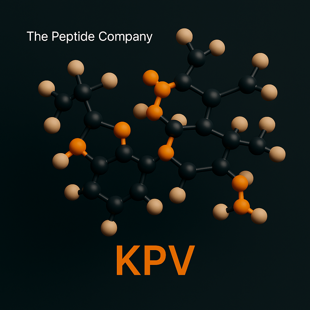
Inflammation is the body’s first response to damage or stress — an essential defense process that, when unresolved, becomes the foundation for chronic disease and tissue degeneration. The search for molecules that can resolve inflammation without suppressing immunity has led researchers to a naturally occurring tripeptide fragment known as KPV.KPV, short for Lysine-Proline-Valine, is a minimal yet powerful sequence derived from the larger melanocortin hormone α-melanocyte-stimulating hormone (α-MSH). Despite its simplicity, KPV demonstrates remarkable anti-inflammatory, wound-healing, and epithelial-protective activity across multiple biological systems.Following the immune-balancing precision of Thymosin Alpha-1, KPV represents the next logical step — focusing on inflammatory control and repair at the tissue interface.
What Is KPV?
KPV is a bioactive tripeptide naturally released during the breakdown of α-MSH, a hormone best known for its roles in pigmentation and immune modulation. Researchers first isolated the KPV fragment while studying α-MSH’s anti-inflammatory effects and discovered that this tiny segment alone retains potent biological activity.Unlike larger peptides, KPV’s short length provides:• High stability under physiological conditions• Strong receptor affinity for melanocortin-1 (MC1R)• Minimal immunogenicity and easy diffusion across epithelial barriersIt functions as a localized anti-inflammatory messenger, particularly within the skin, gastrointestinal tract, and mucosal linings.
Mechanism of Action
KPV’s activity is mediated primarily through the melanocortin-1 receptor (MC1R), a G-protein-coupled receptor expressed on keratinocytes, macrophages, and epithelial cells.Its mechanisms include:• Downregulation of pro-inflammatory cytokines such as IL-1β, TNF-α, and IL-6• Inhibition of NF-κB transcriptional activity, preventing chronic inflammatory gene expression• Upregulation of anti-inflammatory mediators including IL-10• Promotion of epithelial repair, cell migration, and collagen deposition• Stabilization of tight-junction proteins in gut and skin barriersIn contrast to corticosteroids or immunosuppressants, KPV doesn’t blunt the immune system. It promotes a resolution phase of inflammation — encouraging balance rather than suppression.
Research Highlights
1. Skin Barrier Restoration
Studies in dermatologic and wound-healing models demonstrate that topical or localized KPV reduces redness, edema, and pro-inflammatory cytokine expression, enhances keratinocyte proliferation and collagen synthesis, and accelerates wound closure and tissue remodeling. These findings have made KPV a molecule of interest in burn recovery, psoriasis, and cosmetic wound-healing research.
2. Gut Mucosa and Inflammatory Bowel Models
In gastrointestinal research, KPV has shown potential to reduce colonic inflammation in ulcerative colitis models, decrease infiltration of neutrophils and macrophages, and preserve intestinal barrier integrity through tight-junction reinforcement.
3. Systemic Anti-Inflammatory Potential
Pre-clinical work indicates that KPV can act beyond local tissues: reducing systemic inflammation markers in endotoxin-induced models and supporting metabolic and immune recovery under oxidative stress.
Cellular Pathways Overview
| Function | Primary Pathway | Observed Research Effect |
| Cytokine Suppression | NF-κB inhibition | Reduction in IL-1β, IL-6, TNF-α |
| Receptor Activation | MC1R signaling | Promotes anti-inflammatory gene expression |
| Barrier Protection | Tight-junction stabilization | Restores epithelial integrity |
| Wound Repair | MAPK / PI3K pathways | Accelerates healing and tissue regeneration |
| Oxidative Defense | Nrf2 activation (indirect) | Enhances redox balance |
Synergy and Comparison
KPV functions well as part of a multi-pathway research model for immune and tissue optimization.Conceptual pairings include:• GHK-Cu — synergistic effects on collagen repair and tissue remodeling• LL-37 — complementary antimicrobial and immune-balancing activity• Thymosin Alpha-1 — higher-order immune recalibration paired with inflammation resolution• Glutathione — redox and detoxification support during inflammatory recovery
Research Use and Safety
In the literature, KPV has been evaluated both topically and systemically. Typical concentrations in experimental models range from 100 µg to several milligrams per administration, depending on the study design.Across publications, no significant adverse events have been reported at research-level concentrations. However, KPV remains a research-only molecule and is not approved for clinical or consumer use.All mentions of KPV in this article are for educational and in-vitro research discussion only.
Summary: Precision Anti-Inflammation
KPV represents a new category of anti-inflammatory regulation — one that works with the immune system rather than against it. It demonstrates how targeted peptide fragments can deliver complex biological effects using elegant simplicity: three amino acids capable of redirecting inflammatory pathways, protecting tissues, and accelerating recovery.As research evolves, KPV continues to embody a central principle of modern bioactive design — resolve, don’t suppress.
Key Concepts
• Tripeptide fragment of α-MSH with independent biological activity• Modulates inflammation through MC1R and NF-κB signaling• Promotes epithelial repair and barrier stability• Serves as a bridge between immune modulation and regeneration research
References (Selection)
1. Brzoska T, et al. J Invest Dermatol. (2008).2. Catania A, et al. Ann N Y Acad Sci. (2010).3. Getting SJ, et al. Br J Pharmacol. (2001).4. Zhao H, et al. Peptides. (2021).5. Wang L, et al. J Inflamm Res. (2022).
Educational & Research Disclaimer
This content is for educational and research purposes only. No medical advice or product claims are implied. All compounds discussed are not approved for human or clinical use and are intended for in-vitro laboratory research only.
——————————
Selected References
- PMID: 19765659 — α-MSH peptide signaling and MC1R modulation
- PMID: 19406120 — Anti-inflammatory actions of melanocortin peptides
- PMID: 16239978 — NF-κB suppression via melanocortin pathways
- PMID: 21610182 — Peptide-induced epithelial repair and barrier protection
- Frontiers in Immunology — Melanocortin system and inflammation resolution
- Journal of Peptide Science — Bioactive tripeptides and immunomodulation
FAQ:
What is KPV?
KPV is a tripeptide fragment of α-MSH studied for its anti-inflammatory and tissue-protective properties. It is used exclusively in research settings.
How does KPV work in research?
KPV modulates MC1R and NF-κB signaling pathways, which help regulate inflammation and support epithelial repair in experimental models.
Is KPV approved for human use?
No. KPV is a research-only molecule and is not approved for clinical or consumer use.
What are researchers studying KPV for?
Current research includes inflammation control, epithelial barrier support, tissue protection, and immune modulation.
How is KPV typically studied?
Studies evaluate KPV using in-vitro assays or experimental models to examine its effects on inflammatory pathway responses.
Does KPV have known side effects?
Published research reports minimal adverse effects at research-level concentrations, but no clinical safety data exists.
Related Research Compounds
Thymosin Alpha-1 (Tα1): Immune Resilience and the Science of Thymic Restoration
LL-37: The Antimicrobial Peptide and Innate Immunity Blueprint
KPV Capsules 250mcg (60 ct)
KPV 250mcg capsules are a research compound studied for anti-inflammatory signaling, NF-κB pathway modulation, and epithelial barrier integrity research. For research use only.
A New Class of Bioactive Molecules
Mitochondria are traditionally described as the “powerhouses” of the cell, responsible for ATP production and energy balance. In recent years, a new perspective has emerged: mitochondria also behave as endocrine signaling hubs, releasing peptides that regulate systemic metabolism, stress responses, and cellular adaptation.
Among these peptides, MOTS-c has generated significant scientific interest. Unlike most bioactive peptides encoded in the cell nucleus, MOTS-c is encoded directly within mitochondrial DNA (mtDNA) — a rare feature that places it at the intersection of metabolism, exercise physiology, and cellular resilience.
MOTS-c connects longevity science, metabolic regulation, and mitochondrial stress response research — making it the ideal entry point into the “Mitochondrial & Metabolic Optimization” phase of your content series.
What Is MOTS-c?
MOTS-c (Mitochondrial Open Reading Frame of the 12S rRNA type-c) is a 16-amino-acid peptide encoded by the mitochondrial genome. First identified in 2015, MOTS-c challenges the traditional belief that peptide-coding sequences exist only in the nucleus.
MOTS-c has been observed to:
• Enhance metabolic flexibility
• Increase glucose utilization
• Improve insulin sensitivity
• Activate stress-response pathways
• Support mitochondrial homeostasis
Because mitochondria respond dynamically to nutrients, exercise, and oxidative stress, MOTS-c is increasingly viewed as a key coordinator of adaptive metabolism.
Mechanism of Action
MOTS-c regulates nutrient sensing and cellular stress pathways through several core mechanisms.
1. AMPK Activation
MOTS-c activates AMPK, the body’s primary energy sensor. This leads to:
• Increased glucose uptake
• Enhanced fatty acid oxidation
• Improved insulin sensitivity
• Suppressed energy-consuming anabolic pathways
2. Exercise-Mimetic Effects
MOTS-c is released during physical stress and enhances:
• Muscular glucose transport
• Endurance capacity
• Stress tolerance
3. Nuclear Translocation
Under metabolic stress, MOTS-c can translocate into the nucleus and regulate gene transcription related to:
• Stress resistance
• Antioxidant defense
• Metabolic efficiency
4. Folate & Methionine Pathways
MOTS-c influences one-carbon metabolism and redox balance — essential for DNA repair, methylation, and detoxification.
Research Highlights
1. Metabolic Health
MOTS-c enhances glucose uptake, increases insulin sensitivity, and improves metabolic flexibility.
2. Weight Regulation
Supports fatty acid oxidation and counters metabolic slowdown during caloric surplus.
3. Exercise Performance
Improves endurance, fatigue resistance, and mitochondrial biogenesis signals in research models.
4. Stress Resilience
Activates antioxidant genes and stress-response pathways through AMPK and nuclear signaling.
5. Age-Related Decline
MOTS-c levels decrease with age; restoring them in research settings improves metabolic and physical performance.
Synergistic Combinations (Research Context)
MOTS-c integrates naturally with other metabolic or mitochondrial research molecules:
• 5-Amino-1MQ — complements MOTS-c by enhancing NAD+ availability
• SS-31 — supports mitochondrial membrane stability
• NAD+ precursors — synergize with AMPK and redox pathways
• Glutathione — supports antioxidant demands during mitochondrial output
These molecules form the basis of the “Mitochondrial Optimization” research cluster.
Research Use and Safety
MOTS-c has been evaluated in cellular, animal, and metabolic studies.
Key points:
• No significant toxicity at research levels
• Dose-dependent metabolic effects
• Increasingly studied for metabolic syndrome and aging research
• Not approved for medical, clinical, or consumer use
All discussions refer strictly to research-only contexts.
Summary
MOTS-c represents a new era of mitochondrial biology and metabolic regulation. Its ability to improve metabolic flexibility, enhance stress responses, and support cellular energy balance makes it central to longevity and performance research.
As interest in mitochondrial peptides expands, MOTS-c stands out as a key regulator of cellular adaptation and resilience.
References (Selection)
1. Lee C, et al. Cell Metabolism. (2015).
2. Reynolds JC, et al. Aging Cell. (2021).
3. Zarse K, et al. Aging. (2019).
4. Kim KH, et al. Nat Commun. (2018).
5. Cobb LJ, et al. J Cachexia Sarcopenia Muscle. (2016).
Educational & Research Disclaimer
This content is for educational and research purposes only. No medical advice or product claims are implied. Compounds discussed are not approved for human or clinical use and are intended for in-vitro laboratory research only.
————————
FAQ:
What is MOTS-c in research?
MOTS-c is a mitochondrial-encoded peptide studied for its roles in metabolic regulation, stress adaptation, and cellular resilience in preclinical models.
What pathways does MOTS-c influence in studies?
Research suggests MOTS-c interacts with AMPK activation, mitochondrial signaling, and cellular energy regulation, especially under metabolic or oxidative stress.
Is MOTS-c considered a therapeutic compound?
No. MOTS-c is a research molecule used exclusively in laboratory and in-vitro models. It is not approved for medical, dietary, or clinical use.
How is MOTS-c typically used in research environments?
Researchers investigate MOTS-c in cell assays and animal models to study energy balance, metabolic signaling, and stress responses. All use must follow lab protocols.
Does MOTS-c impact exercise or performance in research studies?
Some preclinical studies explore MOTS-c’s effect on exercise tolerance and metabolic flexibility, but these findings are experimental and not applicable to human use.
How should MOTS-c be stored in a laboratory?
Labs typically store MOTS-c lyophilized in a cool, dry environment protected from light, following their institutional handling and stability procedures.
Is MOTS-c safe for human consumption or self-administration?
No. MOTS-c sold by The Peptide Company is for laboratory and in-vitro use only. It is not for human use, self-administration, or clinical application.
Selected References
PMID: 26562337 — Mitochondrial-encoded peptides and metabolic regulation
PMID: 25738465 — MOTS-c activation of AMPK and cellular energy pathways
PMID: 31447076 — Exercise-induced MOTS-c and metabolic homeostasis
PMID: 33414491 — Mitochondrial-derived peptides in stress adaptation
PMID: 35408335 — MOTS-c signaling in metabolic flexibility and resilience
Frontiers in Endocrinology — Mitochondrial peptides in energy regulation
Nature Communications — MOTS-c and nuclear signaling under metabolic stress
Related Research Compounds:
NMN: NAD⁺ Precursor Biology, Cellular Metabolism, and Mitochondrial Research
A New Direction in Cellular Energy Control
Cellular metabolism is shaped by a network of nutrient-sensing pathways that regulate energy production, fat storage, and mitochondrial efficiency. While much attention has focused on AMPK activation, NAD⁺ metabolism, and mitochondrial peptides, another key regulatory node is emerging: nicotinamide N-methyltransferase (NNMT).
NNMT is an enzyme involved in methylation balance and NAD⁺ availability. When NNMT becomes overactive, it disrupts metabolic flexibility and contributes to weight gain, insulin resistance, and reduced cellular energy.
5-Amino-1MQ is a small-molecule research compound studied for its ability to inhibit NNMT, rebalance metabolic signaling, and support healthier NAD⁺ dynamics. As part of the “Mitochondrial & Metabolic Optimization” series, it follows naturally after MOTS-c by highlighting how metabolism can be regulated through intracellular enzymatic control.
What Is 5-Amino-1MQ?
5-Amino-1MQ (5-amino-1-methylquinolinium) is a potent NNMT inhibitor evaluated in metabolic, obesity, and cellular energy research. Unlike peptide-based molecules, 1MQ is a small organic compound with high cellular penetration.
NNMT plays a role in:
• methylation balance
• NAD⁺ recycling
• lipid storage
• metabolic rate
• insulin sensitivity
When NNMT is elevated, it diverts methyl groups, alters nicotinamide availability, and disrupts metabolic processes. 1MQ’s purpose in research is to rebalance this pathway.
Mechanism of Action
1. NNMT Inhibition
NNMT converts nicotinamide into 1-methylnicotinamide (MNA), which cannot be recycled into NAD⁺. When NNMT is active:
• NAD⁺ levels fall
• Lipid accumulation increases
• Methylation imbalance occurs
5-Amino-1MQ blocks this process, resulting in:
• Increased nicotinamide
• Greater NAD⁺ synthesis potential
• Improved oxidative metabolism
2. NAD⁺ Regulation
By preventing NAD⁺ depletion, 1MQ supports:
• sirtuin activity
• mitochondrial function
• DNA repair signaling
• redox balance
3. Enhanced Metabolic Efficiency
Observed effects include:
• reduced adipocyte size
• decreased lipid storage
• improved glucose tolerance
4. Methylation Balance
NNMT consumes methyl donors. Inhibiting NNMT improves SAM availability and stabilizes gene expression patterns involved in metabolism.
Research Highlights
1. Fat Mass Reduction
5-Amino-1MQ reduces adipocyte size and systemic fat accumulation in research models.
2. Improved Glucose Handling
Demonstrates enhanced insulin sensitivity and glucose uptake.
3. Increased NAD⁺ Availability
Supports metabolic efficiency, mitochondrial function, and redox balance.
4. Anti-Obesity Effects
Helps regulate lipid storage and increases energy expenditure.
Synergistic Combinations (Research Context)
• MOTS-c — complements AMPK activation and metabolic adaptation
• SS-31 — supports mitochondrial membrane health
• NAD⁺ precursors — synergize with 1MQ’s nicotinamide preservation
• Glutathione — supports redox balance under increased metabolic output
Research Use and Safety
5-Amino-1MQ has been evaluated in metabolic and cellular research models.
Key points:
• No significant toxicity reported at research levels
• Metabolic benefits are dose-dependent
• Human studies remain limited
• Not approved for medical or consumer use
All mentions refer strictly to research-only contexts.
Summary
5-Amino-1MQ represents a promising direction in metabolic regulation by targeting NNMT, a key enzyme that influences NAD⁺ availability, methylation balance, and lipid metabolism.
Through NNMT inhibition and NAD⁺ preservation, 1MQ supports improved metabolic efficiency, reduced fat accumulation, and enhanced cellular energy expenditure.
References (Selection)
1. Kraus D, et al. Nature Medicine. (2014).
2. Ulanovskaya OA, et al. Nature Chemical Biology. (2013).
3. Stromsdorfer KL, et al. J Biol Chem. (2016).
4. Kannt A, et al. Drug Discovery Today. (2021).
Educational & Research Disclaimer
This content is for educational and research purposes only. No medical advice or product claims are implied. Compounds discussed are not approved for human or clinical use and are intended for in-vitro laboratory research only.
———————-
FAQ:
What is 5-Amino-1MQ in research?
5-Amino-1MQ is a small-molecule NNMT inhibitor studied for its effects on cellular metabolism, NAD+ regulation, and energy efficiency in preclinical models.
How does 5-Amino-1MQ work in laboratory studies?
Research indicates that 5-Amino-1MQ inhibits nicotinamide N-methyltransferase (NNMT), a metabolic enzyme that influences NAD+ turnover, energy expenditure, and methylation balance in cells.
Is 5-Amino-1MQ intended for human or medical use?
No. 5-Amino-1MQ from The Peptide Company is intended exclusively for laboratory and in-vitro research. It is notapproved for human consumption, supplementation, or therapeutic use.
What research applications involve 5-Amino-1MQ?
5-Amino-1MQ is studied in metabolic models to explore weight regulation, adipocyte energy mobilization, and NAD+-dependent cellular pathways. All investigations remain preclinical.
Does 5-Amino-1MQ affect NAD+ levels in studies?
Many studies examine how NNMT inhibition may modulate intracellular NAD+ availability and downstream signaling, but these findings are experimental and not connected to medical claims.
How is 5-Amino-1MQ handled in research labs?
Researchers typically store and handle the compound under standard small-molecule laboratory protocols, including dry, cool conditions and protection from light.
Can 5-Amino-1MQ be self-administered?
No. Research compounds on this site are not intended for any form of self-administration. All work must be conducted by qualified personnel in controlled research settings.
Related Research Compounds:
GLP-1 Pathway Peptides: Comparative Research on Semaglutide, Tirzepatide & Retatrutide
Frag 176–191: Growth Hormone–Derived Fragment and Lipolytic Research Mechanisms
NMN: NAD⁺ Precursor Biology, Cellular Metabolism, and Mitochondrial Research
References
PMID: 25555209 — NNMT regulation and metabolic energy balance
PMID: 29284151 — NNMT inhibition and adipocyte metabolism
PMID: 28087616 — NAD+ turnover and cellular signaling
PMID: 31727847 — Enzyme pathways involved in NAD+-dependent regulation
PMID: 33785733 — Small-molecule modulation of metabolic efficiency
Frontiers in Pharmacology — NNMT inhibitors and metabolic research
Journal of Biological Chemistry — NAD+ biosynthesis and enzymatic regulation

5-Amino-1MQ 50mg Capsules (60 ct)
5-Amino-1MQ 50mg capsules are a research compound studied for NNMT pathway modulation, metabolic efficiency signaling, and energy balance regulation. For research use only.
A New Frontier in Mitochondrial Medicine
Mitochondria regulate cellular energy, stress responses, redox balance, and metabolic flexibility. As these systems decline with age or metabolic stress, mitochondrial dysfunction becomes a driver of fatigue, impaired tissue repair, and systemic metabolic decline.
SS-31, also known as Elamipretide or MTP-131, is a mitochondria-targeted tetrapeptide designed to selectively target the inner mitochondrial membrane and interact with cardiolipin. By stabilizing mitochondrial structure and reducing oxidative stress, SS-31 has become a leading research tool for studying mitochondrial restoration.
What Is SS-31?
SS-31 is a synthetic mitochondria-targeted tetrapeptide with the sequence D-Arg-Dmt-Lys-Phe-NH₂.
It can:
• Cross mitochondrial membranes
• Concentrate at the inner mitochondrial membrane
• Bind electrostatically to cardiolipin
• Support mitochondrial architecture and ATP production
Unlike general antioxidants, SS-31 modulates ROS production at the source while preserving physiological signaling.
Mechanism of Action
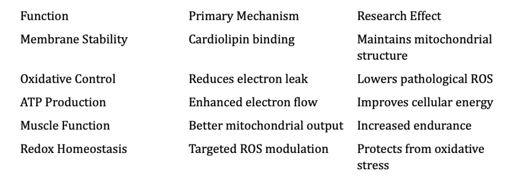
1. Cardiolipin Stabilization
SS-31 binds directly to cardiolipin, helping maintain membrane curvature, respiratory chain structure, and ATP synthesis efficiency.
2. Reduced ROS Production
By stabilizing membrane architecture, SS-31 reduces “electron leak” that generates excess ROS. This improves redox balance while maintaining physiological signaling.
3. Enhanced ATP Generation
With more efficient electron flow, SS-31 supports improved mitochondrial energy output and muscular performance.
4. Protection Against Mitochondrial Stress
SS-31 has been evaluated in models of ischemia-reperfusion injury, age-related decline, metabolic syndrome, and mitochondrial diseases.
Research Highlights
1. Muscle Function and Fatigue
Improves skeletal muscle endurance, mitochondrial respiration, and ATP turnover.
2. Cardiac Performance
Supports healthier mitochondrial morphology, reduces oxidative stress, and improves cardiac output in research settings.
3. Age-Related Mitochondrial Decline
Reverses cardiolipin oxidation, restores respiratory chain stability, and enhances endurance in aging models.
4. Mitochondrial Disorders
Improves mitochondrial structure and oxygen utilization in studies of mitochondrial pathology.
Cellular Pathways Overview
Synergistic Combinations (Research Context)
• MOTS-c — enhances mitochondrial signaling and AMPK activation
• 5-Amino-1MQ — improves NAD⁺ availability and metabolic pathways
• NAD⁺ precursors — support sirtuin activity and mitochondrial energy
• Glutathione — reinforces redox balance during mitochondrial output
Research Use and Safety
SS-31 has undergone extensive preclinical and early clinical evaluation.
Key points:
• Generally well-tolerated in research environments
• Effects are most pronounced under mitochondrial stress
• Not approved for clinical or consumer use
All descriptions refer strictly to laboratory and research-only contexts.
Summary
SS-31 represents a shift in mitochondrial science toward targeted membrane stabilization. By binding cardiolipin, reducing electron leak, and supporting ATP generation, SS-31 restores a fundamental layer of cellular energy production.
Its effects on muscle function, cardiac resilience, and age-related mitochondrial decline position SS-31 as a central molecule in metabolic and longevity research.
References (Selection)
1. Birk AV, et al. J Am Soc Nephrol. (2013).
2. Szeto HH. Pharmacology & Therapeutics. (2014).
3. Zhao K, et al. J Biol Chem. (2004).
4. Kloner RA, et al. Circulation. (2015).
5. Dai DF, et al. Aging Cell. (2017).
Educational & Research Disclaimer
This content is for educational and research purposes only. No medical advice or product claims are implied. Compounds discussed are not approved for human or clinical use and are intended for in-vitro laboratory research only.
———————–
FAQ:
What is SS-31 (Elamipretide) in research?
SS-31 is a mitochondria-targeted tetrapeptide studied for promoting mitochondrial protection, stabilizing cardiolipin, and supporting cellular energy output in preclinical models.
How does SS-31 interact with mitochondria in studies?
Research shows SS-31 localizes to the inner mitochondrial membrane, where it binds cardiolipin and may help reduce oxidative stress and improve electron transport efficiency.
Is SS-31 considered a therapeutic compound?
No. SS-31 from The Peptide Company is intended strictly for laboratory and in-vitro research use and is not approved for human, medical, or clinical use.
What research applications involve SS-31?
SS-31 is explored in cell and animal models examining mitochondrial dysfunction, bioenergetics, oxidative stress, membrane stability, and aging-related metabolic decline.
Does SS-31 improve ATP production in studies?
Some preclinical work suggests SS-31 may improve mitochondrial coupling efficiency and ATP output, though these findings remain research-only and non-medical.
How is SS-31 typically handled in the lab?
Researchers store SS-31 lyophilized in cool, light-protected conditions and reconstitute it following standard laboratory procedures and institutional guidelines.
Can SS-31 be self-administered?
No. SS-31 is not for human use, self-administration, or consumption of any kind. It is for controlled research environments only.
Related Research Compounds:
MOTS-c: The Mitochondrial-Encoded Peptide for Metabolic Regulation and Cellular Resilience
NMN: NAD⁺ Precursor Biology, Cellular Metabolism, and Mitochondrial Research
SS-31 10mg
SS-31 10mg is a research compound studied for mitochondrial targeting, bioenergetic efficiency, and cardiolipin stabilization pathway mechanisms. For research use only.
The Body’s Built-In Defense Signal
Long before adaptive immunity learns to recognize specific pathogens, the body relies on a fast, powerful system known as innate immunity. At the center of this system is a family of antimicrobial peptides — small molecules capable of destroying pathogens, guiding immune cell responses, and initiating tissue repair.Among these, LL-37 is the most extensively studied. Derived from the human cathelicidin precursor hCAP18, LL-37 is a 37-amino-acid peptide with broad-spectrum antimicrobial activity and potent immunomodulatory effects. It serves as both a first-response defender and a coordinator of tissue regeneration, making it one of the most versatile molecules in innate immune biology.As the next step in your “Regeneration & Immunity” series, LL-37 builds on the themes established by Thymosin Alpha-1 (immune calibration) and KPV (anti-inflammation and repair) — completing the triad of immune balance, inflammation control, and direct pathogen defense.
What Is LL-37?
LL-37 is the only known human cathelicidin-derived peptide, produced primarily by neutrophils, epithelial cells (skin, lungs, gut), macrophages, and dendritic cells.It becomes active when the precursor protein hCAP18 is cleaved into its functional form, LL-37. This peptide is present at infection sites, mucosal surfaces, and damaged tissues — essentially anywhere the body requires rapid protection.Its biological spectrum includes antibacterial activity, antiviral and antifungal effects, immune signaling, chemotaxis, and wound healing.
Mechanism of Action
LL-37 functions through a multi-layered set of biological mechanisms.1. Direct Antimicrobial Activity:LL-37 disrupts microbial membranes by binding to negatively charged lipid bilayers, forming pores or destabilizing membrane integrity, and leading to rapid pathogen lysis.2. Immune Cell Activation and Chemotaxis:It interacts with receptors such as FPR2, TLR2, TLR4, and P2X7, helping recruit immune cells and enhance antigen presentation.3. Modulation of Inflammation:LL-37 downregulates excessive cytokine release, balances NF-κB activity, and enhances appropriate acute inflammation.4. Wound Healing, Repair, and Angiogenesis:LL-37 promotes keratinocyte migration, fibroblast activity, collagen formation, and VEGF signaling — playing a significant role in tissue regeneration.
Research Highlights
1. Broad-Spectrum Antimicrobial Defense:LL-37 has demonstrated strong activity across bacterial, viral, and fungal pathogens, with rapid membrane disruption as its primary mechanism.2. Lung and Respiratory Immunity:In airway research, LL-37 supports mucosal defense, viral clearance, and controlled inflammation.3. Skin Regeneration:LL-37 accelerates epithelial closure, enhances angiogenesis, reduces biofilm formation, and supports regulated repair mechanisms.4. Antiviral and Immunomodulatory Behavior:LL-37 can disrupt viral envelopes, enhance interferon signaling, and strengthen innate antiviral defense.
Cellular Pathways Overview
| Function | Target Pathway | Research Effect |
| Antimicrobial Defense | Membrane permeabilization | Direct pathogen lysis |
| Immune Activation | FPR2, TLR2, TLR4 | Increased innate immune responsiveness |
| Chemotaxis | GPCR signaling | Recruitment of leukocytes |
| Tissue Repair | EGFR, MAPK, VEGF | Accelerated healing and angiogenesis |
| Inflammation Modulation | NF-κB regulation | Controlled cytokine output |
Synergy and Research Context
LL-37 integrates naturally into multi-pathway research models involving:• KPV — inflammation control and epithelial repair support• Thymosin Alpha-1 — immune modulation and balance• GHK-Cu — regenerative effects and collagen remodeling• Glutathione — oxidative balance during immune activation and tissue repairTogether, these molecules form a conceptual network around immune activation, inflammation resolution, and tissue regeneration.
Research Use and Safety
LL-37 has been studied across many biological systems, but its effects are highly concentration-dependent. Low to moderate concentrations support defense and repair, while high concentrations may induce excess inflammation or cytotoxicity in vitro.No significant toxicity has been observed in regulated research dosing ranges. LL-37 remains a research-only compound, not approved for human or consumer use.All mentions of LL-37 in this article are for educational and in-vitro research discussion only.
Summary
LL-37 represents one of nature’s most efficient biological designs — a molecule capable of killing pathogens, guiding immune cells, regulating inflammation, and accelerating tissue repair.As interest in host defense peptides grows, LL-37 stands out as a blueprint for next-generation antimicrobial and regenerative research.
References (Selection)
1. Sørensen OE, et al. Nat Rev Immunol. (2006).2. Vandamme D, et al. Cell Mol Life Sci. (2012).3. Dürr UHN, et al. Biochim Biophys Acta. (2006).4. Nell MJ, et al. J Leukoc Biol. (2006).5. Heilborn JD, et al. J Invest Dermatol. (2003).
Educational & Research Disclaimer
This content is for educational and research purposes only. No medical advice or product claims are implied. All compounds discussed are not approved for human or clinical use and are intended for in-vitro laboratory research only.
—————–
FAQ
What is LL-37 in research?
LL-37 is a human-derived antimicrobial peptide studied for its roles in innate immunity, host defense, epithelial barrier function, and pathogen response in preclinical models.
How does LL-37 function in laboratory studies?
Research shows LL-37 interacts with microbial membranes, modulates cytokine signaling, and influences immune cell activation, making it a key peptide in host-defense exploration.
Is LL-37 considered a therapeutic product?
No. LL-37 from The Peptide Company is provided strictly for laboratory and in-vitro research use. It is not a therapy, drug, supplement, or product for human use.
What research applications involve LL-37?
LL-37 is explored in models of infection defense, wound repair, microbiome regulation, epithelial integrity, inflammation modulation, and innate immune signaling.
Does LL-37 have antimicrobial activity in studies?
Yes. LL-37 has been shown in preclinical research to disrupt bacterial membranes and modulate pathogen-related immune responses. These findings are experimental only.
How is LL-37 typically stored in research settings?
Researchers store LL-37 lyophilized in cool, dry, stable environments away from light and reconstitute it under institutional laboratory protocols.
Can LL-37 be applied or administered by consumers?
No. LL-37 is not intended for any form of self-administration. It is for controlled laboratory and in-vitro research environments only.
Related Research Compounds:
Thymosin Alpha-1 (Tα1): Immune Resilience and the Science of Thymic Restoration
KPV: The Anti-Inflammatory Tripeptide and Cellular Repair Mechanism
References
PMID: 12711666 — Human cathelicidin LL-37 antimicrobial activity
PMID: 17034334 — LL-37 in innate immunity and host defense
PMID: 19348957 — Epithelial barrier modulation by LL-37
PMID: 32503546 — LL-37 and immune-cell signaling interactions
PMID: 25485019 — Antimicrobial peptides and pathogen membrane disruption
Frontiers in Immunology — Host-defense peptides in innate immune pathways
Nature Reviews Microbiology — Human antimicrobial peptide mechanisms
LL-37 – 5MG | High-Purity Research Compound
LL-37 is a synthetic antimicrobial peptide studied for its role in innate immune signaling, antimicrobial defense mechanisms, and inflammatory pathway modulation in research models. For research use only.
Short peptide bioregulators—also known as cytomedins—are ultrashort amino acid sequences that influence gene expression, chromatin dynamics, and intracellular signaling in specific tissues. Pancragen is associated with pancreatic endocrine and exocrine regulatory pathways, making it a research tool for studying transcriptional regulation and cellular homeostasis in pancreatic tissue models.
What Are Short Peptide Bioregulators?
Short peptide bioregulators consist of 2–4 amino acids and are derived from highly conserved regulatory motifs found within tissue-specific proteins. Their ultrashort structure enables cellular and nuclear penetration, where they may interact with DNA-binding proteins, transcription factors, and intracellular signaling complexes. They function differently from classical receptor-binding peptides, operating primarily within the cytoplasm and nucleus.
What Is Pancragen?
Pancragen is a pancreatic-targeting short peptide bioregulator studied in models involving endocrine β-cells and exocrine acinar cells. Research examines its relationship to gene expression normalization, protein synthesis pathways, cellular stress responses, and targeted transcriptional regulation in pancreatic tissue.
Structural Overview
Pancragen, like other bioregulators, consists of an ultrashort peptide sequence that mimics naturally occurring motifs found within pancreatic regulatory proteins. Its small molecular size enables it to diffuse across cell membranes and potentially interact with nuclear DNA-associated proteins, influencing transcriptional activity and RNA expression.
Mechanism of Action (Research Context)
Pancragen’s mechanisms are based on intracellular and nuclear regulatory interactions rather than receptor-mediated activity. Research models highlight potential roles in gene expression modulation, β-cell transcriptional pathways, exocrine enzyme regulation, chromatin-associated signaling, and peptide-protein interactions that support pancreatic tissue stability.
Pancragen in Pancreatic Tissue Research
Pancragen appears in studies examining β-cell identity markers, endocrine homeostasis pathways, protein synthesis regulation, stress-response genes, and tissue-specific gene-expression networks. Its role in pancreatic research centers around transcriptional support, metabolic stability, and cellular differentiation signals.
Summary
Pancragen is a pancreatic-specific short peptide bioregulator studied for its potential influence on gene expression, transcriptional regulation, and intracellular stability in pancreatic endocrine and exocrine models. Its ultrashort size and regulatory focus make it a unique research tool within the broader field of tissue-specific peptide biology.
Educational & Research Disclaimer
This article is for educational and scientific research purposes only. No therapeutic claims, clinical guidance, or usage recommendations are provided. Compounds referenced are not approved for human use and are intended solely for controlled laboratory research.
FAQ:
What is Pancragen in research?
Pancragen is a short peptide bioregulator studied for its potential influence on pancreatic cellular activity, gene expression, and endocrine-related pathways in controlled laboratory models. It is provided as a lyophilized research compound for in-vitro use only.
How does Pancragen function in laboratory studies?
Research suggests Pancragen may interact with nuclear structures and DNA, helping support gene regulation involved in pancreatic tissue health, metabolic signaling, and cellular repair processes in experimental settings.
Is Pancragen considered a therapeutic product?
No. Pancragen supplied by The Peptide Company is not a therapy, drug, supplement, or clinical product. It is intended for laboratory and in-vitro research environments only.
What research applications involve Pancragen?
Pancragen is explored in studies relating to pancreatic tissue preservation, age-related cellular changes, metabolic pathways, endocrine function, and gene-regulation activity under controlled experimental conditions.
Does Pancragen have biological activity in studies?
In preclinical literature, Pancragen has been examined for potential influences on cellular repair markers, metabolic gene activity, and pancreatic tissue homeostasis. These findings are experimental only and not indicative of clinical outcomes.
How is Pancragen typically stored in research settings?
Pancragen is generally stored as a dry, stable powder away from heat and light. Once reconstituted, it is kept refrigerated as required by laboratory protocol and used only within controlled in-vitro research workflows.
Can Pancragen be self-administered or used by consumers?
No. Pancragen is not intended for self-administration or consumer use. It is strictly for institutional research, academic experimentation, and in-vitro laboratory applications only.
Related Research Compounds
Bronchogen: Short Peptide Bioregulator for Bronchial and Pulmonary Tissue Research
Cardiogen: Short Peptide Bioregulator for Cardiac and Myocardial Tissue Research
ProstaMax : Short Peptide Bioregulator for Prostate Tissue Regulatory Research
References (Selection)
PMID: 2469555 — Peptide bioregulators and regulation of pancreatic cellular functions
PMID: 25519238 — Gene-expression modulation in bioregulatory peptide models
PMID: 20370450 — Short peptides and nuclear interactions in metabolic tissues
PMID: 21504972 — Peptide-induced regulation of age-related cellular changes
PMID: 29264985 — Experimental peptide signaling in endocrine and metabolic pathways

Introduction
Frag 176–191 is a synthetic peptide representing the C-terminal region of the human growth hormone (GH) molecule. It does not activate the GH receptor and does not stimulate IGF-1 production. Instead, it exhibits fragment-specific behavior in adipocyte research models, particularly involving lipolytic and metabolic signaling pathways. This article examines Frag 176–191’s structural basis, receptor-independent activity, and relevance in GH-fragment research.
What Is Frag 176–191?
Frag 176–191 is a 15–amino-acid peptide derived from the lipolytic region of GH. It excludes domains required for GH receptor binding, making it useful for studying GH fragment–specific activity without engaging the GH → IGF-1 endocrine axis. It is frequently used in adipocyte and metabolic research models to explore lipid mobilization and energy regulation.
Structural Overview
Full-length human GH is 191 amino acids long. The region spanning residues 176–191 has been identified as central to GH’s lipolytic signaling. Frag 176–191 isolates this region, eliminating all GHR-binding components. Modified versions of this fragment often incorporate substitutions that increase stability and protect against rapid enzymatic degradation.
Mechanism of Action (Research Context)
Frag 176–191 is notable for its non–GH receptor-mediated behavior. It influences adipocyte metabolic pathways including cAMP accumulation, PKA activation, and downstream regulation of hormone-sensitive lipase (HSL) and adipose triglyceride lipase (ATGL). Studies also show interactions with AMPK signaling and mitochondrial fatty acid oxidation pathways in metabolic models.
Frag 176–191 vs Full-Length GH (Mechanistic Only)
The fragment’s short structure and absence of GH receptor binding separate its behavior from the classical GH → IGF-1 endocrine axis. While full-length GH activates JAK2/STAT5 and IGF-1 production, Frag 176–191 operates through cAMP–PKA and lipolytic enzyme regulation. This makes it a valuable tool in studies that require GH-related lipolysis without systemic anabolic signaling.
Cellular Pathways Associated With Frag 176–191
Key pathways associated with Frag 176–191 include:• cAMP → PKA activation • Regulation of HSL and ATGL • AMPK pathway involvement • β-oxidation and mitochondrial signaling • Suppression of lipogenic pathways These pathways define the fragment’s relevance in adipocyte metabolic research models.
Research Applications
Frag 176–191 is used for studying GH fragment–specific lipolysis, adipocyte metabolism, endocrine-independent GH motif signaling, lipid mobilization, and metabolic regulation circuits. It is frequently referenced alongside AOD‑9604, Tesamorelin, CJC‑1295, and GLP-1 pathway research in comparative metabolic studies.
Summary
Frag 176–191 isolates the lipolytic region of the GH protein while removing GH receptor signaling and IGF-1 axis involvement. Its receptor-independent metabolic behavior makes it suitable for studying adipocyte activity, lipid breakdown mechanisms, and GH fragment functional specificity in controlled research environments.
Frag 176–191 vs Full-Length GH (Research Comparison)
| Property | Full-Length GH | Frag 176–191 |
| Length | 191 amino acids | 15 amino acids |
| Receptor Binding | Activates GH receptor | Does not activate GH receptor |
| IGF-1 Axis Activation | Yes | None |
| Mechanism | JAK2–STAT5 endocrine pathway | cAMP–PKA, HSL/ATGL pathways |
| Research Use | Endocrine, metabolic, anabolic studies | Lipolysis, adipocyte-specific models |
FAQ:
What is Frag 176–191 in research?
Frag 176–191 is a laboratory peptide fragment derived from the C-terminal region of growth hormone. It is studied for its role in experimental models of lipolysis regulation, energy utilization, and metabolic signaling pathways.
How does Frag 176–191 function in laboratory studies?
In preclinical research, Frag 176–191 has been shown to influence pathways associated with fat-cell metabolism and lipid mobilization without activating traditional growth hormone–related receptors. These findings remain experimental only.
Is Frag 176–191 considered a therapeutic compound?
No. Frag 176–191 provided by The Peptide Company is for laboratory and in-vitro research use only. It is not a therapy, drug, supplement, or product for human or clinical use.
What research applications involve Frag 176–191?
Researchers explore Frag 176–191 in controlled studies related to lipid turnover, fat-cell signaling, metabolic pathways, and mitochondrial energy mechanisms.
Does Frag 176–191 show lipolytic activity in studies?
Preclinical data suggest Frag 176–191 may influence markers of lipolysis and adipocyte activity in experimental systems. These observations apply only to controlled research environments.
How is Frag 176–191 typically handled in laboratory settings?
It is supplied as a lyophilized powder and is generally stored away from heat, moisture, and light. After reconstitution, it is kept refrigerated according to laboratory protocol.
Can Frag 176–191 be administered or used by consumers?
No. Frag 176–191 is not intended for self-administration or consumer use. It is exclusively for institutional lab research and in-vitro experimentation.
Related Research Compounds
GLP-1 Pathway Peptides: Comparative Research on Semaglutide, Tirzepatide & Retatrutide
References
PMID: 10872804 — Growth hormone fragment research and metabolic activity
PMID: 17185307 — Lipolytic pathways influenced by GH-derived fragments
PMID: 16597689 — Mechanisms of adipocyte regulation in peptide studies
PMID: 17579216 — Experimental metabolic signaling induced by peptide derivatives
PMID: 19041303 — C-terminal GH fragments and lipid-cell pathway interactions
Introduction
Short peptide bioregulators—also known as cytomedins—are ultrashort amino acid sequences studied for their role in regulating gene expression, cellular differentiation, chromatin structure, and intracellular signaling. Bronchogen is associated with bronchial and pulmonary epithelial research, with studies examining its effects on epithelial integrity, mucosal barrier dynamics, and cytokine expression.
What Are Short Peptide Bioregulators?
Bioregulators consist of 2–4 amino acids and are derived from naturally occurring regulatory proteins. Their small size allows them to enter cells and interact with nuclear and cytoplasmic targets. Research indicates their involvement in transcriptional regulation, intracellular signaling pathways, and tissue-specific gene expression.
What Is Bronchogen?
Bronchogen is a short peptide bioregulator linked to bronchial and pulmonary epithelial cell function. Studies explore its influence on epithelial gene expression, mucosal barrier characteristics, cytokine normalization, and bronchial tissue homeostasis in research models.
Structural Overview
Short peptide bioregulators typically contain 2–4 amino acids and mimic conserved intracellular motifs found in tissue-regulatory proteins. Bronchogen’s sequence corresponds to motifs associated with epithelial maintenance pathways. Its size facilitates access to the cytoplasm and nucleus, allowing potential interactions with DNA-associated proteins.
Mechanism of Action (Research Context)
Bronchogen influences nuclear and cytoplasmic regulatory pathways related to bronchial epithelial function. Research models note its relationship with gene expression modulation, tight-junction protein dynamics, cytokine-expression profiles, and tissue-specific differentiation pathways.
Bronchogen in Respiratory Tissue Research
Bronchogen appears in studies involving epithelial barrier maintenance, mucosal defense modeling, bronchial cellular stress responses, and tissue-specific genomic regulation. Researchers use it to explore respiratory epithelial behavior under various stressors and environmental stimuli.
Summary
Bronchogen is a short peptide bioregulator studied for its relationship with bronchial epithelial regulation. Its ultrashort structure, intracellular penetration, and influence on nuclear pathways make it valuable for exploring epithelial gene expression, mucosal barrier function, and airway tissue homeostasis in research environments.
FAQ:
What is Bronchogen in research?
Bronchogen is a short peptide bioregulator (sequence Ala-Glu-Asp-Leu; AEDL) primarily studied for its effects on bronchial and pulmonary cell activity, gene regulation, and tissue signaling in laboratory models.
How does Bronchogen function in laboratory studies?
Experimental work suggests Bronchogen may interact with DNA, stabilize chromatin structure, influence gene expression in bronchial epithelial cells, and modulate inflammatory pathways in lung-tissue models.
Is Bronchogen considered a therapeutic product?
No. Bronchogen provided by The Peptide Company is strictly for laboratory and in-vitro use. It is not a therapy, drug, supplement, or product for human or clinical use.
What research applications involve Bronchogen?
Bronchogen is explored in controlled experimental settings involving bronchial epithelium regeneration, pulmonary tissue signaling, age-related decline in lung tissue, and inflammatory cell modulation.
Does Bronchogen influence lung-tissue inflammation or structure in studies?
Preclinical data (in animal models) suggest that Bronchogen may reduce neutrophilic inflammation in bronchial lavage fluid, support epithelial integrity, and increase markers such as surfactant protein B in pulmonary models.
How is Bronchogen typically handled in research settings?
It is supplied as a lyophilized powder, stored away from heat and light, and after reconstitution is refrigerated. Usage is limited to controlled research environments, not human administration.
Can Bronchogen be used by consumers or self-administered?
No. Bronchogen is for institutional lab research and in-vitro modeling only, not for consumer use or self-administration.
Related Research Compounds:
Cardiogen: Short Peptide Bioregulator for Cardiac and Myocardial Tissue Research
ProstaMax : Short Peptide Bioregulator for Prostate Tissue Regulatory Research
PMID: 30199201 — Peptide modulation of lung inflammation and epithelial function
PMID: 21597687 — Nuclear penetration and DNA interaction of short regulatory peptides
PMID: 22098268 — Chromatin stabilization influenced by peptide AEDL sequences
PMID: 25828794 — Peptide-based modulation of bronchial epithelial cell activity
PMID: 34202042 — Peptide regulation of gene expression and cellular signaling
Introduction
Sermorelin is a 29-amino-acid fragment of endogenous GHRH used in research to study growth hormone (GH) pulsatility, endocrine timing, and receptor-specific signaling. It preserves the natural binding region of GHRH and produces a short-lived, physiologically aligned GHRH signal.
What Is Sermorelin?
Sermorelin consists of the first 29 amino acids of human GHRH. This segment contains the full receptor-binding region necessary to activate the GHRH receptor (GHRHR) on pituitary somatotroph cells, producing a controlled GH pulse in research models.
Structural Overview
Endogenous GHRH has 44 amino acids. The 1-29 region contains the biologically active binding sequence. Sermorelin isolates this region, allowing researchers to study receptor-specific activation with rapid degradation similar to native GHRH.
Mechanism of Action
Sermorelin binds the GHRH receptor, activating Gs protein signaling, increasing cAMP, and activating PKA. This triggers calcium influx, GH vesicle exocytosis, and downstream IGF-1 pathway activation. Because feedback loops remain intact, GH pulses remain physiologically regulated.
Research Applications
Sermorelin is used to study GH pulse patterns, GHRH-somatostatin interactions, IGF-1 downstream signaling, circadian timing, and comparative analog studies. It serves as the baseline reference molecule for other GHRH analogues such as CJC-1295 and Tesamorelin.
Context With Other GHRH Analogues
Sermorelin is often compared mechanistically with CJC-1295 (short and long-acting forms) and Tesamorelin to evaluate how structural length, stability, and receptor engagement influence GH pulsatility in controlled experiments.
Summary
Sermorelin provides a physiologic, short-duration GHRH signal ideal for studying GH pulsatility, feedback regulation, and endocrine timing. Its rapid degradation, receptor specificity, and intact feedback loops make it a valuable research tool for GH/IGF axis analysis.
Sermorelin vs Endogenous GHRH (Research Comparison)
| Property | Endogenous GHRH | Sermorelin (GHRH 1-29) |
| Length | 44 amino acids | 29 amino acids |
| Binding Region | N-terminal region | Identical N-terminal binding region |
| Stability | Longer due to additional sequence | Shorter, rapidly degraded |
| Signaling Style | Pulsatile | Pulsatile, research analogue |
FAQ:
What is Sermorelin in research?
Sermorelin is a synthetic fragment of the growth hormone-releasing hormone (GHRH) molecule studied in experimental models for its effects on stimulating growth hormone (GH) pulsatility, endocrine regulation, and neuroendocrine axis signaling. It is provided for laboratory research and in-vitro use only.
How does Sermorelin function in laboratory studies?
In research, Sermorelin interacts with GHRH receptors at the pituitary, triggering episodic GH release and supporting physiological pulsatile GH secretion rather than continuous GH levels. These findings remain experimental and apply to controlled study settings.
Is Sermorelin considered a therapeutic product?
No. The Sermorelin referenced here (by The Peptide Company) is for laboratory and in-vitro research use only. It is not a therapy, drug, supplement, or product for human or clinical use.
What research applications involve Sermorelin?
Researchers explore Sermorelin in controlled models of GH-axis regulation, aging-related somatotropic decline, GH-pulse amplitude and frequency dynamics, and endocrine/neuroendocrine signaling.
Does Sermorelin influence GH pulses in studies?
Yes — preclinical and clinical studies show Sermorelin can restore or enhance GH pulse amplitude, support rhythmic GH release, and influence downstream IGF-1 levels in controlled research settings.
How is Sermorelin typically handled in research settings?
As a lyophilized powder, Sermorelin should be stored away from light and moisture. When reconstituted, it should be refrigerated and used in institutional laboratory or in-vitro protocols only.
Can Sermorelin be used by consumers or self-administered?
No. Sermorelin is not intended for self-administration or consumer use. It is strictly reserved for institutional laboratory research or in-vitro experimentation.
Related Research Compounds
IGF-1 Analogues: LR3 and DES Structural Variations and Receptor Binding in Research Models
CJC-1295: GHRH Analog, DAC Conjugation, and Growth Hormone Pulsatility in Research
References (Selection)
PMID: 18031173 — Sermorelin: a review of its use in the diagnosis and regulation of GH secretion
PMID: 1688413 — Brain effects of growth hormone-releasing hormone (sermorelin) in older adults
PMID: 7108996 — Role of GHRH/ghrelin axis on growth hormone release and pulsatility
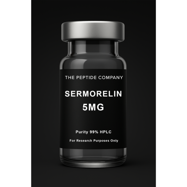
Sermorelin 5mg
Sermorelin 5mg is a research compound studied as a GHRH fragment for growth hormone pulsatility, pituitary axis modulation, and endocrine regulation. For research use only.