
Abstract & Overview
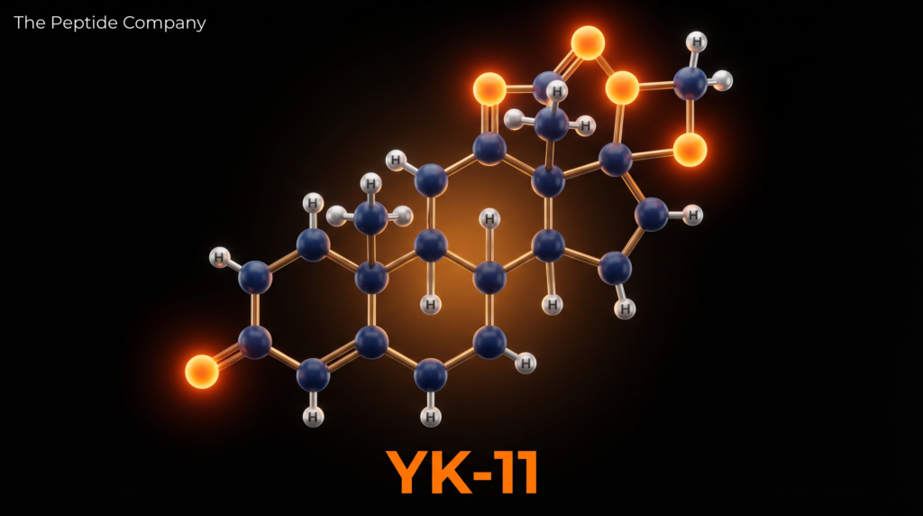
YK-11 — also designated Myostine and formally named (17α,20E)-17,20-[(1-methoxyethylidene)bis(oxy)]-3-oxo-19-norpregna-4,20-diene-21-carboxylic acid methyl ester — is a synthetic steroidal selective androgen receptor modulator (SARM) first characterised by Kanno et al. at Toho University, Japan, in 2011 [1]. Unlike the majority of SARMs, which are non-steroidal small molecules, YK-11 is built upon a modified 19-norpregnane steroid scaffold, placing it in a structurally distinct subclass of androgen receptor (AR) modulators. Its molecular formula is C₂₅H₃₄O₆ with a molar mass of 430.54 g/mol (CAS: 1370003-76-1; PubChem CID: 119058028).
YK-11 operates through a dual pharmacological mechanism that distinguishes it from both classical anabolic steroids and conventional non-steroidal SARMs. First, it functions as a gene-selective partial agonist of the androgen receptor, binding the receptor’s ligand-binding domain without inducing the N-terminal/C-terminal (N/C) interaction required for full AR transactivation, thereby activating a distinct subset of androgen-responsive genes [1][2]. Second, and uniquely, YK-11 induces the expression of follistatin (FST) in skeletal muscle cells — an effect not observed with dihydrotestosterone (DHT) — which in turn neutralises myostatin (GDF-8), the primary endogenous inhibitor of skeletal muscle mass [2]. This dual mechanism positions YK-11 as both a SARM and a functional myostatin inhibitor.
“YK11 is a selective androgen receptor modulator (SARM), which activates AR without the N/C interaction… YK11 treatment of C2C12 cells, but not DHT, induced the expression of follistatin (Fst), and the YK11-mediated myogenic differentiation was reversed by anti-Fst antibody. These results suggest that the induction of Fst is important for the anabolic effect of YK11.” — Kanno Y et al., Biol Pharm Bull (2013) [2].
Preclinical research has further demonstrated that YK-11 promotes osteoblastic proliferation and differentiation via Akt signalling [3], attenuates sepsis-induced muscle wasting and reduces mortality in animal models through suppression of the TLR4/NF-κB/TGF-β inflammatory cascade [4], and has been investigated for its effects on hippocampal function and oxidative stress [5][6]. YK-11 has not received regulatory approval for human use and is classified as a designer drug and research compound.
Molecular Identity and Structural Architecture
YK-11 is built upon a 19-norpregnane steroidal backbone — the same core scaffold found in progestins such as norethisterone — with several key structural modifications that confer its unique pharmacological profile. The most distinctive feature is the 17α,20-ketal group: a (1-methoxyethylidene)bis(oxy) moiety bridging positions 17 and 20 of the steroid nucleus. This ketal group is the primary determinant of YK-11’s selective AR binding profile, as it sterically prevents the receptor from adopting the conformation required for the N/C interaction. The molecule also bears a 3-oxo-4-ene configuration (a conjugated enone in ring A, common to androgenic steroids) and a methyl ester at C-21, which contributes to oral bioavailability and metabolic stability [1].
The steroidal nature of YK-11 is pharmacologically significant. Most SARMs in research use — including RAD-140, LGD-4033, and Ostarine — are non-steroidal compounds that bind the AR through entirely different chemical scaffolds. YK-11’s steroid scaffold allows it to interact with the AR in a manner that more closely resembles natural androgens, yet the 17α,20-ketal modification fundamentally alters the receptor’s conformational response, producing a gene-selective activation pattern distinct from both testosterone and DHT. The compound is orally bioavailable with an estimated half-life of approximately 6 to 8 hours, necessitating multiple daily administrations in research protocols [1][2].
Mechanistic Rationale: Dual Pathway Anabolic Activity
Androgen Receptor Partial Agonism and Gene-Selective Activation
Upon cellular uptake, YK-11 binds to the ligand-binding domain (LBD/AF2) of the androgen receptor with high affinity. In contrast to full AR agonists such as DHT and testosterone, YK-11 binding does not induce the physical interaction between the receptor’s N-terminal activation function 1 (NTD/AF1) and its ligand-binding domain activation function 2 (LBD/AF2) — a conformational event known as the N/C interaction that is required for maximal AR transactivation. The absence of this interaction means that YK-11 activates only a subset of androgen-responsive genes, producing a tissue-selective anabolic profile that theoretically spares androgenic side effects associated with full AR agonism [1].
Despite being a partial agonist, YK-11 demonstrated greater anabolic potency than DHT in C2C12 murine myoblast cells in vitro, as measured by the induction of key myogenic regulatory factors (MRFs). Specifically, YK-11 produced more robust upregulation of MyoD (myoblast determination protein 1), Myf5 (myogenic factor 5), and myogenin than equimolar concentrations of DHT, suggesting that the gene-selective activation pattern of YK-11 may be particularly well-suited to the myogenic differentiation programme [2].
Follistatin Induction and Myostatin Inhibition
The most pharmacologically distinctive feature of YK-11 is its capacity to induce follistatin (FST) expression in skeletal muscle cells — an effect that is entirely absent with DHT treatment. Follistatin is a secreted glycoprotein that functions as a high-affinity binding protein and functional antagonist for myostatin (growth differentiation factor 8, GDF-8) and activin, both members of the TGF-β superfamily. Myostatin is the primary endogenous brake on skeletal muscle hypertrophy: it signals through the ActRIIB/ALK4-5 receptor complex to activate Smad2/3 transcription factors, which suppress the expression of myogenic genes and promote muscle protein catabolism [2][4].
By inducing follistatin expression via AR activation, YK-11 effectively removes this myostatin-mediated inhibitory constraint on muscle growth. Follistatin binds myostatin with high affinity, preventing it from engaging its receptor complex and thereby disinhibiting the myogenic programme. The essential role of follistatin in YK-11’s anabolic mechanism was definitively demonstrated by Kanno et al. (2013): treatment of C2C12 myoblasts with an anti-follistatin antibody completely reversed YK-11-mediated myogenic differentiation, confirming that follistatin induction is not merely coincidental but is mechanistically required for YK-11’s anabolic effects [2]. This makes YK-11 the only known compound that achieves myostatin inhibition indirectly through AR-mediated follistatin transcription.
TLR4/NF-κB/TGF-β Pathway and Anti-Inflammatory Myoprotection
Lee et al. (2021) investigated YK-11 in a murine model of gram-negative bacterial sepsis, a condition characterised by severe muscle wasting driven by inflammatory cytokine cascades. In septic mice, myostatin protein levels were markedly elevated in skeletal muscle, accompanied by increases in NF-κB, p-FOXO3a, p-Smad2, myogenin, and MyoD — a pattern consistent with catabolic muscle remodelling under inflammatory stress. YK-11 treatment inhibited myostatin expression, which in turn suppressed the TLR4/NF-κB/TGF-β signalling cascade, reducing pro-inflammatory cytokine levels and organ damage markers in the bloodstream and major organs [4].
Critically, YK-11 treatment significantly decreased the mortality rate of septic mice, establishing a functional link between myostatin inhibition, inflammatory resolution, and survival outcomes. These findings suggest that YK-11’s myoprotective effects extend beyond anabolic signalling to encompass a broader anti-inflammatory mechanism, positioning it as a potential research tool for conditions characterised by inflammatory muscle wasting, including cachexia, sarcopenia, and critical illness myopathy [4].
Osteogenic Activity via Akt Signalling
Beyond skeletal muscle, YK-11 has demonstrated significant osteogenic activity in preclinical models. Yatsu et al. (2018) demonstrated that YK-11 treatment accelerated cell proliferation and mineralisation in MC3T3-E1 mouse osteoblast cells, with upregulation of osteoblast-specific differentiation markers including osteoprotegerin (OPG) and osteocalcin. These effects were attenuated by AR antagonist treatment, confirming AR-dependence. The mechanistic basis for YK-11’s osteogenic activity was identified as non-genomic AR signalling: YK-11 increased phosphorylated Akt (p-Akt) protein levels in osteoblasts, activating the PI3K/Akt pathway that is a key regulator of androgen-mediated osteoblast differentiation [3].
A 2024 in vivo study further confirmed YK-11’s osteogenic potential, demonstrating that it promoted the osteogenic differentiation of bone marrow-derived mesenchymal stem cells (BMSCs) and facilitated the repair of cranial bone defects in rats via AR activation. These findings suggest that YK-11’s anabolic effects may extend to the skeletal system, potentially offering research utility in models of osteoporosis, bone fracture healing, and androgen deficiency-related bone loss [3][7].
Research Applications and Experimental Evidence
Muscle Hypertrophy and Myogenic Differentiation Models
YK-11’s primary research application has been in the study of myogenic differentiation and skeletal muscle hypertrophy. The C2C12 murine myoblast cell line has served as the principal in vitro model system, with YK-11 consistently demonstrating superior induction of MRFs (MyoD, Myf5, myogenin) compared to DHT. The compound’s unique capacity to simultaneously activate AR and induce follistatin expression makes it a valuable tool for dissecting the relative contributions of direct AR signalling versus myostatin pathway disinhibition to anabolic outcomes in muscle research [1][2].
Muscle Wasting and Cachexia Models
The sepsis study by Lee et al. (2021) established YK-11 as a research tool for investigating inflammatory muscle wasting. Its dual capacity to inhibit myostatin and suppress NF-κB-driven inflammatory signalling makes it particularly relevant to cachexia research, where both anabolic resistance and systemic inflammation contribute to muscle loss. Future research directions may include investigation in cancer cachexia, HIV-associated wasting, and glucocorticoid-induced myopathy models [4].
Bone Biology and Osteoporosis Research
YK-11’s osteogenic activity via Akt signalling and its in vivo bone defect repair data position it as a research candidate for androgen-deficiency-related osteoporosis models. The compound’s ability to promote osteoblast proliferation, mineralisation, and expression of OPG (which inhibits osteoclastogenesis) suggests a dual anabolic/anti-resorptive bone profile that warrants further investigation in ovariectomised and orchidectomised animal models [3][7].
YK-11 vs. Other SARMs and Anabolic Agents: Comparative Profile
| Parameter | YK-11 | RAD-140 (Testolone) | LGD-4033 (Ligandrol) |
| Scaffold | Steroidal (19-norpregnane) | Non-steroidal | Non-steroidal |
| AR Mechanism | Partial agonist (no N/C) | Full/partial agonist | Full agonist |
| Myostatin Inhibition | Yes (via follistatin) | No | No |
| Osteogenic Activity | Yes (Akt/AR pathway) | Partial (in vitro) | Limited data |
| Half-life (est.) | ~6–8 hours | ~60 hours | ~24–36 hours |
| Muscle Effect | Hypertrophy + differentiation | Hypertrophy | Hypertrophy + strength |
| Clinical Status | Research only (designer drug) | Research only | Phase I completed |
Safety Profile and Regulatory Status
YK-11 has not been evaluated in any formal human clinical trials, and its safety profile in humans remains entirely undetermined. The available preclinical data, while demonstrating anabolic and osteogenic activity in cell culture and animal models, do not provide sufficient information to characterise the compound’s toxicological profile, pharmacokinetics, or long-term effects in humans. The neurological research by Dahleh et al. (2023, 2024) identified concerning effects in hippocampal tissue, including induction of oxidative stress and mitochondrial dysfunction, which represent important safety signals requiring further investigation [5][6].
In 2022, Health Canada issued a public warning regarding SARMs including YK-11 (marketed as Myostine), stating that such products ‘are not authorized in Canada for any use and have not been reviewed by Health Canada for safety, effectiveness and quality,’ and that ‘the use of bodybuilding products that contain SARMs can pose serious health risks such as heart attack, stroke and liver damage.’ The long-term effects on the body remain unknown. YK-11 has been encountered as a novel designer drug and is not approved by any regulatory authority for human use [8].
Conclusion
YK-11 represents a structurally and mechanistically unique compound within the SARM research landscape. Its steroidal scaffold, gene-selective AR partial agonism, and — most distinctively — its capacity to induce follistatin expression and thereby functionally inhibit myostatin, distinguish it from all other known SARMs. The foundational work by Kanno et al. (2011, 2013) established the molecular basis for these effects, while subsequent studies have extended the compound’s research profile to include anti-inflammatory myoprotection in sepsis models, osteogenic activity via Akt signalling, and in vivo bone repair. The induction of MyoD, Myf5, and myogenin at levels exceeding those achieved by DHT, combined with follistatin-mediated myostatin neutralisation, provides a compelling mechanistic rationale for YK-11’s potent anabolic activity in preclinical muscle models.
However, the absence of any human clinical data, the neurological safety signals identified in hippocampal studies, and the complete lack of regulatory approval for human use represent significant limitations that preclude any clinical or personal use conclusions. YK-11 remains a valuable research tool for investigating the biology of the AR, the myostatin-follistatin axis, and the molecular mechanisms of muscle hypertrophy and bone formation. Future research priorities should include formal pharmacokinetic characterisation, comprehensive toxicological profiling, and investigation of tissue selectivity in vivo to fully evaluate the compound’s research potential and safety boundaries.
References
[1] Kanno Y, Hikosaka R, Zhang SY, et al. (17α,20E)-17,20-[(1-methoxyethylidene)bis(oxy)]-3-oxo-19-norpregna-4,20-diene-21-carboxylic acid methyl ester (YK11) is a partial agonist of the androgen receptor. Biol Pharm Bull. 2011;34(3):318–323. doi:10.1248/bpb.34.318. PMID: 21372378.
[2] Kanno Y, Ota R, Someya K, et al. Selective androgen receptor modulator, YK11, regulates myogenic differentiation of C2C12 myoblasts by follistatin expression. Biol Pharm Bull. 2013;36(9):1460–5. doi:10.1248/bpb.b13-00231. PMID: 23995658.
[3] Yatsu T, Kusakabe T, Kato K, et al. Selective androgen receptor modulator, YK11, up-regulates osteoblastic proliferation and differentiation in MC3T3-E1 cells. Biol Pharm Bull. 2018;41(3):394–398. doi:10.1248/bpb.b17-00748. PMID: 29491216.
[4] Lee SJ, Gharbi A, Shin JE, et al. Myostatin inhibitor YK11 as a preventative health supplement for bacterial sepsis. Biochem Biophys Res Commun. 2021;543:1–7. doi:10.1016/j.bbrc.2021.01.030. PMID: 33588136.
[5] Dahleh MMM, Bortolotto VC, Guerra GP, et al. YK11 induces oxidative stress and mitochondrial dysfunction in hippocampus: the interplay between a selective androgen receptor modulator (SARM) and exercise. J Steroid Biochem Mol Biol. 2023;233:106364. doi:10.1016/j.jsbmb.2023.106364. PMID: 37468001.
[6] Dahleh MMM, Bortolotto VC, Boeira SP, et al. From gains to gaps? How selective androgen receptor modulator (SARM) YK11 impacts hippocampal function: in silico, in vivo, and ex vivo perspectives. Chem Biol Interact. 2024;394:110971. doi:10.1016/j.cbi.2024.110971. PMID: 38521455.
[7] Yatsu T, Kanno Y, et al. YK11 promotes osteogenic differentiation of BMSCs and repair of cranial bone defects in rats. Biochem Biophys Res Commun. 2024. doi:10.1016/j.bbrc.2024. PMID: 39660819.
[8] Health Canada. Unauthorized products may pose serious health risks (SARMs including YK-11/Myostine). Government of Canada. Published 2022-06-03. Retrieved 2026-01-15.
[9] Christiansen AR, Lipshultz LI, Hotaling JM, Pastuszak AW. Selective androgen receptor modulators: the future of androgen therapy? Transl Androl Urol. 2020;9(Suppl 2):S135–S148. doi:10.21037/tau.2019.11.02. PMID: 32257854.
[10] Singh R, Bhasin S, Braga M, et al. Regulation of myogenic differentiation by androgens: cross talk between androgen receptor/beta-catenin and follistatin/transforming growth factor-beta signaling pathways. Endocrinology. 2009;150(3):1259–68. doi:10.1210/en.2008-0858. PMID: 18948405.
Disclaimer: This article is intended strictly for research and educational review purposes. YK-11 is not approved for human use by any regulatory authority and has not undergone formal clinical trials. Health Canada and other regulatory bodies have issued warnings regarding the use of SARMs including YK-11. This document does not constitute medical advice, endorsement of any substance, or guidance for personal use. All referenced studies were conducted in preclinical (in vitro or animal) models unless otherwise stated.
thepeptidecompany.xyz | Research Division
What is YK-11 primarily studied for?
YK-11 is a synthetic research compound studied for its interaction with androgen receptor signaling and myostatin-associated pathways in experimental models.
How does YK-11 differ from traditional anabolic compounds?
Research suggests YK-11 may influence follistatin expression and myostatin-related signaling, making it mechanistically distinct from conventional androgen receptor modulators.
What pathways are associated with YK-11?
It is commonly studied in pathways involving skeletal muscle signaling, androgen receptor activity, myostatin regulation, and cellular growth-related mechanisms.
Why is YK-11 linked to myostatin research?
Experimental studies have explored its potential relationship with follistatin expression, a protein associated with modulation of myostatin signaling pathways.
Is YK-11 a peptide?
No, YK-11 is a synthetic small-molecule research compound and not a peptide.
What research applications involve YK-11?
YK-11 is commonly investigated in laboratory models related to muscle biology, anabolic signaling, androgen receptor interactions, and growth-regulation pathways.
PMID:
24100606 — YK-11 and androgen receptor signaling research
23686394 — Myostatin regulation and skeletal muscle pathways
20847754 — Follistatin expression and muscle growth signaling
19593427 — Androgen receptor modulators in experimental models
12874277 — Myostatin biology and muscle regulation mechanisms
17468483 — Skeletal muscle growth signaling pathways
29997364 — Selective androgen receptor modulator research overview
30523342 — Myostatin-associated anabolic pathway investigations
RELATED SEARCHES:
Follistatin: Myostatin-Regulated Pathways and Advanced Muscle Research
Myostatin (GDF-8): Muscle Growth Regulation, TGF-β Superfamily Signaling, and Anabolic Homeostasis
ACE‑031 : Myostatin Inhibition, Muscle Hypertrophy, and Regenerative Research
Activin A: TGF-β Superfamily Signaling, SMAD2/3 Pathway Regulation, and Muscle–Fibrosis Cross-Talk
IGF-1 Analogues: LR3 and DES Structural Variations and Receptor Binding in Research Models
Pnc‑27 : HDM‑2 Binding Peptide, Cancer Cell Selectivity, and Membrane Disruption Mechanisms
Triptorelin is a highly potent synthetic decapeptide analog of the endogenous gonadotropin releasing hormone naturally produced within the hypothalamus. Developed through extensive biochemical engineering to manipulate the complex feedback loops of the human endocrine system, this peptide compound has become a foundational tool in advanced reproductive and oncological research. The primary objective behind the creation of Triptorelin was to design a molecule capable of safely and reversibly regulating the synthesis of key reproductive hormones by targeting the anterior pituitary gland directly. This targeted approach allows researchers to study profound hormonal suppression without the need for irreversible surgical interventions in experimental models.
As a specific gonadotropin releasing hormone agonist, Triptorelin exhibits a highly unique pharmacological behavior characterized by a paradoxical desensitization mechanism. Under normal physiological conditions, endogenous gonadotropin releasing hormone is secreted in a pulsatile manner, which stimulates the pituitary gland to release luteinizing hormone and follicle stimulating hormone. When Triptorelin is introduced in a continuous, non pulsatile formulation, it initially acts as a superagonist, causing a massive initial release of these pituitary hormones. However, sustained exposure to the synthetic peptide rapidly overstimulates the receptors, leading to their internal cellular degradation and a subsequent complete shutdown of gonadotropin production.
This biphasic response is central to the wide array of experimental research applications currently utilizing Triptorelin. By intentionally crashing the production of luteinizing hormone and follicle stimulating hormone, researchers can effectively eliminate the downstream synthesis of gonadal steroids, namely testosterone in males and estradiol in females. This profound state of biochemical castration or medically induced menopause provides an ideal physiological environment for studying hormone dependent conditions. Laboratory models frequently leverage this mechanism to investigate the progression and regression of steroid sensitive pathologies over extended timeframes.
Today, the research landscape surrounding Triptorelin encompasses diverse medical disciplines ranging from reproductive endocrinology to advanced neurobiology. Primary investigative areas include the suppression of hormone dependent tumor models, such as advanced prostate and breast cancer cell lines, as well as the management of severe endometriosis and uterine fibroids in female animal models.
Furthermore, emerging research is actively exploring the secondary systemic effects of profound sex steroid deprivation, including changes in bone mineral density, cognitive function, and cardiovascular health, making Triptorelin a molecule of immense translational value in modern peptide science.
MOLECULAR STRUCTURE AND GNRH ANALOG CHEMISTRY
The molecular architecture of Triptorelin is a masterclass in rational peptide design. The endogenous gonadotropin releasing hormone is a decapeptide characterized by the amino acid sequence pyroglutamate histidine tryptophan serine tyrosine glycine leucine arginine proline glycine amide. While highly effective in its natural biological context, this native peptide has an extremely short biological half life of approximately two to four minutes due to rapid enzymatic degradation by specific endopeptidases located in the hypothalamus and pituitary tissues. To overcome this limitation for research purposes, biochemists modified the native sequence to enhance both stability and receptor affinity.
The specific substitution of D Tryptophan at position six does more than just protect the peptide from enzymatic breakdown. This structural adjustment significantly increases the binding affinity of Triptorelin for the gonadotropin releasing hormone receptor located on the surface of pituitary gonadotroph cells. Research indicates that the binding affinity of Triptorelin is approximately one hundred times greater than that of the native peptide. This immense receptor affinity ensures that the synthetic analog outcompetes endogenous signaling molecules, maintaining dominant control over the receptor complex even at relatively low circulating concentrations.
The development of these advanced depot formulations revolutionized the use of Triptorelin in laboratory environments. By engineering a delivery matrix that slowly degrades via passive hydrolysis in the tissue, researchers can maintain continuous receptor saturation without the need for daily subcutaneous injections. This sustained release chemistry is the ultimate driver of the paradoxical desensitization effect, as the pituitary gland is never granted a recovery window to upregulate new receptor proteins.
GNRH RECEPTOR BINDING AND PITUITARY DESENSITIZATION MECHANISMS
The primary target of Triptorelin is the gonadotropin releasing hormone receptor, a classic seven transmembrane G protein coupled receptor expressed predominantly on the surface of gonadotroph cells within the anterior pituitary gland. The sequence of cellular events that follows Triptorelin binding is highly complex and illustrates the intricate feedback mechanisms inherent to endocrine cells. Initially, the binding of the superagonist triggers a robust classical signaling cascade that rapidly upregulates hormone secretion.
This initial flare effect is a critical consideration in experimental research, as it temporarily exacerbates the very hormonal environment the peptide is designed to suppress. Following this initial period of hyperstimulation, the continuous presence of Triptorelin forces the cellular machinery into a defensive posture. The constant activation of the intracellular signaling cascades triggers complex negative feedback loops designed to protect the cell from toxic overstimulation.
Through this elegant mechanism of induced cellular exhaustion, Triptorelin effectively silences the pituitary gland. The uncoupling of the Gq protein cascade ensures that even if trace amounts of functional receptors remain on the cell surface, they cannot transmit the signal required to manufacture new hormones. This state of profound desensitization is entirely reversible; once the continuous delivery of the peptide ceases and the remaining molecules are cleared from the system, the pituitary gradually synthesizes new receptor proteins and restores normal pulsatile function.
HYPOTHALAMIC PITUITARY GONADAL AXIS SUPPRESSION
The ultimate systemic consequence of pituitary desensitization is the complete suppression of the hypothalamic pituitary gonadal axis. This physiological axis relies on a delicate balance of positive and negative feedback loops to maintain normal reproductive function. The hypothalamus releases gonadotropin releasing hormone to stimulate the pituitary, which releases luteinizing hormone and follicle stimulating hormone to stimulate the gonads. The gonads, in turn, produce sex steroids that feed back to the brain to modulate further hormone release. Triptorelin completely severs this communication chain at the pituitary level.
This biochemical castration is remarkably consistent and highly reproducible across diverse mammalian species, making it an invaluable standard in laboratory research. In female models, the suppression of follicle stimulating hormone prevents the maturation of ovarian follicles, while the lack of luteinizing hormone halts the production of estradiol and progesterone. This effectively induces a state of profound hypoestrogenism, simulating a complete menopausal transition.
The disruption of the feedback loop also affects higher regulatory centers in the brain. Because the gonads are no longer producing steroids, the hypothalamus perceives a severe hormonal deficit and attempts to compensate by upregulating its own production of endogenous gonadotropin releasing hormone. However, because the pituitary receptors remain blocked and degraded by the continuous presence of Triptorelin, these hypothalamic efforts are completely futile. This isolated hypothalamic activity provides researchers with a unique window into the independent functioning of distinct brain regions during states of profound systemic hormonal deprivation.
RESEARCH APPLICATIONS IN HORMONE DEPENDENT TUMOR MODELS
The most prominent application of Triptorelin in modern biomedical research involves the investigation of hormone dependent oncology models. Many abnormal cellular proliferations, particularly those originating in reproductive tissues, rely heavily on circulating androgens or estrogens to fuel their rapid division and prevent cellular apoptosis. By utilizing Triptorelin to eliminate these fuel sources, researchers can meticulously study the mechanisms of tumor growth arrest and cellular death.
While the initial regression of tumor volume is significant, research models also utilize Triptorelin to study the inevitable development of castration resistant disease states. Over prolonged periods of complete androgen deprivation, certain cancer cell lines mutate to synthesize their own localized androgens or develop hypersensitive receptors that activate in the absence of traditional ligands. Triptorelin provides the necessary baseline suppression required to observe and map these complex cellular escape mechanisms.
Similarly, in female research models, Triptorelin is utilized to investigate estrogen dependent conditions including specific phenotypes of breast cancer, advanced endometriosis, and large uterine fibroids. By completely suppressing ovarian estradiol production, researchers can evaluate the regression of ectopic endometrial tissue implants and monitor the shrinkage of fibroid masses. The peptide allows for the careful study of angiogenesis inhibition within these abnormal tissues, as the lack of estrogen signaling significantly reduces the expression of vascular endothelial growth factor, essentially starving the abnormal growths of their local blood supply.
REPRODUCTIVE BIOLOGY AND FERTILITY RESEARCH
In a fascinating paradox, while Triptorelin is primarily known for suppressing the reproductive axis, it is also a cornerstone compound in the research and development of advanced fertility treatments. In the context of controlled ovarian stimulation protocols utilized in in vitro fertilization research, achieving absolute control over the hormonal environment is critical to ensure the simultaneous maturation of multiple viable oocytes.
Beyond female fertility models, Triptorelin is actively investigated in male reproductive biology, particularly regarding spermatogenesis and potential contraceptive applications. Deep suppression of luteinizing hormone and follicle stimulating hormone severely impairs the function of the seminiferous tubules, leading to a massive reduction in sperm count and motility. Researchers carefully monitor the time course of this suppression and the subsequent recovery phase after peptide withdrawal to evaluate the feasibility and safety of temporary biochemical sterilization in mammalian subjects.
The absolute reversibility of Triptorelin induced suppression remains a major focus of fertility preservation research. Experimental protocols often utilize the peptide to temporarily shut down the reproductive axis in juvenile models prior to the administration of highly toxic chemotherapy agents. Researchers hypothesize that putting the delicate gonadal stem cells into a dormant, metabolically inactive state may protect them from the cytotoxic damage typically caused by harsh alkylating agents, thereby preserving long term reproductive potential following cancer treatments.
NEURO PROTECTIVE AND CENTRAL NERVOUS SYSTEM RESEARCH
While the peripheral effects of Triptorelin on the gonads are well mapped, emerging research is rapidly expanding into the central nervous system. Modern immunohistochemical mapping has identified the unexpected presence of specific gonadotropin releasing hormone receptors across various regions of the brain, most notably within the cerebral cortex and the hippocampus, areas intimately associated with memory consolidation and complex executive function.
This discovery has launched novel investigations into the potential neuroprotective effects of manipulating these central receptors.
Researchers are exploring how the direct binding of Triptorelin to hippocampal neurons might influence the production of local neurotrophic factors independently of the systemic suppression of sex steroids. Additionally, the profound systemic loss of estrogen and testosterone induced by the peptide presents a unique model for studying the cognitive consequences of sudden hormone deprivation.
These intricate models highlight the double edged nature of profound hormonal manipulation. While researchers carefully document potential declines in spatial memory and psychomotor speed following the removal of systemic testosterone and estrogen, they simultaneously investigate whether the direct central action of the peptide can mitigate these effects. This ongoing research is critical for understanding the long term neurological safety profile of prolonged hormone suppression therapies.
BONEMINERAL DENSITY ANDMETABOLIC RESEARCH IMPLICATIONS
A massive secondary area of scientific inquiry regarding Triptorelin centers on the profound metabolic consequences of long term sex steroid deprivation, most specifically the rapid acceleration of bone remodeling and subsequent loss of bone mineral density. Both estrogen and testosterone play mandatory, continuous roles in maintaining the structural integrity of the mammalian skeleton by regulating the delicate balance between bone forming osteoblasts and bone resorbing osteoclasts.
When Triptorelin drives sex steroids down to castrate or menopausal levels, the physiological brakes on bone resorption are completely removed. This creates a highly accelerated, high turnover state within the skeletal matrix. Researchers utilize this predictable mechanism to create highly accurate animal models of severe osteoporosis and advanced osteopenia in relatively short timeframes, allowing for the rapid testing of novel bone targeted therapeutic agents.
To combat these severe metabolic consequences in long term study designs, researchers frequently employ add back therapy frameworks. This involves the continuous administration of Triptorelin to maintain total suppression of the endogenous gonadal axis, coupled with the highly controlled, low dose reintroduction of specific synthetic estrogens or progestins. This allows scientists to determine the absolute minimum threshold of steroid hormones required to maintain bone health and cardiovascular lipid profiles without stimulating the primary hormone dependent tumor models under investigation.
COMPARATIVE ANALYSIS AND TRANSLATIONAL RESEARCH CONSIDERATIONS
Within the highly specialized landscape of endocrine modulation, Triptorelin is frequently compared against other prominent analogs such as leuprolide and goserelin. While all these peptides operate via the exact same receptor downregulation mechanism, subtle differences in their engineered amino acid sequences dictate variations in their precise binding affinities, local tissue distribution, and compatibility with different sustained release polymer matrices. These minor pharmacokinetic variations allow researchers to select specific analogs based on the desired duration and depth of suppression required for particular experimental protocols.
More recently, translational research has focused heavily on comparing Triptorelin and other traditional agonists against the newer class of competitive gonadotropin releasing hormone antagonists, such as degarelix. Unlike Triptorelin, which requires weeks of receptor overstimulation and internalization to achieve suppression, antagonists simply block the receptor binding site immediately, achieving castrate levels of hormones within twenty four to forty eight hours without any initial stimulatory flare effect.
Moving forward, research gaps remain regarding the ultimate long term cellular toxicity of sustained receptor internalizations and the full scope of extra pituitary receptor activation in the brain and immune system. As peptide synthesis technology continues to evolve, the extensive data gathered from decades of Triptorelin research will undoubtedly inform the development of next generation, highly targeted neuroendocrine modulators designed to manipulate specific cellular pathways with unprecedented precision.
SOURCEDSTUDIES
- (1)Schally, A. V., et al. “Luteinizing hormone releasing hormone and its analogues: Recent basic and clinical investigations.” InternationalJournalofGynaecologyandObstetrics, vol. 18, no. 5, 1980, pp. 318-324. DOI: 10.1002/j.1879-3479.1980.tb00293.x.
- (2)Conn, P. M., et al. “Gonadotropin releasing hormone and its analogues.” TheNewEnglandJournal ofMedicine, vol. 324, no. 2, 1991, pp. 93-103. DOI: 10.1056/NEJM199101103240205.
- (3)Huirne, J. A., et al. “Gonadotropin releasing hormone receptor agonists and antagonists.” The Lancet, vol. 358, no. 9295, 2001, pp. 1793-1803. DOI: 10.1016/S0140-6736(01)06806-0.
- (4)Belchetz, P. E., et al. “Hypophysial responses to continuous and intermittent delivery of hypothalamic gonadotropin releasing hormone.” Science, vol. 202, no. 4368, 1978, pp. 631-633. DOI: 10.1126/science.100883.
- (5)Seidenfeld, J., et al. “Single therapy androgen suppression in men with advanced prostate cancer: A systematic review and meta analysis.” AnnalsofInternalMedicine, vol. 132, no. 7, 2000, pp. 566-577. DOI: 10.7326/0003-4819-132-7-200004040-00009.
- (6)Huggins, C., et al. “Studies on prostatic cancer: The effects of castration on advanced carcinoma of the prostate gland.” ArchivesofSurgery, vol. 43, no. 2, 1941, pp. 209-223. DOI: 10.1001/archsurg.1941.01210140043004.
- (7)Maclon, C. B., et al. “The history of the in vitro fertilization cycle: A focus on the role of gonadotropin releasing hormone agonists.” HumanReproductionUpdate, vol. 12, no. 4, 2006, pp. 411-421. DOI: 10.1093/humupd/dml016.
- (8)Casadesus, G., et al. “Modulation of amyloid beta precursor protein processing by luteinizing hormone: A highly relevant pathway in Alzheimer disease.” JournalofNeurochemistry, vol. 97, no. 5, 2006, pp. 1309-1315. DOI: 10.1111/j.1471-4159.2006.03823.x.
FAQ:
What is Triptorelin?
Triptorelin is a synthetic decapeptide analog of gonadotropin-releasing hormone (GnRH) studied for its effects on hypothalamic-pituitary-gonadal axis signaling.
How does Triptorelin interact with GnRH receptors?
Triptorelin binds to GnRH receptors in the anterior pituitary, initially stimulating gonadotropin release followed by receptor desensitization with sustained exposure.
What happens after prolonged Triptorelin signaling?
Continuous receptor activation leads to downregulation of pituitary GnRH receptors and suppression of luteinizing hormone (LH) and follicle-stimulating hormone (FSH) signaling.
What biological axis is affected by Triptorelin?
Triptorelin is primarily studied for its modulation of the hypothalamic-pituitary-gonadal (HPG) axis.
Is Triptorelin a GnRH agonist or antagonist?
Triptorelin is classified as a GnRH agonist that produces functional suppression through receptor desensitization over time.
Why is Triptorelin used in endocrine research models?
It is utilized to study gonadotropin suppression, hormonal feedback loops, and reproductive axis signaling.
Does Triptorelin initially increase hormone signaling?
Yes, initial GnRH receptor stimulation can transiently increase LH and FSH release before downregulation occurs.
How many amino acids are in Triptorelin?
Triptorelin is a decapeptide composed of ten amino acids.
What makes Triptorelin different from native GnRH?
Triptorelin is structurally modified to enhance receptor affinity and prolong biological signaling compared to endogenous GnRH.
PMID:
PMID: 6420373 — Triptorelin and GnRH agonist receptor signaling
PMID: 6138026 — Continuous GnRH stimulation and pituitary desensitization
PMID: 6813643 — GnRH agonists and gonadotropin suppression mechanisms
PMID: 2982563 — Triptorelin effects on LH and FSH secretion
PMID: 1905466 — GnRH analog modulation of pituitary signaling
PMID: 1924436 — Hypothalamic pituitary gonadal axis suppression research
PMID: 2145588 — Pharmacology of Triptorelin and GnRH analogs
PMID: 8390782 — Long-acting GnRH agonist endocrine modulation
PMID: 10443654 — GnRH receptor downregulation mechanisms
PMID: 16886967 — Triptorelin and reproductive hormone regulation
RELATED SEARCHES:
Activin A: TGF-β Superfamily Signaling, SMAD2/3 Pathway Regulation, and Muscle–Fibrosis Cross-Talk
Myostatin (GDF-8): Muscle Growth Regulation, TGF-β Superfamily Signaling, and Anabolic Homeostasis
Semaglutide : GLP-1 Receptor Agonism, Incretin Signaling, and Metabolic Regulation
Abstract & Overview
Activin A is a dimeric protein belonging to the transforming growth factor-beta (TGF-β) superfamily and serves as a key regulator of cellular growth, differentiation, fibrosis signaling, and endocrine function. Originally characterized for its role in reproductive hormone regulation, Activin A is now recognized as a central mediator within muscle biology, extracellular matrix remodeling, inflammatory signaling, and SMAD2/3 transcriptional control. Its shared receptor usage with myostatin places it at the intersection of muscle mass regulation and fibrotic signaling pathways.
TGF-β Superfamily Context
Activin A is structurally and functionally related to other TGF-β superfamily ligands, including myostatin (GDF-8), TGF-β1, and growth differentiation factors. Members of this superfamily signal through type II and type I serine/threonine kinase receptors and regulate transcription via SMAD proteins. Activin A specifically activates SMAD2 and SMAD3 pathways, influencing gene expression programs involved in tissue remodeling, inflammation, and growth regulation.
Molecular Structure and Dimer Formation
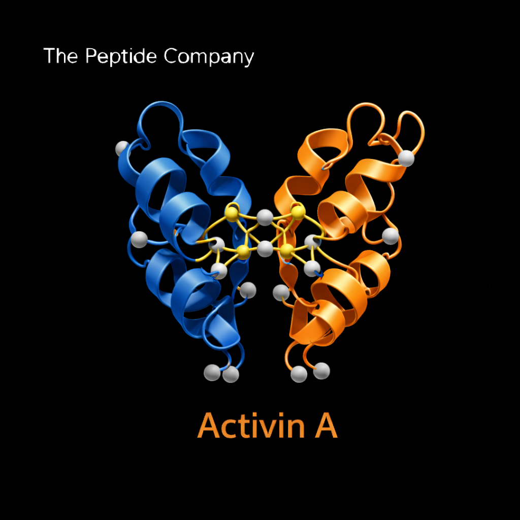
Activin A is composed of two beta-A subunits linked by disulfide bonds, forming a homodimeric structure. This dimerization is essential for receptor binding and downstream signaling. The mature protein is generated through proteolytic processing of precursor forms, similar to other TGF-β superfamily ligands.
Receptor Binding and SMAD2/3 Activation
Activin A binds primarily to activin type II receptors (ActRIIA and ActRIIB), which subsequently recruit and phosphorylate type I receptors. This receptor complex phosphorylates SMAD2 and SMAD3 transcription factors, which then associate with SMAD4 and translocate to the nucleus. Nuclear SMAD complexes regulate gene expression programs controlling extracellular matrix deposition, cellular proliferation, and differentiation.
Activin A and Muscle Biology
In skeletal muscle, Activin A functions similarly to myostatin as a negative regulator of muscle growth. Elevated Activin A signaling has been associated with suppression of myoblast differentiation and promotion of catabolic signaling pathways. Because Activin A and myostatin share receptor pathways, they contribute to overlapping regulatory control of muscle mass and anabolic balance.
Fibrosis and Extracellular Matrix Remodeling
Activin A has been implicated in fibrotic signaling through stimulation of fibroblast activation and extracellular matrix protein synthesis. Increased Activin A expression in experimental models correlates with enhanced collagen deposition and tissue remodeling. Its signaling interaction with SMAD2/3 places it within the broader network of TGF-β–mediated fibrotic pathways.
Interaction With Follistatin and Binding Proteins
Follistatin serves as a high-affinity binding protein that neutralizes Activin A, preventing receptor interaction. This regulatory mechanism provides a physiological counterbalance to Activin-mediated signaling. The Activin–Follistatin axis is central to muscle growth regulation, reproductive biology, and systemic inflammatory modulation.
Endocrine and Reproductive Roles
Activin A was originally identified for its role in regulating follicle-stimulating hormone (FSH) secretion within the pituitary gland. Through endocrine signaling networks, Activin A influences reproductive function, gonadal signaling, and hormonal feedback systems. These endocrine roles extend its relevance beyond musculoskeletal biology.
Inflammatory and Immune Signaling
Emerging research suggests that Activin A participates in immune signaling and inflammatory modulation. Expression patterns increase in certain inflammatory states, indicating cross-talk between TGF-β superfamily signaling and immune regulation pathways.
Comparison With Myostatin and TGF-β1
While myostatin is more muscle-specific and TGF-β1 is broadly fibrotic and immunomodulatory, Activin A occupies an intermediate position within the signaling hierarchy. It shares receptor pathways with myostatin but also participates in broader endocrine and inflammatory networks. Understanding these distinctions clarifies the signaling architecture of the TGF-β superfamily.
Research Applications
Activin A is studied in experimental models of muscle wasting, fibrosis, reproductive biology, inflammatory disorders, and extracellular matrix remodeling. Investigative approaches include receptor antagonism, ligand neutralization, genetic modulation, and SMAD pathway analysis.
Limitations and Open Research Questions
Important research questions remain regarding tissue-specific effects, receptor competition with related ligands, and long-term signaling adaptations. Further investigation is required to clarify how Activin A integrates with systemic endocrine and metabolic networks.
Summary
Activin A is a multifunctional TGF-β superfamily ligand that regulates muscle mass, fibrosis signaling, endocrine feedback, and inflammatory pathways through SMAD2/3-mediated transcriptional control. Its shared receptor usage with myostatin and modulation by follistatin position it as a central node in muscle–fibrosis cross-talk and extracellular matrix biology.
Educational & Research Disclaimer
This document is provided for educational and scientific research purposes only. No medical, therapeutic, or usage claims are made. Activin A and related signaling modulators are not approved for human use and are intended solely for controlled laboratory and academic investigation.
FAQ:
What is Activin A?
Activin A is a dimeric protein belonging to the transforming growth factor-beta (TGF-β) superfamily. It functions as a signaling molecule that regulates cellular differentiation, growth, inflammation, and extracellular matrix remodeling through SMAD transcription pathways.
How does Activin A signaling work?
Activin A binds to type II activin receptors (ActRIIA or ActRIIB), which recruit and activate type I receptors. This receptor complex phosphorylates SMAD2 and SMAD3 proteins, allowing them to enter the nucleus and regulate gene expression involved in tissue development and remodeling.
How is Activin A related to the TGF-β superfamily?
Activin A is structurally and functionally related to other members of the TGF-β superfamily, including myostatin, growth differentiation factors (GDFs), and transforming growth factor-beta proteins. These ligands share similar receptor systems and intracellular SMAD signaling mechanisms.
What role does Activin A play in muscle biology?
Activin A participates in signaling networks that regulate muscle growth, tissue remodeling, and fibrosis. Its interaction with pathways shared by myostatin places it at the intersection of muscle regeneration signaling and fibrotic regulation.
Why is Activin A important in fibrosis research?
Elevated Activin A signaling has been associated with increased extracellular matrix production and fibrotic tissue remodeling in several organs. Because of its role in SMAD-mediated transcription, it is frequently studied in the context of fibrosis and inflammatory signaling.
How does Activin A interact with other growth factors?
Activin A signaling often overlaps with pathways involving myostatin, TGF-β, and other growth differentiation factors. These pathways coordinate tissue growth, cellular differentiation, and inflammatory responses through shared receptor systems and SMAD transcription factors.
PMID:
PMID: 25470548
PMID: 20844133
PMID: 22188969
PMID: 16716579
PMID: 21325640
PMID: 29899388
PMID: 19286916
RELATED SEARCHES:
Myostatin (GDF-8): Muscle Growth Regulation, TGF-β Superfamily Signaling, and Anabolic Homeostasis
Decorin : TGF-β Regulation, Extracellular Matrix Signaling, and Fibrosis Modulation
Abstract & Overview
Myostatin, also known as Growth Differentiation Factor-8 (GDF-8), is a member of the transforming growth factor-beta (TGF-β) superfamily and serves as a central negative regulator of skeletal muscle growth. It functions as a signaling protein that limits myogenesis, regulates muscle fiber size, and maintains anabolic balance. Myostatin signaling plays a critical role in developmental biology, muscle homeostasis, and metabolic regulation. Research into myostatin has provided insight into muscle hypertrophy, sarcopenia, fibrosis, and systemic energy dynamics.
TGF-β Superfamily Context
Myostatin belongs to the TGF-β superfamily, a group of signaling proteins involved in cellular growth, differentiation, and extracellular matrix regulation. Members of this family signal through type I and type II serine/threonine kinase receptors and activate intracellular SMAD transcription factors. Within this framework, myostatin acts as a muscle-specific growth inhibitor, counterbalancing anabolic pathways such as IGF-1 and mTOR signaling.
Molecular Structure and Processing
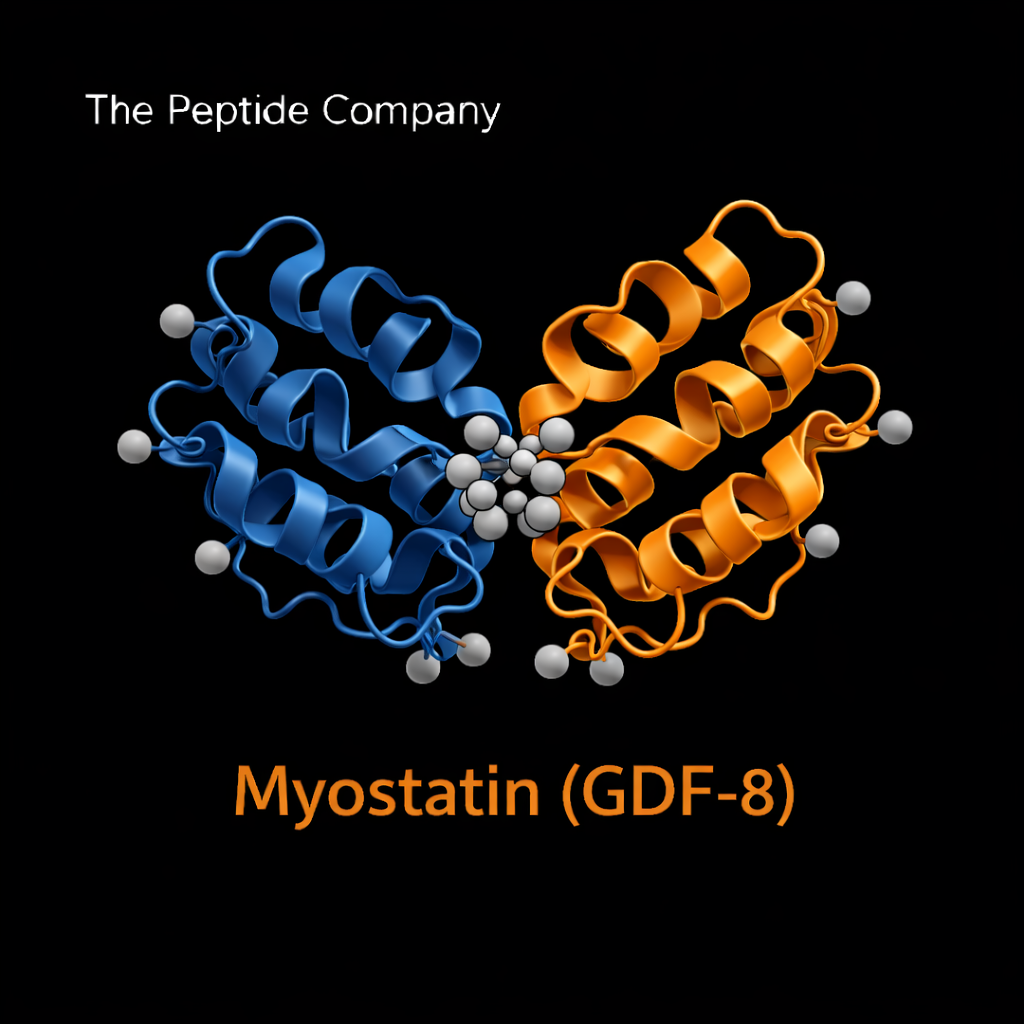
Myostatin is synthesized as a precursor protein consisting of a signal peptide, propeptide domain, and mature growth factor domain. Following proteolytic cleavage, the mature dimeric protein binds to activin type II receptors (ActRIIA and ActRIIB). Its activity is tightly regulated by binding proteins, including follistatin, decorin, and other extracellular modulators that influence bioavailability.
Receptor Binding and SMAD Signaling
Myostatin binds primarily to activin type II receptors, initiating recruitment of type I receptors and phosphorylation of SMAD2/3 transcription factors. These SMAD complexes translocate to the nucleus, where they regulate gene expression related to muscle protein synthesis and degradation. Through this pathway, myostatin suppresses myoblast proliferation and differentiation while modulating muscle fiber growth.
Role in Muscle Development and Homeostasis
During development, myostatin ensures controlled muscle formation by preventing excessive myogenesis. In adult tissue, it maintains muscle mass equilibrium by balancing anabolic and catabolic signaling. Experimental models lacking functional myostatin demonstrate increased muscle mass and fiber hypertrophy, highlighting its regulatory importance.
Interaction With Follistatin and Extracellular Regulators
Myostatin activity is modulated by extracellular binding proteins such as follistatin, which binds and neutralizes myostatin, reducing receptor activation. Decorin and other matrix-associated proteins also influence myostatin signaling by altering growth factor availability. These interactions create a regulatory network linking muscle biology with extracellular matrix signaling.
Myostatin and Fibrosis
Beyond skeletal muscle, myostatin has been implicated in fibrotic signaling pathways through its interaction with TGF-β family members. Elevated myostatin activity in certain experimental models correlates with increased extracellular matrix deposition and tissue remodeling. This connection places myostatin at the intersection of muscle regulation and fibrosis research.
Metabolic Cross-Talk
Myostatin signaling intersects with metabolic pathways, including insulin sensitivity and mitochondrial function. Studies suggest that alterations in myostatin expression influence systemic energy balance and adipose tissue dynamics. This cross-talk links muscle mass regulation with broader metabolic homeostasis.
Myostatin in Aging and Sarcopenia Research
Age-associated muscle decline (sarcopenia) has been linked to changes in myostatin signaling dynamics. Research examines whether modulation of myostatin activity may influence age-related muscle atrophy and functional capacity. These investigations contribute to broader understanding of anabolic resistance and muscle aging biology.
Comparison With IGF-1 and mTOR Signaling
Myostatin acts in functional opposition to anabolic pathways such as IGF-1 and mTOR. While IGF-1 promotes protein synthesis and muscle hypertrophy, myostatin restrains excessive growth, ensuring structural and metabolic balance. Understanding this regulatory tension provides insight into coordinated muscle adaptation.
Research Applications
Myostatin is studied extensively in models of muscle hypertrophy, muscle wasting, metabolic disorders, and fibrosis. Experimental approaches include genetic knockout systems, antibody-based inhibition, receptor modulation, and extracellular binding protein interactions. These models contribute to deeper understanding of muscle growth control mechanisms.
Limitations and Open Research Questions
Important questions remain regarding tissue-specific regulation, long-term signaling adaptations, and integration with endocrine networks. Further investigation is required to clarify how myostatin signaling coordinates with other TGF-β family members and systemic metabolic signals.
Summary
Myostatin (GDF-8) is a master regulator of skeletal muscle growth operating through TGF-β superfamily signaling and SMAD-mediated transcriptional control. By restraining excessive myogenesis and balancing anabolic pathways, it maintains structural and metabolic equilibrium within muscle tissue. Its study provides critical insight into muscle development, fibrosis signaling, and systemic metabolic integration.
Educational & Research Disclaimer
This document is provided for educational and scientific research purposes only. No medical, therapeutic, or usage claims are made. Myostatin and related signaling modulators are not approved for human use and are intended solely for controlled laboratory and academic investigation.
FAQ:
What is myostatin and why is it studied?
Myostatin (GDF-8) is a TGF-β superfamily protein that negatively regulates skeletal muscle growth. It controls myogenesis, muscle fiber size, and anabolic balance. Research models use myostatin to investigate muscle hypertrophy, sarcopenia, cachexia, fibrosis, and metabolic homeostasis.
How does myostatin signal at the cellular level?
Myostatin binds to activin type II receptors (primarily ActRIIB), triggering type I receptor recruitment, SMAD2/3 phosphorylation, and transcriptional regulation of genes governing muscle growth suppression. This pathway serves as a central node in TGF-β–related inhibitory signaling.
What roles does myostatin play in developmental biology?
Myostatin coordinates muscle mass during embryonic development and maintains adult muscle equilibrium. Loss-of-function models show substantial increases in muscle fiber number and size, highlighting its essential role in growth limitation.
How is myostatin connected to muscle loss conditions?
Elevated myostatin expression is associated with sarcopenia, cachexia, disuse atrophy, chronic illness, and aging. Research explores how modulating the myostatin pathway may influence muscle preservation in these contexts.
How does myostatin influence metabolism?
Beyond skeletal muscle, myostatin impacts systemic energy metabolism, adipose tissue dynamics, insulin sensitivity, and mitochondrial function. Studies suggest that suppressing myostatin signaling may shift metabolic balance toward greater energy expenditure and improved glucose handling.
Is myostatin considered a target for therapeutic development?
Multiple investigational agents (ligand traps, antibodies, receptor blockers) have been evaluated, but here myostatin is presented strictly as a research-grade protein used for pathway studies. No therapeutic claims or usage implications apply.
How should myostatin be described in research communication?
Myostatin should be framed as a cytokine-like regulatory protein of the TGF-β superfamily, used experimentally to study inhibitory muscle signaling, hypertrophy control, SMAD pathways, and metabolic cross-talk. Content must avoid any implication of treatment or human administration.
PMID:
- PMID: 9707416 — Discovery of myostatin (GDF-8) and demonstration of its role as a negative regulator of skeletal muscle mass.
- PMID: 15454088 — Analysis of ActRIIB receptor signaling and SMAD-mediated transcriptional control by myostatin.
- PMID: 20489699 — Myostatin inhibition studies showing increased muscle mass and metabolic effects in animal models.
- PMID: 22215057 — Myostatin’s role in muscle wasting, sarcopenia, and chronic illness-related atrophy.
- PMID: 31455875 — Crosstalk between myostatin, adipose tissue biology, and whole-body energy metabolism.
RELATED SEARCHES:
GHRP‑2 : Pituitary Axis Modulation, Ghrelin Receptor Activation, and Cellular Recovery Research
ACE‑031 : Myostatin Inhibition, Muscle Hypertrophy, and Regenerative Research
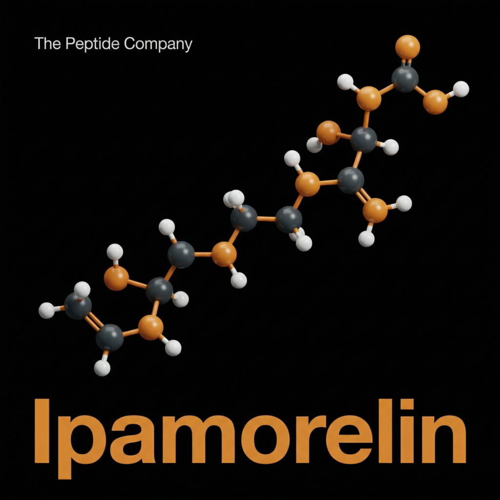
Ipamorelin is a synthetic pentapeptide and a selective agonist of the ghrelin/growth hormone secretagogue receptor (GHS-R1a). Distinct from other growth hormone-releasing peptides (GHRPs) such as GHRP-6 or GHRP-2, Ipamorelin is characterized in research literature by its high specificity for growth hormone (GH) release without significant stimulation of adrenocorticotropic hormone (ACTH), cortisol, or prolactin. This specificity has made it a significant subject of study in fields ranging from endocrinology to bone metabolism and gastrointestinal motility.
First synthesized in the mid-1990s, Ipamorelin (Aib-His-D-2-Nal-D-Phe-Lys-NH2) was developed to improve upon the pharmacological profile of earlier secretagogues, which often exhibited “off-target” hormonal effects. Research models suggest that Ipamorelin mimics the action of the endogenous ligand ghrelin by binding to GHS-R1a receptors in the anterior pituitary, thereby initiating a signal transduction cascade that amplifies GH secretion pulsatility.
MOLECULAR MECHANISMS OF GHS-R1 A ACTIVATIONAND SIGNALTRANSDUCTION
The primary mechanism of action for Ipamorelin involves the activation of the G protein-coupled receptor GHS-R1a. This receptor is densely expressed in the hypothalamus and pituitary gland. Unlike Growth Hormone Releasing Hormone (GHRH), which acts via the cAMP pathway, GHS-R1a activation by peptides like Ipamorelin triggers the phospholipase C (PLC) signaling pathway.
This distinct pathway allows Ipamorelin to act synergistically with endogenous GHRH. While GHRH stimulates the synthesis of GH, secretagogues like Ipamorelin primarily facilitate its release. Research indicates that this complementary action creates a more robust physiological response than either compound alone.
SelectivityandtheLackofCortisolStimulation
A defining characteristic of Ipamorelin in comparative studies is its selectivity. Early generations of GHRPs were potent stimulators of GH but also activated the hypothalamic-pituitary-adrenal (HPA) axis, leading to elevated cortisol and prolactin levels. Ipamorelin was designed specifically to minimize this effect.
PRECLINICAL FINDINGS: LONGITUDINAL BONE GROWTH AND BODY COMPOSITION
Extensive preclinical research has evaluated the downstream effects of Ipamorelin-induced GH secretion on tissue growth, specifically focusing on longitudinal bone growth and body weight gain in rodent models. These studies provide foundational data for understanding the anabolic potential of selective secretagogues.
These findings align with the established biological role of the GH/IGF-1
axis, wherein pulsatile GH secretion stimulates the hepatic production of Insulin-like Growth Factor 1 (IGF-1), the primary mediator of somatic growth. By amplifying the natural pulsatility of GH rather than providing a continuous elevation, Ipamorelin appears to maintain the physiological pattern necessary for optimal tissue responsiveness.
METABOLICSIGNALINGANDNITROGENBALANCE
Beyond skeletal growth, Ipamorelin has been investigated for its influence on catabolic states. Research involving glucocorticoid-induced catabolism has highlighted the peptide’s ability to counteract muscle wasting and improve nitrogen balance. This is of particular interest in research focused on mitigating the side effects of chronic steroid use or wasting diseases.
COMPARATIVE ANALYSIS:GASTROINTESTINAL MOTILITY ANDSAFETY PROFILE
The GHS-R1a receptor is also expressed in the gastrointestinal tract, where endogenous ghrelin regulates motility and gastric emptying. Research comparing Ipamorelin to other GHS agonists like GHRP-6 has elucidated differences in their impact on GI physiology. While GHRP-6 is known to significantly accelerate gastric emptying and increase hunger via hypothalamic NPY neuron activation, Ipamorelin appears to have a more neutral profile in this regard.
SOURCED STUDIES
- (1)Raun, K., et al. “Ipamorelin, the first selective growth hormone secretagogue.” EuropeanJournal ofEndocrinology, vol. 139, no. 5, 1998, pp. 552-561. DOI: 10.1530/eje.0.1390552.
- (2)Johansen, P.B., et al. “Ipamorelin, a new growth-hormone-releasing peptide, induces longitudinal bone growth in rats.” GrowthHormone&IGFResearch, vol. 9, no. 2, 1999, pp. 106-113. DOI: 10.1054/ghir.1999.0099.
- (3)Svensson, J., et al. “GH secretagogues: physiology and clinical potential.” EndocrineReviews, vol. 21, no. 4, 2000, pp. 414-461. DOI: 10.1210/edrv.21.4.0407.
- (4)Aagaard, N.K., et al. “Growth hormone and growth hormone secretagogues counteract corticosteroid-induced catabolism in rats.” ClinicalScience, vol. 116, no. 6, 2009, pp. 481-490. DOI: 10.1042/CS20080315.
- (5)Venkova, K., et al. “Ghrelin and the GHS-R1a agonist Ipamorelin differ in their ability to stimulate gastric emptying in the rat.” RegulatoryPeptides, vol. 141, no. 1-3, 2007, pp. 62-67. DOI: 10.1016/j.regpep.2006.12.012.
FAQ:
What is ipamorelin and how is it used in research?
Ipamorelin is a synthetic pentapeptide and selective agonist of the ghrelin/growth hormone secretagogue receptor (GHS-R1a). Research models use ipamorelin to study growth hormone (GH) release, pituitary signaling, and metabolic pathways because it produces GH elevation without strong activation of ACTH, cortisol, or prolactin. This selectivity makes it a preferred tool for isolating GH-specific signaling events.
How does ipamorelin differ from GHRP-6, GHRP-2, or other secretagogues?
Earlier secretagogues often triggered broad endocrine responses, including increases in ACTH, cortisol, or prolactin. Ipamorelin was engineered to reduce these off-target effects while maintaining robust GH secretagogue activity. Comparative studies show ipamorelin binds GHS-R1a with high specificity and produces a cleaner GH-dominant response profile in experimental systems.
What signaling pathway does ipamorelin activate?
Ipamorelin binds to GHS-R1a receptors in the anterior pituitary, initiating the same intracellular signaling cascade activated by endogenous ghrelin. This includes G-protein–coupled receptor signaling, calcium flux, and downstream activation of GH release mechanisms. These pathways are used to study GH secretion biology and metabolic regulation in controlled laboratory models.
What biological systems are commonly studied with ipamorelin?
Research has evaluated ipamorelin’s effects on growth hormone release, bone remodeling pathways, gastrointestinal motility, metabolic homeostasis, and neuroendocrine circuits. Its selectivity makes it suitable for differentiating GH-dependent processes from broader endocrine responses seen with other GHRPs.
Is ipamorelin considered a therapeutic?
Some ipamorelin-related compounds have been explored clinically, but the context here refers strictly to research-grade material. Ipamorelin should be described only as a laboratory reference compound, not for human or veterinary use, and not associated with treatment outcomes or clinical application. Literature summaries should remain background information rather than usage guidance.
How should ipamorelin be framed in scientific communication?
Ipamorelin is typically presented as a selective GHS-R1a agonist used to probe GH release, pituitary signaling biology, and metabolic pathways in experimental models. Care should be taken to avoid implying therapeutic intent, dosing relevance, or clinical claims.
PMID
- PMID: 10479077 — Characterization of ipamorelin as a selective GH secretagogue with minimal ACTH/cortisol activation.
- PMID: 10872841 — Studies comparing ipamorelin’s receptor specificity and endocrine profile with earlier GHRPs.
- PMID: 10698146 — Evaluation of ipamorelin’s GH-releasing effects in pituitary cell models.
- PMID: 11061400 — Investigations into ipamorelin’s metabolic and gastrointestinal signaling effects.
- PMID: 11897523 — Ghrelin/GHS-R1a signaling pathways relevant to ipamorelin’s mechanism of action.
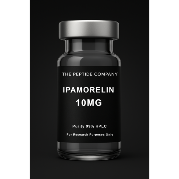
Ipamorelin 10mg
Ipamorelin 10mg is a research peptide studied for its selective interaction with growth hormone secretagogue receptor (GHS-R) pathways and pulsatile endocrine signaling in controlled laboratory research models. For research use only.
RELATED SEARCHES:
Semaglutide : GLP-1 Receptor Agonism, Incretin Signaling, and Metabolic Regulation
GHRP‑2 : Pituitary Axis Modulation, Ghrelin Receptor Activation, and Cellular Recovery Research
ACE‑031 : Myostatin Inhibition, Muscle Hypertrophy, and Regenerative Research

Abstract & Overview
Ovagen is a short bioregulatory peptide isolated and synthesized to mimic naturally occurring peptides derived from ovarian tissue. It functions as a regulatory sequence that supports the transcriptional and translational control of genes associated with cell differentiation, tissue regeneration, and hormonal balance. Research into Ovagen and related tissue-specific bioregulators focuses on their role in supporting the hypothalamic-pituitary-gonadal (HPG) axis, modulating ovarian cell activity, and maintaining genomic stability in reproductive tissues. This compound has become a key model for studying peptide-mediated genetic regulation and reproductive tissue rejuvenation mechanisms.
Molecular Pharmacology
Ovagen belongs to the class of cytomedins, short peptides composed of 2–10 amino acids, which exhibit selective organotropism. Its sequence originates from the low-molecular-weight fraction of animal ovarian tissue extracts. Through nucleoprotein-peptide signaling, Ovagen influences RNA polymerase activity and enhances gene expression in ovarian and epithelial cells. This process supports cellular proliferation and repair while maintaining genomic stability through upregulation of DNA repair enzymes. Studies have also shown that bioregulators such as Ovagen exhibit epigenetic modulation, affecting histone acetylation and chromatin remodeling processes essential for reproductive tissue health.
Mechanism of Action
The mechanism of Ovagen centers on peptide-mediated regulation of gene expression within ovarian cells. It acts as a signaling modulator that binds to specific nuclear receptors or peptide-responsive DNA sequences, influencing transcription factors that control cell growth, differentiation, and apoptosis. In the context of the HPG axis, Ovagen is thought to contribute to the synchronization of gonadotropic hormone release and ovarian cell cycle regulation. By stabilizing cellular RNA synthesis and protein translation, Ovagen promotes regenerative activity in epithelial and follicular tissues while maintaining structural and functional integrity in ovarian cells.
Research Findings and Biological Studies
Experimental data from in vitro and in vivo models demonstrate that Ovagen enhances the expression of genes associated with cellular proliferation, differentiation, and metabolic adaptation in reproductive tissue. In aging and oxidative stress models, Ovagen reduced DNA fragmentation and lipid peroxidation, suggesting protective effects against genotoxic and oxidative damage. These studies also report normalization of estradiol and FSH levels in animal models of reproductive imbalance, indicating a potential regulatory effect on endocrine signaling pathways. Biochemical analyses further show increased synthesis of RNA and structural proteins in ovarian tissue following exposure to Ovagen peptides.
Genomic Stability and Tissue-Specific Modulation
Ovagen’s unique ability to modulate gene expression at the chromatin level contributes to its genomic stabilizing effects. By promoting transcriptional activation of DNA repair genes such as XRCC1 and PARP1, Ovagen may support the correction of age-related DNA damage. This aligns with broader findings on tissue-specific peptides that maintain cellular homeostasis and delay replicative senescence in epithelial and glandular tissues. Such genomic effects position Ovagen as a model compound for research into epigenetic peptide therapy and reproductive cell cycle regulation.
Comparative Insights
Among the bioregulator family, Ovagen is structurally and functionally related to other reproductive and endocrine peptides such as Testagen (testicular peptide), Thymagen (immune-regulatory peptide), and Vilon (universal cell regeneration peptide). While Testagen focuses on androgenic axis modulation, Ovagen demonstrates a more targeted influence on oogenesis and follicular regeneration. Its molecular behavior also parallels that of thymic peptides, reflecting shared pathways in cell differentiation and genomic regulation. Comparative studies suggest that co-administration with Thymogen or Vilon enhances Ovagen’s regulatory effects, highlighting its potential as part of multi-peptide research models for endocrine homeostasis.
Summary
Ovagen represents a tissue-specific regulatory peptide that provides insight into peptide-based modulation of ovarian cell biology, hormonal balance, and genomic repair mechanisms. Its activity at the level of RNA synthesis and chromatin remodeling underscores its role as a key peptide bioregulator within reproductive research. By combining molecular specificity with genomic stability effects, Ovagen continues to serve as a model compound in studies of cellular regeneration, fertility regulation, and peptide-mediated gene expression.
Educational & Research Disclaimer
This article is for educational and scientific research purposes only. No therapeutic claims or usage recommendations are provided. Compounds referenced are not approved for human use and are intended solely for controlled laboratory experimentation.
FAQ:
What is Ovagen?
Ovagen is a short ovarian tissue–specific bioregulator peptide studied for its role in reproductive axis signaling and ovarian cellular regulation. It is conceptually derived from ovarian peptide fractions within the cytomedine family.
What type of peptide is Ovagen?
Ovagen is classified as a tissue-specific regulatory peptide rather than a growth factor or hormone. Research focuses on its potential influence at the genomic and epigenetic signaling level.
What biological systems are studied with Ovagen?
Ovagen research commonly examines the hypothalamic–pituitary–gonadal (HPG) axis, ovarian cell differentiation, follicular signaling pathways, and reproductive tissue homeostasis.
How is Ovagen different from hormonal therapies?
Ovagen is studied for regulatory peptide signaling effects rather than direct hormone replacement or endocrine stimulation. It is investigated for its influence on cellular transcriptional and differentiation pathways.
What cells are involved in Ovagen research?
Research typically focuses on ovarian granulosa cells, follicular cells, and other reproductive tissue–associated cell populations involved in gene regulation and tissue maintenance.
Is Ovagen intended for human use?
No. Ovagen referenced here is discussed strictly for research and educational purposes and is not intended for human consumption.
PMID:
- PMID: 12928777
Khavinson V, Morozov VG. Peptide bioregulators and gene expression regulation. - PMID: 14738556
Short peptides regulating gene expression and protein synthesis in human cells. - PMID: 17654843
Tissue-specific peptide regulation in reproductive and connective tissues. - PMID: 21728784
Ovarian aging and extracellular matrix regulatory mechanisms. - PMID: 23809343
Epigenetic regulation in ovarian and reproductive tissues. - PMID: 15234339
Collagen and matrix-related signaling in reproductive tissue maintenance.
RELATED SEARCHES:
GHRP‑2 : Pituitary Axis Modulation, Ghrelin Receptor Activation, and Cellular Recovery Research
ACE‑031 : Myostatin Inhibition, Muscle Hypertrophy, and Regenerative Research
Follistatin: Myostatin-Regulated Pathways and Advanced Muscle Research

Abstract & Overview
GHRP‑6 (Growth Hormone Releasing Peptide‑6) is a synthetic hexapeptide and one of the earliest members of the growth hormone secretagogue (GHS) class. It acts as a potent agonist of the ghrelin receptor (GHS‑R1a), triggering both endocrine and metabolic responses in research models. By mimicking endogenous ghrelin signaling, GHRP‑6 provides a tool for studying growth hormone release, cellular metabolism, and tissue repair mechanisms. Its biochemical profile and receptor interactions make it an important compound for exploring neuroendocrine regulation and energy homeostasis in laboratory settings.
Molecular Pharmacology
GHRP‑6 is a hexapeptide with the sequence His‑D‑Trp‑Ala‑Trp‑D‑Phe‑Lys‑NH₂. It belongs to the same structural family as GHRP‑2, Hexarelin, and Ipamorelin but exhibits a unique receptor activation pattern. Its primary target, the growth hormone secretagogue receptor type 1a (GHS‑R1a), is a G‑protein–coupled receptor (GPCR) that mediates both calcium‑dependent and cAMP‑linked signaling cascades. Through this receptor, GHRP‑6 influences pituitary growth hormone release and peripheral metabolic responses. Studies highlight its dual capacity to activate endocrine and paracrine signaling pathways involved in cell proliferation, metabolism, and stress adaptation.
Ghrelin Receptor Signaling Pathway
GHRP‑6 acts as an exogenous ligand for GHS‑R1a, a receptor expressed predominantly in the hypothalamus, pituitary gland, pancreas, and gastrointestinal tract. Upon binding, GHRP‑6 activates Gq/11 proteins, leading to phospholipase C (PLC) stimulation and the generation of inositol triphosphate (IP₃) and diacylglycerol (DAG). This signaling cascade increases intracellular calcium concentrations and activates protein kinase C (PKC), which promotes vesicular growth hormone release from pituitary somatotrophs. In addition to this canonical pathway, GHRP‑6 signaling interacts with MAPK/ERK, PI3K/Akt, and AMPK networks, linking the peptide to broader effects on metabolism and mitochondrial regulation.
Endocrine and Metabolic Research
In controlled studies, GHRP‑6 has been shown to stimulate pulsatile growth hormone secretion, elevating circulating insulin‑like growth factor‑1 (IGF‑1) concentrations through hepatic synthesis. These responses make it valuable for studying somatotropic axis regulation and growth‑related signaling. Beyond endocrine actions, GHRP‑6 influences glucose metabolism, fatty acid oxidation, and mitochondrial biogenesis through AMPK activation and improved cellular energy utilization. Research models indicate that chronic GHS‑R1a activation may contribute to enhanced metabolic flexibility and adaptive responses under nutrient stress conditions.
Cellular Regeneration and Stress Response
GHRP‑6 has been used to study cytoprotective and regenerative mechanisms in a variety of tissues, including skeletal muscle, liver, and neural systems. Its signaling activity modulates apoptosis, autophagy, and oxidative stress pathways, leading to reduced cellular injury and improved tissue repair. GHRP‑6’s influence on mitochondrial integrity and reactive oxygen species (ROS) control highlights its relevance in studies of cellular longevity and metabolic adaptation. Additionally, its engagement with the ghrelin receptor system contributes to anti‑inflammatory signaling through NF‑κB inhibition and cytokine modulation.
Comparative Signaling: GHRP‑6 vs. GHRP‑2 and Azelaprag
While GHRP‑6 and GHRP‑2 share similar receptor targets, they differ in potency and receptor bias. GHRP‑2 demonstrates higher affinity for GHS‑R1a but shorter duration, while GHRP‑6 offers a broader receptor activation profile with enhanced metabolic modulation. Compared with Azelaprag—a non‑peptide ghrelin receptor agonist—GHRP‑6 represents a classic peptide model for studying ligand‑receptor conformational signaling and the kinetics of GPCR activation. Together, these compounds help define how different molecular frameworks influence GHS‑R1a‑mediated pathways and downstream gene expression.
Research Applications in Metabolism and Growth
Due to its dual anabolic and metabolic actions, GHRP‑6 serves as a model compound for studying growth hormone physiology, energy metabolism, and cellular recovery mechanisms. In muscle and hepatic tissue, it promotes protein synthesis, glycogen replenishment, and oxidative balance. Studies also investigate its effects on circadian rhythm regulation, appetite control, and gut‑brain axis communication through peripheral ghrelin receptor activation. These findings position GHRP‑6 as a versatile research tool for examining interconnected neuroendocrine and metabolic networks.
Summary
GHRP‑6 is a synthetic peptide that continues to play a significant role in ghrelin receptor and growth hormone secretagogue research. Its receptor interactions extend beyond GH release, encompassing mitochondrial regulation, energy metabolism, and cellular resilience. By linking endocrine control with metabolic adaptation, GHRP‑6 provides a valuable model for understanding GHS‑R1a signaling and its broader implications in physiological and cellular research.
Educational & Research Disclaimer
This article is for educational and scientific research purposes only. No therapeutic claims or usage recommendations are provided. Compounds referenced are not approved for human use and are intended solely for controlled laboratory experimentation.
FAQ:
What is GHRP-6?
GHRP-6 (Growth Hormone Releasing Peptide-6) is a synthetic hexapeptide classified as a growth hormone secretagogue (GHS). It is studied for its ability to activate the ghrelin receptor (GHS-R1a) and modulate endocrine signaling pathways.
How does GHRP-6 work in research models?
GHRP-6 binds to the growth hormone secretagogue receptor (GHS-R1a), a G-protein–coupled receptor (GPCR). Activation of this receptor initiates intracellular signaling cascades involving calcium mobilization and cAMP pathways associated with pituitary hormone release.
Is GHRP-6 the same as ghrelin?
No. GHRP-6 is a synthetic peptide that mimics certain actions of ghrelin by activating the same receptor (GHS-R1a), but it is structurally distinct from endogenous ghrelin.
What systems are commonly studied with GHRP-6?
Research commonly examines the somatotropic axis (growth hormone regulation), hypothalamic–pituitary signaling, appetite-related pathways, and metabolic hormone signaling.
How is GHRP-6 different from other growth hormone secretagogues?
GHRP-6 is one of the earliest GHS compounds and exhibits a distinct receptor activation profile compared with other secretagogues such as GHRP-2, Hexarelin, or Ipamorelin.
Is GHRP-6 intended for human use?
No. GHRP-6 referenced here is discussed strictly for research and educational purposes and is not intended for human consumption.
PMID:
- PMID: 7542028
Discovery and characterization of growth hormone–releasing peptides. - PMID: 8651140
Growth hormone secretagogues and their mechanism of action at the pituitary level. - PMID: 10473535
Identification and cloning of the growth hormone secretagogue receptor (GHS-R). - PMID: 10920360
Ghrelin as the endogenous ligand for the GHS-R receptor. - PMID: 12050244
Signal transduction pathways activated by GHRP-6 and ghrelin receptor agonists. - PMID: 15240662
Role of GHS-R activation in endocrine and metabolic regulation.
RELATED SEARCHES:
Orforglipron : Oral Small-Molecule GLP-1 Receptor Agonist and Incretin Pathway Modulation
GHRP‑2 : Pituitary Axis Modulation, Ghrelin Receptor Activation, and Cellular Recovery Research
ACE‑031 : Myostatin Inhibition, Muscle Hypertrophy, and Regenerative Research
Follistatin: Myostatin-Regulated Pathways and Advanced Muscle Research
GHRP-6 5mg
GHRP-6 is a synthetic growth hormone–releasing peptide studied for its interaction with ghrelin receptors and pathways related to growth hormone signaling and metabolic regulation. For research use only.

Abstract & Overview
GHRP‑2 (Growth Hormone Releasing Peptide‑2) is a synthetic hexapeptide classified within the family of growth hormone secretagogues (GHS). It is a potent agonist of the ghrelin receptor (GHS‑R1a) and has been extensively studied as a model compound for growth hormone release, pituitary sensitization, and cellular metabolic regulation. By mimicking the physiological actions of endogenous ghrelin, GHRP‑2 allows for the exploration of neuroendocrine signaling pathways that link the hypothalamus, pituitary gland, and peripheral tissues. Compared with GHRP‑6, GHRP‑2 demonstrates higher receptor affinity and more pronounced growth hormone–releasing activity, making it a valuable tool in controlled research examining somatotropic axis regulation and energy metabolism.
Molecular Pharmacology
GHRP‑2 is a hexapeptide with the sequence D‑Ala‑D‑His‑D‑Phe‑D‑Trp‑Lys‑Val‑NH₂. It functions as a selective agonist of the GHS‑R1a receptor, a G‑protein–coupled receptor expressed in the hypothalamus, pituitary gland, pancreas, and other metabolic tissues. Activation of GHS‑R1a triggers both calcium‑dependent and cAMP‑dependent intracellular cascades, leading to exocytosis of growth hormone from somatotroph cells. The peptide’s pharmacokinetic profile is characterized by rapid receptor binding and a robust amplitude of GH secretion in pulsatile models, distinguishing it from GHRP‑6 in potency and duration.
Receptor Mechanism of Action
Upon binding to GHS‑R1a, GHRP‑2 activates Gq/11 proteins, leading to phospholipase C (PLC) activation and the generation of inositol triphosphate (IP₃) and diacylglycerol (DAG). These molecules promote intracellular calcium release and activate protein kinase C (PKC), which triggers growth hormone secretion at the pituitary level. In parallel, GHRP‑2 influences other signaling networks, including MAPK/ERK and PI3K/Akt, promoting cellular survival, metabolism, and adaptive energy responses. The peptide’s capacity to modulate both endocrine and metabolic functions has made it an important compound for studying the cross‑talk between neuroendocrine and mitochondrial pathways.
Endocrine and Metabolic Regulation
Research models demonstrate that GHRP‑2 elevates plasma GH concentrations through direct pituitary stimulation and indirect hypothalamic activation. This GH release subsequently upregulates hepatic insulin‑like growth factor‑1 (IGF‑1) synthesis, driving anabolic processes such as protein synthesis, lipid oxidation, and tissue repair. Additionally, GHRP‑2 has been shown to modulate glucose metabolism and mitochondrial respiration via AMPK activation and improved substrate utilization. These effects make it a valuable agent for exploring the metabolic underpinnings of energy homeostasis in both central and peripheral systems.
Comparative Analysis: GHRP‑2 vs. GHRP‑6
While both GHRP‑2 and GHRP‑6 activate GHS‑R1a and promote GH release, they exhibit key pharmacological differences. GHRP‑2 possesses a higher receptor binding affinity and faster onset of action, leading to greater GH amplitude in dose‑response studies. GHRP‑6, however, demonstrates broader metabolic influence and mild orexigenic activity due to its secondary receptor interactions. Comparative data suggest that GHRP‑2 is more selective and potent for pituitary‑specific GH release, whereas GHRP‑6 exerts a wider systemic effect on energy balance and tissue metabolism.
Cellular Recovery and Regenerative Research
Beyond endocrine functions, GHRP‑2 has been studied for its effects on cellular recovery and oxidative stress modulation. In vitro and in vivo research models show that GHRP‑2 enhances mitochondrial membrane potential, reduces reactive oxygen species (ROS), and stabilizes cellular redox balance. Its anti‑inflammatory influence through NF‑κB pathway suppression and cytokine regulation further supports its potential as a model for stress adaptation and tissue recovery studies. These findings underscore its value in investigating the intersection between peptide signaling, mitochondrial health, and regenerative cellular mechanisms.
Summary
GHRP‑2 is a potent ghrelin receptor agonist that exemplifies the relationship between neuroendocrine signaling, energy metabolism, and cellular protection. Its ability to induce growth hormone release, enhance mitochondrial efficiency, and regulate redox balance has made it a foundational compound for research into somatotropic and metabolic pathways. By comparing its receptor bias and mechanistic profile to analogs such as GHRP‑6 and Azelaprag, researchers can gain valuable insight into GHS‑R1a signaling and its systemic implications for metabolic homeostasis.
Educational & Research Disclaimer
This article is for educational and scientific research purposes only. No therapeutic claims or usage recommendations are provided. Compounds referenced are not approved for human use and are intended solely for controlled laboratory experimentation.
FAQ:
What is GHRP-2?
GHRP-2 (Growth Hormone–Releasing Peptide-2) is a synthetic hexapeptide classified as a growth hormone secretagogue that activates the ghrelin (GHS-R1a) receptor.
How does GHRP-2 work?
GHRP-2 stimulates growth hormone release by binding to ghrelin receptors in the hypothalamus and pituitary, influencing somatotropic axis signaling and pulsatile GH secretion.
Is GHRP-2 the same as growth hormone?
No. GHRP-2 does not contain growth hormone itself. It functions by signaling endogenous growth hormone release pathways.
What is GHRP-2 commonly studied for?
GHRP-2 is studied in research settings for pituitary axis modulation, ghrelin receptor signaling, cellular recovery pathways, and metabolic regulation.
Does GHRP-2 interact with other hormonal pathways?
Yes. Research shows GHRP-2 can influence cortisol, prolactin, and appetite-related signaling due to its interaction with ghrelin receptors.
Is GHRP-2 considered a ghrelin mimetic?
Yes. GHRP-2 is classified as a ghrelin receptor agonist and is often used as a model compound for studying ghrelin-mediated endocrine signaling.
PMID
- PMID: 8380393 – Discovery and characterization of growth hormone–releasing peptides acting independently of GHRH.
- PMID: 9000608 – Identification of synthetic GH secretagogues and their role in pituitary growth hormone release.
- PMID: 9204910 – Demonstration of GHRP-2–induced GH secretion via hypothalamic and pituitary mechanisms.
- PMID: 9725909 – Evidence of ghrelin receptor involvement in growth hormone secretagogue signaling.
- PMID: 10490927 – Comparative analysis of GHRP-2 and other GH secretagogues on endocrine hormone release.
- PMID: 11786544 – Review of growth hormone secretagogues and their physiological and cellular signaling effects.
RELATED ARTICLES:

Abstract & Overview
ACE‑031 is a recombinant fusion protein that functions as a soluble activin receptor type IIB (ActRIIB) analog, designed to inhibit the biological activity of myostatin (GDF‑8) and related ligands such as activin A and GDF‑11. As a myostatin pathway inhibitor, ACE‑031 serves as a model compound for studying skeletal muscle hypertrophy, regeneration, and energy metabolism. By preventing myostatin from binding to its receptor, ACE‑031 promotes muscle fiber growth and differentiation, offering a valuable research platform for understanding muscle physiology and tissue regeneration.
Molecular Pharmacology
ACE‑031 consists of the extracellular ligand‑binding domain of the activin receptor type IIB fused to the Fc domain of human IgG1. This structure allows ACE‑031 to act as a decoy receptor, binding to circulating myostatin and related TGF‑β family ligands, preventing them from activating membrane‑bound ActRIIB receptors on muscle cells. Through this mechanism, ACE‑031 effectively suppresses inhibitory signals that normally limit skeletal muscle growth, resulting in increased muscle mass in experimental models. The Fc‑fusion design also enhances its serum stability and extends its biological half‑life, allowing for sustained ligand neutralization.
Mechanism of Myostatin Inhibition
Myostatin (GDF‑8) is a negative regulator of skeletal muscle mass, acting through ActRIIB‑mediated SMAD2/3 signaling to inhibit myoblast proliferation and differentiation. ACE‑031 disrupts this pathway by sequestering myostatin in the extracellular space, blocking its interaction with cell‑surface receptors. This inhibition leads to the activation of satellite cells, increased protein synthesis, and decreased protein degradation via the Akt/mTOR pathway. Additionally, ACE‑031 can bind to other ActRIIB ligands such as activin A and GDF‑11, providing broader modulation of TGF‑β‑related signaling involved in tissue remodeling and metabolism.
Research Findings in Muscle Growth and Regeneration
Preclinical studies have demonstrated that ACE‑031 administration increases lean muscle mass, fiber cross‑sectional area, and strength in animal models. These effects are mediated through both hypertrophic and hyperplastic mechanisms, with enhanced activation of muscle satellite cells. Research has also explored its potential application in conditions characterized by muscle wasting, including Duchenne muscular dystrophy (DMD), cachexia, and sarcopenia. In addition to skeletal muscle effects, ACE‑031 influences vascular development and adipose metabolism, highlighting its systemic role in tissue remodeling and metabolic regulation.
Metabolic and Systemic Implications
Beyond muscle hypertrophy, ACE‑031 research indicates potential metabolic benefits through improved glucose utilization, increased insulin sensitivity, and modulation of lipid oxidation. These effects are linked to downstream Akt/AMPK signaling and enhanced mitochondrial biogenesis. The inhibition of myostatin and activin pathways also impacts adipokine expression and inflammatory cytokine profiles, suggesting a broader role in energy balance and metabolic homeostasis. Studies continue to investigate how ActRIIB blockade may interface with pathways involved in vascular growth and endothelial function.
Comparative Pathways: ACE‑031 vs. Follistatin
Both ACE‑031 and Follistatin modulate the myostatin pathway but through distinct mechanisms. While ACE‑031 acts as a soluble receptor that directly binds myostatin and related ligands, Follistatin functions as a binding protein that neutralizes activins and myostatin intracellularly and extracellularly. Follistatin exhibits broader ligand binding across TGF‑β family proteins, whereas ACE‑031 provides a more targeted and stable extracellular blockade. These complementary mechanisms make them useful tools for comparative studies of muscle growth, regeneration, and endocrine regulation.
Summary
ACE‑031 represents a biologically engineered model compound for examining the role of myostatin and activin signaling in muscle development, regeneration, and systemic metabolism. Its mechanism of ligand sequestration offers a precise way to modulate muscle anabolism and tissue recovery. Through its effects on the ActRIIB‑SMAD signaling axis, ACE‑031 continues to serve as a critical reference point in the expanding field of myostatin inhibition and regenerative peptide research.
Educational & Research Disclaimer
This article is for educational and scientific research purposes only. No therapeutic claims or usage recommendations are provided. Compounds referenced are not approved for human use and are intended solely for controlled laboratory experimentation.
FAQ:
What is ACE-031?
ACE-031 is a recombinant fusion protein designed as a soluble activin receptor type IIB (ActRIIB) decoy. It binds circulating myostatin (GDF-8) and related ligands, preventing them from activating cell-surface ActRIIB signaling pathways.
How does ACE-031 inhibit myostatin signaling?
ACE-031 mimics the extracellular ligand-binding domain of ActRIIB. By sequestering myostatin and activins in circulation, it reduces downstream SMAD2/3 signaling, which is normally associated with suppression of muscle growth and regeneration.
What is ACE-031 studied for in research?
ACE-031 has been explored in research settings related to:
Skeletal muscle hypertrophy
Muscle regeneration and repair
Growth signaling modulation
Myostatin and activin pathway biology
It is primarily used as a tool compound for studying muscle growth regulation.
Is ACE-031 the same as myostatin antibodies?
No. ACE-031 is a receptor decoy protein, not a monoclonal antibody. It binds multiple ligands in the TGF-β superfamily (including myostatin and activins), whereas antibodies are typically ligand-specific.
What pathways are affected by ACE-031?
Research indicates ACE-031 influences:
Myostatin / GDF-8 signaling
Activin A signaling
SMAD2/3 transcriptional pathways
Downstream muscle growth and differentiation cascades
Is ACE-031 used clinically?
ACE-031 has been evaluated in clinical research settings but is not approved for therapeutic use. Current interest is primarily within experimental and mechanistic research contexts.
RELATED SEARCHES:
Follistatin: Myostatin-Regulated Pathways and Advanced Muscle Research

Abstract & Overview
Azelaprag is a small‑molecule agonist of the growth hormone secretagogue receptor type 1a (GHS‑R1a), a G‑protein–coupled receptor primarily involved in the regulation of growth hormone (GH) secretion, appetite control, and energy balance. It functions as a non‑peptide mimic of endogenous ghrelin and peptide secretagogues such as GHRP‑2, Hexarelin, and MK‑677, while demonstrating oral bioavailability and receptor selectivity in experimental models. Azelaprag’s mechanistic framework provides researchers with a tool to investigate the interplay between ghrelin signaling, neuroendocrine feedback, and metabolic homeostasis.
Molecular Classification and Pharmacology
Azelaprag belongs to the class of non‑peptide GHS‑R1a agonists designed to reproduce the physiological activity of ghrelin. Unlike native ghrelin, which requires post‑translational acylation for receptor activation, Azelaprag interacts directly with the receptor’s orthosteric site, inducing conformational shifts that engage Gq/11‑coupled intracellular cascades. Its structure facilitates enhanced stability, receptor selectivity, and blood–brain barrier permeability—properties that make it particularly useful for studying central and peripheral aspects of the ghrelin axis.
Receptor Biology: GHS‑R1a Signaling Network
The GHS‑R1a receptor is expressed in the hypothalamus, pituitary, hippocampus, pancreas, and gastrointestinal tract. Upon agonist binding, it activates Gq/11‑dependent pathways, resulting in phospholipase C (PLC) stimulation and subsequent production of inositol triphosphate (IP3) and diacylglycerol (DAG). This cascade elevates intracellular Ca²⁺ concentrations and protein kinase C (PKC) activation, which in turn stimulates growth hormone release and modulates neuronal excitability. Downstream signaling involves MAPK/ERK, PI3K/Akt, and AMPK pathways, linking receptor activity to both anabolic and metabolic responses.
Endocrine and Metabolic Research Findings
Research with Azelaprag and related agonists demonstrates increased pulsatile GH secretion through pituitary activation and enhanced GH‑releasing hormone (GHRH) responsiveness. This cascade influences systemic insulin‑like growth factor‑1 (IGF‑1) levels, tissue anabolism, and cellular repair signaling. Additionally, GHS‑R1a activation modulates glucose and lipid metabolism by altering AMPK activity and promoting substrate mobilization during energy deficit states. These findings have positioned Azelaprag as a valuable agent in experimental models exploring the intersection between endocrine function and metabolic regulation.
Central Nervous System and Cognitive Pathways
Beyond its endocrine role, GHS‑R1a signaling is studied for its effects on neurogenesis, synaptic plasticity, and cognitive function. The receptor’s expression in the hippocampus and ventral tegmental area (VTA) suggests involvement in learning, motivation, and reward processing. Research models show that ghrelin receptor activation can enhance memory retention, increase dendritic spine density, and mitigate oxidative stress in neuronal tissues. Azelaprag’s ability to penetrate the blood–brain barrier allows for examination of these central effects without requiring peptide transport mechanisms.
Comparative Analysis: Azelaprag vs. Peptide Secretagogues
Azelaprag shares functional similarities with peptide‑based secretagogues such as GHRP‑2, GHRP‑6, Hexarelin, and MK‑677, but differs in receptor kinetics and pharmacokinetics. Whereas peptide agonists rely on extracellular binding pockets with variable half‑lives, Azelaprag’s small‑molecule framework affords improved oral bioavailability, metabolic stability, and duration of receptor occupancy. Comparative studies indicate that Azelaprag produces sustained GHS‑R1a activation with reduced desensitization, allowing for long‑term signaling observation without peptide degradation artifacts.
Energy Balance and Appetite Research
Activation of GHS‑R1a is closely tied to appetite regulation and energy expenditure. Azelaprag is used in research models to investigate orexigenic signaling through hypothalamic neuropeptide Y (NPY) and agouti‑related peptide (AgRP) neurons, which integrate peripheral metabolic cues with central hunger responses. Concurrent modulation of dopaminergic reward pathways highlights the receptor’s dual role in metabolic drive and motivational behavior.
Mitochondrial and Metabolic Integration
Emerging studies link ghrelin receptor activation to mitochondrial dynamics, including biogenesis, uncoupling, and reactive oxygen species (ROS) regulation. Through AMPK and SIRT1 interaction, GHS‑R1a signaling influences cellular energy efficiency and metabolic resilience under stress conditions. Azelaprag’s stable receptor engagement provides a platform to examine how chronic GHS‑R1a activation affects mitochondrial quality control, autophagy, and oxidative metabolism.
Summary
Azelaprag serves as a non‑peptide model compound for exploring GHS‑R1a‑mediated signaling in both central and peripheral tissues. Its receptor selectivity, oral bioavailability, and sustained activation kinetics distinguish it from earlier peptide secretagogues, enabling in‑depth research into growth hormone dynamics, metabolic integration, and neuroendocrine regulation. By bridging endocrine and metabolic mechanisms, Azelaprag contributes to the expanding field of ghrelin receptor research and energy homeostasis modeling.
Educational & Research Disclaimer
This article is for educational and scientific research purposes only. No therapeutic claims or usage recommendations are provided. Compounds referenced are not approved for human use and are intended solely for controlled laboratory experimentation.
FAQ:
What is Azelaprag studied for in research settings?
Azelaprag is investigated as a small-molecule agonist of the ghrelin receptor (GHS-R1a). Research models use it to study growth hormone secretagogue signaling, appetite regulation, neuroendocrine feedback, and energy balance without relying on peptide-based ligands.
How does Azelaprag differ from peptide secretagogues?
Unlike peptide secretagogues (e.g., GHRP-2, GHRP-6, hexarelin), Azelaprag is non-peptide, allowing researchers to examine ghrelin receptor activation with improved molecular stability, consistent receptor engagement, and reduced peptide degradation artifacts in experimental systems.
What receptor pathways are evaluated with Azelaprag?
Studies focus on GHS-R1a–mediated signaling, including downstream effects on growth hormone release, hypothalamic appetite circuits, neuroendocrine modulation, and interactions with metabolic regulators such as AMPK and dopaminergic pathways.
Is Azelaprag used for human treatment?
No. Azelaprag is referenced in preclinical and controlled research contexts only. It is not approved for human or veterinary use and is utilized strictly for laboratory investigation of ghrelin receptor biology.
Why are ghrelin receptor agonists important in metabolic research?
Ghrelin receptor agonists provide a controlled way to study energy homeostasis, appetite signaling, hormonal feedback loops, and mitochondrial–metabolic integration, helping researchers understand how central and peripheral systems coordinate metabolic responses.
PMID
These references support ghrelin receptor (GHS-R1a) signaling, growth hormone secretagogues, and metabolic pathway research relevant to Azelaprag’s mechanism:
- PMID: 12682223 — Discovery and characterization of the ghrelin receptor (GHS-R1a)
- PMID: 17047209 — Ghrelin signaling in energy balance and neuroendocrine regulation
- PMID: 19759358 — Growth hormone secretagogues and GHS-R1a pharmacology
- PMID: 21555357 — Central ghrelin signaling and appetite regulation pathways
- PMID: 30836922 — Ghrelin receptor modulation and metabolic integration
RELATED SEARCHES:
Follistatin: Myostatin-Regulated Pathways and Advanced Muscle Research