

Introduction
Follistatin is a multi-domain extracellular glycoprotein studied for its regulatory influence on TGF-β superfamily ligands—most notably myostatin (GDF-8) and activin A. These ligands shape muscle growth limitation, inflammation, and reproductive biology. Because follistatin binds these ligands and prevents receptor activation, it remains a prominent molecule in advanced muscle-regulation research.
Structural Biology of Follistatin
Follistatin contains three core follistatin domains (FSD1–3), each enriched with cysteine-rich regions and conserved β-sheet structures enabling ligand binding. Two major isoforms exist: FST288, which binds heparan-sulfate proteoglycans and remains membrane-associated, and FST315, which is more soluble and circulates through tissues.
Mechanism of Action: Ligand Sequestration
Follistatin’s primary activity is binding myostatin and activin A, preventing their interaction with ActRIIB receptors. This stops SMAD2/3 phosphorylation and downstream transcription of inhibitory genes, shifting balance toward increased myogenic signaling.
Myogenesis and Muscle Regulatory Biology
Follistatin affects satellite-cell activation, myogenic differentiation, and myotube formation. It enhances expression of myogenic regulatory factors (MRFs) such as MyoD, Myf5, Myogenin, and MRF4, strengthening the foundation of muscle-fiber formation.
Muscle Fiber Architecture
Research models show increased muscle-fiber cross‑sectional area, fast‑twitch fiber gene signatures, and enhanced transcription of contractile machinery such as myosin heavy chains, actin, troponins, and titin.
mTORC1 Signaling Interactions
By neutralizing myostatin and activin, follistatin indirectly increases AKT phosphorylation and mTORC1 signaling. Reduced FOXO activation shifts balance away from protein breakdown pathways and toward protein synthesis.
Connective Tissue and Tendon Research
Follistatin influences collagen transcription, fibroblast proliferation, extracellular matrix turnover, and TGF‑β-linked fibrosis pathways, expanding its relevance beyond skeletal muscle models.
Non-Muscle Tissue Research Themes
Follistatin also regulates reproductive hormone pathways (activin–inhibin systems), modulates inflammatory cytokine activity, and participates in metabolic transcriptional adjustments in non-muscle research models.
Summary
Follistatin is a high‑affinity binding protein for myostatin and activin A. Through ligand sequestration and SMAD-pathway modulation, it influences satellite-cell activation, muscle-fiber hypertrophy, connective-tissue signaling, transcriptional balance, and metabolic stability across research models.
Educational & Research Disclaimer
This article is for educational and scientific research purposes only. No therapeutic claims or usage guidance is provided. Compounds referenced are not approved for human use and are intended solely for controlled laboratory experimentation.
PMID
- PMID: 12805624 – Follistatin–myostatin interaction and muscle regulation
- PMID: 17095501 – Activin/myostatin signaling pathways
- PMID: 19717448 – Follistatin binding proteins and TGF-β superfamily
- PMID: 22798624 – Myostatin inhibition and muscle hypertrophy models
- PMID: 31209268 – Follistatin in skeletal muscle biology
FAQ:
What is follistatin studied for in research models?
Follistatin is studied for its ability to bind and neutralize myostatin and activin, making it a key regulator of muscle growth, cellular differentiation, and tissue remodeling in experimental models.
How does follistatin interact with myostatin pathways?
Follistatin inhibits myostatin by direct binding, preventing myostatin from activating its receptor and downstream SMAD signaling pathways involved in muscle growth suppression.
Is follistatin used in human treatment?
No. Follistatin referenced in this article is discussed strictly in the context of laboratory and preclinical research and is not approved for human use.
Related Searches:
Sermorelin: GHRH Fragment Research and Growth Hormone Pulsatility Models
IGF-1 Analogues: LR3 and DES Structural Variations and Receptor Binding in Research Models
Introduction
Kisspeptin refers to a family of neuropeptides encoded by the KISS1 gene that play a central role in regulating the hypothalamic–pituitary–gonadal (HPG) axis. Discovered through cancer-metastasis research and later identified as a master regulator of reproductive neuroendocrinology, kisspeptin signaling is now a foundational topic in neuroscience and endocrine research.
Molecular Structure and Peptide Variants
Kisspeptin peptides are derived from a common precursor and exist in multiple biologically active forms, including kisspeptin‑54, kisspeptin‑14, kisspeptin‑13, and kisspeptin‑10. All variants share a conserved C‑terminal sequence essential for receptor binding. Research focuses on how peptide length influences stability, diffusion, and signaling dynamics.
KISS1 Receptor Biology (GPR54)
Kisspeptin exerts its effects through the G‑protein–coupled receptor KISS1R (also known as GPR54). This receptor is highly expressed on gonadotropin‑releasing hormone (GnRH) neurons within the hypothalamus. Receptor activation couples primarily to Gq/11 proteins, leading to phospholipase C activation, intracellular calcium release, and downstream transcriptional signaling.
Control of GnRH Pulsatility
Kisspeptin signaling is a primary upstream driver of GnRH pulsatility. Research demonstrates that kisspeptin neurons integrate metabolic, circadian, and stress-related signals to regulate the timing and amplitude of GnRH release. This pulsatile control is essential for downstream secretion of luteinizing hormone (LH) and follicle‑stimulating hormone (FSH).
Integration with Metabolic and Energy Signals
Kisspeptin neurons receive input from metabolic regulators including leptin, insulin, and AMPK‑related pathways. Research explores how energy availability and nutritional status influence reproductive signaling through kisspeptin-mediated neuroendocrine integration.
Sex Steroid Feedback Mechanisms
Kisspeptin signaling mediates both positive and negative feedback effects of sex steroids such as estrogen and testosterone. Distinct populations of kisspeptin neurons within the hypothalamus are involved in feedback regulation, allowing precise control of reproductive hormone cycles in research models.
Developmental and Puberty-Related Research
Kisspeptin is a key factor in the initiation of puberty. Research investigates how developmental changes in kisspeptin expression and receptor sensitivity trigger activation of the HPG axis and long-term reproductive competence.
Extra-Reproductive Signaling Pathways
Beyond reproductive control, kisspeptin and KISS1R are expressed in other tissues including the pancreas, liver, and cardiovascular system. Studies explore potential roles in metabolic regulation, cell migration, and tissue-specific signaling outside the classical HPG axis.
Summary
Kisspeptin is a central neuropeptide regulator of the hypothalamic–pituitary–gonadal axis, integrating metabolic, circadian, and hormonal signals to control reproductive endocrine function. Its receptor-mediated signaling and developmental importance make it a cornerstone of neuroendocrine research.
Educational & Research Disclaimer
This article is for educational and scientific research purposes only. No therapeutic claims or usage recommendations are provided. Compounds referenced are not approved for human use and are intended solely for controlled laboratory experimentation.
PMID:
- PMID: 12124405 – Discovery of kisspeptin and GPR54
- PMID: 15126532 – Kisspeptin regulation of GnRH secretion
- PMID: 15998810 – Central control of reproductive axis
- PMID: 18332454 – Kisspeptin neurons and puberty onset
- PMID: 31117005 – Kisspeptin signaling in reproductive research models
FAQ:
What is kisspeptin in research models?
Kisspeptin is a neuropeptide that activates the GPR54 receptor and serves as a key upstream regulator of the hypothalamic–pituitary–gonadal (HPG) axis.
How does kisspeptin regulate reproductive signaling?
Kisspeptin stimulates gonadotropin-releasing hormone (GnRH) neurons, triggering downstream release of LH and FSH in experimental systems.
Is kisspeptin involved in puberty and fertility research?
Yes. Kisspeptin signaling is essential for puberty onset and reproductive function, making it a central target in neuroendocrine research.
Where is kisspeptin expressed?
Research shows kisspeptin expression in the hypothalamus, placenta, and peripheral tissues involved in reproductive regulation.
How is kisspeptin studied in laboratory research?
Studies examine receptor activation, GnRH neuron firing, hormone pulsatility, and developmental timing in controlled research models.
Related Searches:
Sermorelin: GHRH Fragment Research and Growth Hormone Pulsatility Models
Introduction
Hexarelin is a synthetic hexapeptide belonging to the growth hormone secretagogue (GHS) class. It is widely studied for its interaction with the growth hormone secretagogue receptor (GHS‑R1a) and its downstream signaling effects on endocrine, metabolic, and tissue‑repair pathways. Unlike endogenous growth hormone–releasing hormone, Hexarelin engages ghrelin‑related signaling mechanisms, making it a distinct tool for growth‑axis research.
Molecular Structure and Peptide Class
Hexarelin is composed of six amino acids arranged to optimize binding affinity for the GHS‑R1a receptor. Its compact structure confers resistance to rapid enzymatic degradation compared to native peptides. Research examines how structural modifications influence receptor activation kinetics, signaling bias, and downstream transcriptional effects.
Growth Hormone Secretagogue Receptor Biology
The GHS‑R1a receptor is a G‑protein–coupled receptor expressed in the pituitary, hypothalamus, cardiac tissue, skeletal muscle, and other peripheral tissues. Hexarelin binding activates Gq/11‑mediated signaling, leading to intracellular calcium mobilization, phospholipase C activation, and growth hormone pulse initiation in experimental models.
Endocrine Signaling and Growth Axis Research
Hexarelin is studied for its effects on the hypothalamic–pituitary axis, particularly its role in modulating growth hormone release patterns. Research investigates pulse amplitude, feedback regulation, and interactions with somatostatin signaling. These studies help clarify growth hormone dynamics independent of direct GHRH stimulation.
Cardiovascular and Myocardial Research
Beyond endocrine effects, Hexarelin is examined in cardiovascular research for its influence on myocardial signaling pathways. Studies explore interactions with cardiomyocyte survival mechanisms, calcium handling, and gene expression related to cardiac stress adaptation. These properties distinguish Hexarelin from other GHS compounds in tissue‑specific research contexts.
Metabolic and Tissue-Level Effects
Hexarelin research extends into metabolic regulation, including effects on lipid metabolism, energy balance, and insulin‑related signaling pathways. Tissue‑level studies examine its influence on skeletal muscle protein turnover, connective tissue signaling, and regenerative transcriptional programs.
Receptor Desensitization and Signaling Dynamics
Continuous exposure to GHS compounds can lead to receptor desensitization. Research on Hexarelin investigates receptor internalization, resensitization kinetics, and biased signaling profiles that differentiate acute versus chronic receptor engagement.
Summary
Hexarelin is a growth hormone secretagogue studied for its interaction with the GHS‑R1a receptor, its modulation of growth hormone signaling, and its broader tissue‑level effects on cardiovascular, metabolic, and regenerative pathways. Its distinct receptor dynamics and signaling profile make it a valuable compound in growth‑axis and integrative physiology research.
Educational & Research Disclaimer
This article is for educational and scientific research purposes only. No therapeutic claims or usage recommendations are provided. Compounds referenced are not approved for human use and are intended solely for controlled laboratory experimentation.
PMID:
- PMID: 7510399 – Discovery and GH-releasing activity of hexarelin
- PMID: 10459801 – GHS-R binding and endocrine effects
- PMID: 14523018 – IGF-1 and GH axis modulation
- PMID: 16029934 – Cardiovascular and metabolic research effects
- PMID: 18265864 – Comparison with other GH secretagogues
FAQ:
What is hexarelin in research models?
Hexarelin is a synthetic growth hormone secretagogue that activates the growth hormone secretagogue receptor (GHS-R), stimulating pulsatile GH release in experimental systems.
How does hexarelin differ from GHRH analogs?
Unlike GHRH analogs that act directly on the pituitary, hexarelin works through GHS-R activation, influencing both hypothalamic and pituitary signaling.
Does hexarelin affect IGF-1 levels?
Research shows hexarelin-induced GH release can secondarily influence IGF-1 signaling, depending on study duration and model.
Is hexarelin studied outside of GH signaling?
Yes. Research explores cardiovascular, metabolic, and tissue-protective effects independent of GH release.
How is hexarelin used in laboratory research?
Hexarelin is examined for receptor binding, GH pulsatility, endocrine feedback loops, and comparative secretagogue potency.
Related Searches:
Sermorelin: GHRH Fragment Research and Growth Hormone Pulsatility Models
IGF-1 Analogues: LR3 and DES Structural Variations and Receptor Binding in Research Models
CJC-1295: GHRH Analog, DAC Conjugation, and Growth Hormone Pulsatility in Research

Overview
Melanotan II (MT‑II) is a synthetic cyclic heptapeptide analog of α‑melanocyte‑stimulating hormone (α‑MSH). It activates melanocortin receptors, particularly MC1R on melanocytes, to increase eumelanin synthesis. Interest has centered on pigmentation enhancement, photobiology/photoprotection, neuromodulatory effects (including appetite and sexual arousal pathways), and broad melanocortin signaling.
Mechanism of Action (Research Context)
By agonizing MC1R on melanocytes, MT‑II up‑regulates eumelanin (brown/black melanin) production and can augment the tanning response; eumelanin is associated with improved photoprotection compared with pheomelanin. MT‑II and related analogs can interact with other melanocortin receptors (e.g., MC3/4/5) expressed in the CNS and peripheral tissues, which is consistent with reported effects on appetite and sexual function in experimental settings.
Potential Research Benefits (Reported in Literature)
• Pigmentation enhancement and tanning response
Multiple investigations describe increases in eumelanin content and an exaggerated tanning response to UV light. In controlled settings, α‑MSH analogs increased pigmentation without pathologic findings at exposed sites, and effects were synergistic with UV exposure in some protocols.
• Photoprotection / photobiology
Because eumelanin is photoprotective, increased MC1R signaling is mechanistically linked to reduced UV‑induced damage in skin models. Reviews emphasize the central role of the α‑MSH/MC1R axis in photoprotection and support the rationale for studying MT‑II in this context.
• Appetite modulation (research context)
Melanocortin pathways in the hypothalamus regulate energy balance. Reports and commentaries note decreased appetite in some subjects exposed to MT‑II analogs, consistent with central MC3/MC4 receptor activation; magnitude of effect varies by dose and protocol.
• Sexual arousal / libido effects (research context)
Early randomized, placebo‑controlled experiments reported increases in sexual desire and erectile response in male subjects following MT‑II exposure; subsequent pharmacologic development led to bremelanotide for HSDD in premenopausal populations. These observations support ongoing interest in melanocortin‑mediated sexual function pathways.
Potential Reported Side Effects / Adverse Events
Across trials, observational reports, and safety statements, subjects most commonly report transient flushing, nausea, decreased appetite, yawning/somnolence, headache, and GI upset. Erection‑related events have been described in males (including prolonged erections in rare cases). Dermatologic changes such as new or darkening nevi and freckling have been described in surveillance reports; any pigmentary change warrants clinical evaluation. Regulatory advisories also caution about unapproved products and potential serious risks with unsupervised use.
Reported Findings / Key Points
• Photobiology: increased eumelanin and enhanced tanning response in controlled settings.
• Sexual function signals: increased sexual desire metrics in early MT‑II studies; modern development of bremelanotide built on this pathway.
• Appetite: qualitative reports of reduced appetite are consistent with central melanocortin signaling.
• Tolerability: nausea and flushing are the most frequent short‑term effects; severity is dose‑dependent in early studies.
• Safety notes: pigmentary lesion changes have been reported; unregulated products raise additional risk concerns.
Chemical / Physical Information
• Sequence (example): Ac‑Nle‑c[Asp‑His‑D‑Phe‑Arg‑Trp‑Lys]‑NH₂ (cyclic heptapeptide) • Target receptors: primarily MC1R (pigmentation), with activity at MC3/4/5 (CNS/peripheral effects) • General handling (peptide guidance): store lyophilized material at −20 °C, protect from light/moisture; aliquot reconstituted solutions; avoid repeated freeze–thaw.
Notes on Formats Studied
Published protocols have investigated different experimental formats (e.g., subcutaneous administration). Dosing schedules and endpoints vary; interpret results within each study’s design and population.
Regulatory & Compliance Notes
Regulatory agencies have issued advisories regarding unapproved products marketed for tanning or other purposes. Status and legal frameworks differ by jurisdiction; confirm local requirements before any regulated activity.
References (Selection)
• Dorr RT, et al. Effects of a superpotent melanotropic peptide in volunteers. JAMA Dermatology, 2004. • Wessells H, et al. Melanocortin agonists and sexual function; early MT‑II data. • Kingsberg SA, et al. Bremelanotide for HSDD: phase 3 results (2019). • Böhm M, et al. Chronic MC1R signaling: benefits/risks overview (2024). • DermNet NZ: Melanotan II overview and side effects. • TGA advisory on melanotan products (2025). • Reviews on α‑MSH/MC1R and photoprotection (2024).
Disclaimer
This is only intended for research purposes only. None of this is intended for consumption. This is only for educational purposes.
————————————
Selected References
PMID: 16133878 — Melanotan peptides and melanocortin receptor activation
PMID: 12893986 — Melanocortin pathways in pigmentation and photoprotection
PMID: 18573760 — Melanotan II effects on energy balance and neuroendocrine signaling
PMID: 21717461 — Peptide-based modulation of skin pigmentation and UV defense
Frontiers in Endocrinology — Melanocortin system and neuroregulatory mechanisms
Journal of Peptide Science — Melanocortin-derived peptides and physiological activity
FAQ:
What is Melanotan II?
Melanotan II is a synthetic analog of α-MSH (alpha-melanocyte-stimulating hormone) studied for its effects on pigmentation pathways and melanocortin receptor signaling in research settings.
How does Melanotan II work in research models?
Melanotan II activates melanocortin receptors—primarily MC1R for pigmentation and MC3R/MC4R for autonomic and behavioral responses—allowing researchers to study a range of signaling pathways.
Is Melanotan II approved for human use?
No. Melanotan II discussed here is a research compound and is not approved for clinical or general consumer use.
What are researchers studying Melanotan II for?
Research explores Melanotan II in pigmentation models, photoprotection studies, melanocortin pathway mapping, and autonomic signaling.
Does Melanotan II affect tanning or pigmentation?
In research models, Melanotan II increases melanogenesis via MC1R activation, influencing melanin production.
How is Melanotan II typically evaluated in studies?
Studies use in vitro models, ex vivo skin systems, and experimental animals to examine pigmentation, receptor activity, and downstream signaling markers.
Are there known effects or side effects in research settings?
Research models sometimes report flushing, appetite changes, or autonomic responses, though findings vary by protocol and dosage.
Related Research Compounds
GLP-1 Pathway Peptides: Comparative Research on Semaglutide, Tirzepatide & Retatrutide
Sermorelin: GHRH Fragment Research and Growth Hormone Pulsatility Models
IGF-1 Analogues: LR3 and DES Structural Variations and Receptor Binding in Research Models
CJC-1295: GHRH Analog, DAC Conjugation, and Growth Hormone Pulsatility in Research
Overview
IGF‑1 LR3 (Insulin‑like Growth Factor 1 Long Arg3) is a synthetic analog of endogenous IGF‑1 with an extended half‑life due to modifications at the N‑terminus and a substitution of arginine at position 3. These changes significantly reduce binding to IGF binding proteins, increasing bioavailability and duration of action. IGF‑1 is a potent growth factor involved in cellular proliferation, muscle protein synthesis, and tissue regeneration. LR3 is widely studied in preclinical and laboratory contexts for its extended activity profile.
Mechanism of Action (Research Context)
IGF‑1 LR3 acts primarily through the IGF‑1 receptor (IGF‑1R), a transmembrane tyrosine kinase receptor that activates PI3K/Akt and MAPK pathways. These pathways are crucial for cellular growth, differentiation, and survival. The LR3 modification extends its half‑life from minutes to approximately 20–30 hours, allowing for prolonged receptor activation. This peptide influences myogenesis, satellite cell proliferation, protein synthesis, and tissue repair mechanisms.
Potential Research Benefits (Reported in Literature)
• Muscle growth and repair: IGF‑1 LR3 is frequently studied for its role in stimulating satellite cells, promoting protein synthesis, and supporting muscle hypertrophy in preclinical models.
• Tissue regeneration: Research shows activity in supporting regenerative pathways in skeletal muscle, tendons, and other tissues.
• Enhanced recovery: Prolonged receptor activation has been associated with improved recovery time in models of injury and physical stress.
• Metabolic effects: IGF‑1 influences glucose uptake and insulin sensitivity pathways, making it relevant in metabolic research.
• Neuroprotective properties: Early data suggest IGF‑1 signaling may support neural growth and protection, including modulation of neuroinflammation and apoptosis in experimental settings.
Potential Reported Side Effects / Adverse Events
Reported effects from early human studies and observational contexts include transient hypoglycemia, headache, water retention, joint discomfort, and localized reactions. As IGF‑1 is a potent growth factor, prolonged or uncontrolled exposure may influence cellular proliferation and raise theoretical concerns regarding mitogenic effects. Formal safety and toxicology data remain incomplete.
Reported Findings / Key Points
• IGF‑1 LR3 is a long‑acting analog of IGF‑1 with reduced binding to IGF binding proteins, resulting in increased bioavailability.
• Strongly activates IGF‑1R signaling cascades involved in anabolic and regenerative pathways.
• Preclinical data indicate potential applications in muscle repair, tissue regeneration, and neuroprotection.
• Side effects primarily relate to insulin‑like actions, fluid balance, and mitogenic potential.
• No regulatory approval exists for therapeutic use.
Chemical / Physical Information
• Sequence: 83 amino acids with Arg substitution at position 3 (Long R3 modification) • Approximate molecular weight: ~9111 Da • Class: Synthetic IGF‑1 analog • General handling: store lyophilized at −20 °C, protect from light and moisture; aliquot reconstituted solutions and avoid repeated freeze–thaw cycles.
Notes on Formats Studied
IGF‑1 LR3 has been evaluated in research settings using injectable formulations. No approved human dosing exists. Preclinical protocols vary widely.
Regulatory & Compliance Notes
IGF‑1 LR3 is not approved for therapeutic use by any major health authority. It appears on the WADA Prohibited List under peptide hormones and growth factors. All procurement, storage, and research use must comply with relevant legal and institutional regulations.
References (Selection)
• Le Roith D, et al. ‘Insulin‑like growth factors and their binding proteins.’ Ann N Y Acad Sci. • Shavlakadze T, et al. IGF‑1 signaling in skeletal muscle regeneration. • Velloso CP. Regulation of muscle mass by IGF‑1 signaling pathways. • WADA Prohibited List — Peptide Hormones and Growth Factors.
Disclaimer
This is only intended for research purposes. None of this is intended for human consumption. This is only for educational and informational purposes.
———————————-
Selected References
PMID: 11397942 — IGF-1 LR3 analog activity and extended receptor signaling
PMID: 11875112 — IGF-1–mediated anabolic and regenerative pathways
PMID: 21636535 — Growth-factor signaling in muscle hypertrophy and repair
PMID: 25540140 — Peptide-based modulation of IGF-1 pathways and metabolic effects
Frontiers in Endocrinology — IGF-1 axis regulation and therapeutic applications
Journal of Peptide Science — Growth-factor peptides and anabolic mechanisms
FAQ:
What is IGF-1 LR3?
IGF-1 LR3 is a long-acting analog of insulin-like growth factor-1, designed for extended receptor activity and used in research to study cellular growth, recovery, and metabolic pathways.
How does IGF-1 LR3 work in research studies?
IGF-1 LR3 binds to IGF-1 receptors and downstream signaling pathways such as PI3K-Akt and MAPK, influencing cellular growth, protein synthesis, and nutrient utilization.
Why is IGF-1 LR3 longer-acting than regular IGF-1?
IGF-1 LR3 contains a modified amino-terminal sequence and an extended chain length, reducing its binding to IGF-binding proteins and increasing biological half-life.
What do researchers study IGF-1 LR3 for?
Research explores IGF-1 LR3 in contexts such as muscle cell proliferation, tissue recovery, metabolic function, and cellular signaling efficiency.
Is IGF-1 LR3 approved for medical or consumer use?
No. IGF-1 LR3 discussed here is a research compound and is not approved for therapeutic or general consumer use.
How is IGF-1 LR3 evaluated in laboratory models?
Studies use in vitro cell assays and animal models to measure effects on protein synthesis, nutrient partitioning, growth signaling, and regenerative activity.
What side effects are reported in research models?
Research notes potential insulin-like activity and hypoglycemia-related responses in certain models, but comprehensive human safety is not established.
Related Research Compounds
Sermorelin: GHRH Fragment Research and Growth Hormone Pulsatility Models
IGF-1 Analogues: LR3 and DES Structural Variations and Receptor Binding in Research Models
CJC-1295: GHRH Analog, DAC Conjugation, and Growth Hormone Pulsatility in Research
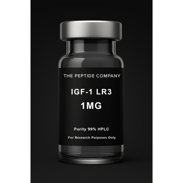
IGF-1 LR3 1mg
IGF-1 LR3 1mg is a research compound studied for insulin-like growth factor signaling, receptor binding dynamics, and cellular growth pathway mechanisms. For research use only.
Tesamorelin is a synthetic peptide analog of growth hormone–releasing hormone (GHRH), designed to enhance stability and receptor affinity. It contains 44 amino acids and mimics the native GHRH sequence responsible for stimulating growth hormone (GH) secretion from the anterior pituitary. In research contexts, Tesamorelin is used to investigate body composition, lipid metabolism, visceral adipose tissue (VAT) modulation, and hepatic fat regulation. It is particularly studied for its ability to modulate GH/IGF-1 axis dynamics, serving as a model compound in metabolic and endocrine research.
Mechanism of Action (Research Context)
Tesamorelin binds to GHRH receptors located on pituitary somatotrophs, activating a Gs-coupled signal transduction cascade that stimulates adenylate cyclase activity. This increases cyclic adenosine monophosphate (cAMP) levels, activating protein kinase A (PKA) and promoting growth hormone synthesis and release. The subsequent rise in GH elevates hepatic production of insulin-like growth factor 1 (IGF-1), which mediates downstream anabolic and lipolytic processes. These include enhanced lipolysis, improved lipid turnover, reduced hepatic lipogenesis, and redistribution of adipose tissue — particularly the reduction of visceral fat stores.
Selected Research Highlights
• Visceral Adipose Tissue (VAT): Multiple studies have documented significant VAT reduction measured via MRI or CT after Tesamorelin administration, suggesting its utility in modulating central fat depots.• Liver Fat and NAFLD Research: Data indicate reductions in hepatic fat fraction and improved markers of steatosis, positioning Tesamorelin as a candidate for studying non-alcoholic fatty liver mechanisms.• Lipid Profile Improvements: Studies report reductions in triglycerides and non-HDL cholesterol levels, contributing to improved metabolic biomarkers.• Body Composition: Increases in lean mass and reductions in central adiposity are frequently observed, indicating selective metabolic repartitioning.• Glucose Homeostasis: Generally stable in normoglycemic research subjects, although impaired glucose tolerance may occur in susceptible models — requiring protocol-based monitoring.
Potential Research Benefits (Reported in Literature)
• Model compound to investigate GH/IGF-1 axis function in metabolic studies• Tool for evaluating visceral adiposity and ectopic fat distribution• Research on GH-mediated lipid oxidation and metabolic rate enhancement• Comparative framework against GLP-1 and GIP-based incretin mechanisms• Investigations into the link between GH axis modulation and hepatic fat metabolism• Potential exploration in muscle anabolic signaling and body composition optimization
Chemical / Physical Information
• Sequence: A 44–amino acid peptide analog of GHRH• Class: GHRH receptor agonist• Molecular Weight: Approximately 5,117 Da• Appearance: White lyophilized powder• Solubility: Water soluble• Storage: Lyophilized at -20 °C, protected from light and moisture; reconstituted solutions should be aliquoted and frozen to prevent repeated freeze–thaw cycles.
Study Design Notes (Research Context)
Research protocols commonly employ subcutaneous administration, with dosing frequencies ranging from daily to every other day, depending on study objectives. Endpoints include MRI-based VAT quantification, hepatic proton-density fat fraction (PDFF) assessment, lipid panel evaluation, IGF-1 measurement, and quality-of-life metrics. Trial durations typically range from 12 to 52 weeks, with continued effects dependent on sustained GH stimulation.
Safety / Tolerability (Reported in Literature)
• Common Observations: Mild injection-site reactions (erythema, pruritus), edema, arthralgia, and transient headache.• Endocrine Responses: Predictable rise in IGF-1 levels proportional to GH induction.• Glucose Effects: Transient glucose intolerance or insulin resistance observed in subsets; standard monitoring protocols mitigate risk.• Cardiometabolic Profile: Generally well tolerated, with no major cardiovascular signal in short-term studies.• Research Screening: Typical exclusion criteria include active malignancy, uncontrolled diabetes, and pregnancy, aligning with GH/IGF-1 biology.
Regulatory & Compliance Notes
Tesamorelin is approved in specific jurisdictions for clinical indications but is otherwise designated as a research-grade compound in laboratory settings. Procurement, handling, and research use must follow all applicable institutional and legal standards. Laboratories should maintain compliance documentation, including certificates of analysis (COAs) and material safety data sheets (MSDS).
References (Selection)
1. Falutz J, et al. (2005). Effects of Tesamorelin, a Growth Hormone–Releasing Factor Analog, in Individuals with Central Fat Accumulation. J Clin Endocrinol Metab.2. Stanley TL, et al. (2019). Tesamorelin Reduces Liver Fat and Fibrosis in Patients with NAFLD. Lancet Diabetes Endocrinol.3. Gelato MC, et al. (2008). Growth Hormone-Releasing Factor Analog Effects on Body Composition and Metabolism. Metabolism.4. Falutz J, et al. (2010). Long-Term Effects of Tesamorelin on Visceral Adipose Tissue. J Clin Endocrinol Metab.5. Stanley TL, et al. (2021). Comparative Metabolic Outcomes Following Tesamorelin in Obesity Research. Obesity (Silver Spring).
Disclaimer
This publication is intended for educational and research purposes only. Tesamorelin is not approved herein for human or veterinary use. All studies and experiments involving Tesamorelin must adhere to institutional biosafety, ethical, and legal requirements governing peptide research. ——————————————
Selected References
PMID: 21098782 — Tesamorelin effects on IGF-1 and metabolic regulation
PMID: 20826578 — GHRH analogs and body composition changes
PMID: 21753056 — Tesamorelin’s impact on visceral adipose tissue reduction
PMID: 25826926 — Long-term metabolic and endocrine outcomes of GHRH analog therapy
Journal of Clinical Endocrinology & Metabolism — GHRH analog mechanisms and applications
Frontiers in Endocrinology — Growth hormone axis modulation in metabolic disease
FAQ:
What is Tesamorelin?
Tesamorelin is a synthetic growth hormone–releasing hormone (GHRH) analog studied for its ability to stimulate endogenous GH secretion and influence metabolic biomarkers in research settings.
How does Tesamorelin work in research?
Tesamorelin binds to GHRH receptors, increasing pulsatile GH release, which may affect IGF-1 levels, lipolysis, and lipid metabolism in experimental models.
Is Tesamorelin approved for human use?
Clinical Tesamorelin exists for specific medical indications, but the Tesamorelin discussed in research contexts is not approved for general consumer use.
What are researchers studying Tesamorelin for?
Research explores Tesamorelin for visceral fat metabolism, GH/IGF-1 pathway activation, metabolic health markers, and body composition changes.
Does Tesamorelin affect IGF-1 levels?
Yes, studies frequently report increases in IGF-1 as a downstream effect of enhanced endogenous GH release.
How is Tesamorelin different from CJC-1295 or Sermorelin?
Tesamorelin is a full GHRH analog with a different structure and receptor interaction, producing distinct GH pulse patterns compared to shorter GHRH fragments or GHRP compounds.
Are there known side effects in Tesamorelin research?
Research models note possible responses such as injection site irritation or transient GH-related effects, though safety varies by study design.
Related Research Compounds
Frag 176–191: Growth Hormone–Derived Fragment and Lipolytic Research Mechanisms
Sermorelin: GHRH Fragment Research and Growth Hormone Pulsatility Models
IGF-1 Analogues: LR3 and DES Structural Variations and Receptor Binding in Research Models
CJC-1295: GHRH Analog, DAC Conjugation, and Growth Hormone Pulsatility in Research
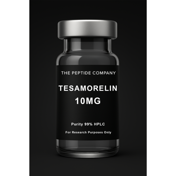
Tesamorelin 10mg
Tesamorelin 10mg is a GHRH analog research compound studied for growth hormone axis signaling, endocrine pathway modulation, and metabolic regulation. For research use only.
Introduction
Sermorelin is a 29-amino-acid fragment of endogenous GHRH used in research to study growth hormone (GH) pulsatility, endocrine timing, and receptor-specific signaling. It preserves the natural binding region of GHRH and produces a short-lived, physiologically aligned GHRH signal.
What Is Sermorelin?
Sermorelin consists of the first 29 amino acids of human GHRH. This segment contains the full receptor-binding region necessary to activate the GHRH receptor (GHRHR) on pituitary somatotroph cells, producing a controlled GH pulse in research models.
Structural Overview
Endogenous GHRH has 44 amino acids. The 1-29 region contains the biologically active binding sequence. Sermorelin isolates this region, allowing researchers to study receptor-specific activation with rapid degradation similar to native GHRH.
Mechanism of Action
Sermorelin binds the GHRH receptor, activating Gs protein signaling, increasing cAMP, and activating PKA. This triggers calcium influx, GH vesicle exocytosis, and downstream IGF-1 pathway activation. Because feedback loops remain intact, GH pulses remain physiologically regulated.
Research Applications
Sermorelin is used to study GH pulse patterns, GHRH-somatostatin interactions, IGF-1 downstream signaling, circadian timing, and comparative analog studies. It serves as the baseline reference molecule for other GHRH analogues such as CJC-1295 and Tesamorelin.
Context With Other GHRH Analogues
Sermorelin is often compared mechanistically with CJC-1295 (short and long-acting forms) and Tesamorelin to evaluate how structural length, stability, and receptor engagement influence GH pulsatility in controlled experiments.
Summary
Sermorelin provides a physiologic, short-duration GHRH signal ideal for studying GH pulsatility, feedback regulation, and endocrine timing. Its rapid degradation, receptor specificity, and intact feedback loops make it a valuable research tool for GH/IGF axis analysis.
Sermorelin vs Endogenous GHRH (Research Comparison)
| Property | Endogenous GHRH | Sermorelin (GHRH 1-29) |
| Length | 44 amino acids | 29 amino acids |
| Binding Region | N-terminal region | Identical N-terminal binding region |
| Stability | Longer due to additional sequence | Shorter, rapidly degraded |
| Signaling Style | Pulsatile | Pulsatile, research analogue |
FAQ:
What is Sermorelin in research?
Sermorelin is a synthetic fragment of the growth hormone-releasing hormone (GHRH) molecule studied in experimental models for its effects on stimulating growth hormone (GH) pulsatility, endocrine regulation, and neuroendocrine axis signaling. It is provided for laboratory research and in-vitro use only.
How does Sermorelin function in laboratory studies?
In research, Sermorelin interacts with GHRH receptors at the pituitary, triggering episodic GH release and supporting physiological pulsatile GH secretion rather than continuous GH levels. These findings remain experimental and apply to controlled study settings.
Is Sermorelin considered a therapeutic product?
No. The Sermorelin referenced here (by The Peptide Company) is for laboratory and in-vitro research use only. It is not a therapy, drug, supplement, or product for human or clinical use.
What research applications involve Sermorelin?
Researchers explore Sermorelin in controlled models of GH-axis regulation, aging-related somatotropic decline, GH-pulse amplitude and frequency dynamics, and endocrine/neuroendocrine signaling.
Does Sermorelin influence GH pulses in studies?
Yes — preclinical and clinical studies show Sermorelin can restore or enhance GH pulse amplitude, support rhythmic GH release, and influence downstream IGF-1 levels in controlled research settings.
How is Sermorelin typically handled in research settings?
As a lyophilized powder, Sermorelin should be stored away from light and moisture. When reconstituted, it should be refrigerated and used in institutional laboratory or in-vitro protocols only.
Can Sermorelin be used by consumers or self-administered?
No. Sermorelin is not intended for self-administration or consumer use. It is strictly reserved for institutional laboratory research or in-vitro experimentation.
Related Research Compounds
IGF-1 Analogues: LR3 and DES Structural Variations and Receptor Binding in Research Models
CJC-1295: GHRH Analog, DAC Conjugation, and Growth Hormone Pulsatility in Research
References (Selection)
PMID: 18031173 — Sermorelin: a review of its use in the diagnosis and regulation of GH secretion
PMID: 1688413 — Brain effects of growth hormone-releasing hormone (sermorelin) in older adults
PMID: 7108996 — Role of GHRH/ghrelin axis on growth hormone release and pulsatility

Sermorelin 5mg
Sermorelin 5mg is a research compound studied as a GHRH fragment for growth hormone pulsatility, pituitary axis modulation, and endocrine regulation. For research use only.
Introduction
Insulin-like Growth Factor-1 (IGF-1) is a central mediator of growth hormone (GH) signaling involved in tissue growth, cellular repair, metabolic function, and proliferation. Two widely examined analogues—IGF-1 LR3 and IGF-1 DES—help researchers study how structural modifications influence receptor activation, signaling duration, and tissue-specific responses. This article outlines IGF-1 biology followed by detailed sections on LR3 and DES, written strictly from a mechanistic, research-based perspective.
What Is IGF 1?
IGF-1 is a peptide hormone produced primarily in the liver in response to GH stimulation. It activates IGF-1 receptors (IGF-1R) and, to a lesser extent, insulin receptors (IR). Its downstream effects operate through the PI3K–AKT, MAPK, and mTOR pathways, influencing metabolism, survival, and growth. Because native IGF-1 is regulated by IGF binding proteins (IGFBPs), analogues offer a controlled way to study IGF-1R-specific activity.
LR3 IGF-1: Structural Modification and Extended Activity
IGF-1 LR3 features a 13-amino-acid N-terminal extension and a substitution of arginine at position 3. These modifications significantly reduce IGFBP binding, increasing receptor availability and extending activity. In research settings, LR3 allows long-duration IGF-1R stimulation and activation of PI3K-AKT, MAPK, and mTOR pathways under reduced IGFBP interference.
IGF-1 DES (1–3 IGF-1): Truncated Analogue With Enhanced Local Activity
IGF-1 DES lacks the first three amino acids of the IGF-1 sequence, further decreasing IGFBP binding. This analogue produces rapid and localized IGF-1R activation with a shorter activity period. Its strong local receptor affinity makes DES useful for studying tissue-specific IGF-1 effects and acute signaling responses.
Mechanistic Comparison
LR3 and DES share the same receptor target (IGF-1R) but differ in duration, IGFBP interaction, and tissue penetration. LR3 provides sustained receptor access and broad exposure, while DES offers intense, localized IGF-1R activation for shorter intervals. Both contribute complementary insights into IGF-1’s biological roles in controlled research environments.
IGF-1R Signaling Overview
IGF-1R engagement activates multiple intracellular pathways:• PI3K → AKT (survival, glucose metabolism) • RAS → MAPK (growth, differentiation) • mTOR (protein synthesis, anabolic signaling) • IRS-1/2 (insulin/IGF cross-talk)These pathways underpin IGF-1’s metabolic and growth-regulating effects in research models.
Summary
IGF‑1 analogues such as LR3 and DES help researchers examine how structural modifications influence receptor engagement, signaling intensity, and tissue specificity. LR3 provides sustained IGF‑1R signaling due to reduced IGFBP interaction, while DES offers rapid, localized receptor activation with a shorter half-life. Together, the analogues support comprehensive study of IGF‑1 pathway biology under different experimental designs.
LR3 vs DES: Mechanistic Comparison (Research Only)
| Parameter | IGF-1 LR3 | IGF-1 DES |
| Structure | 13aa extension + Arg3 substitution | Truncated by 3 amino acids |
| IGFBP Interaction | Reduced | Strongly reduced |
| Receptor Activation | Sustained IGF-1R exposure | Rapid, localized IGF-1R activation |
| Research Focus | Extended signaling duration | Short-duration, high-intensity signaling |
| Half-Life | Longer activity window | Shorter activity window |
Educational & Research Disclaimer
This article is for educational and research purposes only. No medical advice or product claims are made. These compounds are not approved for human use and are intended solely for laboratory research.
FAQ:
What are IGF-1 LR3 and IGF-1 DES in research?
IGF-1 LR3 and IGF-1 DES are synthetic analogues of the native growth-factor peptide Insulin‑like Growth Factor 1 (IGF-1). LR3 includes added amino acids and a substitution to reduce binding to IGF-binding proteins, while DES is a truncated form (missing the first three amino acids). Both are studied for their modified receptor-binding profiles, bioavailability, and signaling activity in experimental models.
How do IGF-1 LR3 and IGF-1 DES function in laboratory studies?
Research shows that LR3 has reduced affinity for IGF-binding proteins (IGFBPs) and a longer half-life, allowing increased receptor interaction and signaling effects compared to native IGF-1. Similarly, DES(1-3)IGF-1 lacks the first three amino acids, exhibits markedly reduced IGFBP binding and enhanced potency in specific systems.
Are IGF-1 LR3 and IGF-1 DES considered therapeutic products?
No. The versions described here are for research use only by The Peptide Company. They are not approved therapies, supplements, or consumer products and are intended for controlled laboratory and in-vitro experimentation only.
What research applications involve IGF-1 LR3 and IGF-1 DES?
These analogues are used in experimental studies investigating tissue-specific growth-factor effects, receptor-binding kinetics, bioavailability outside IGFBP regulation, metabolic and endocrine signaling, and targeted cell-growth models.
Do IGF-1 LR3 and IGF-1 DES have different receptor-binding profiles compared to native IGF-1?
Yes. LR3 has a longer chain and substitution that increases its resistance to IGFBPs and prolongs systemic availability. DES lacks the N-terminal three residues, giving it low IGFBP affinity and potentially higher localized potency in tissue models.
How are IGF-1 LR3 and IGF-1 DES typically stored and handled in research settings?
They are supplied as lyophilized powders, stored in dry, stable conditions away from light and extreme heat. After reconstitution according to lab protocol, they should be properly refrigerated and used only for designated in-vitro or institutional applications.
Can IGF-1 LR3 or IGF-1 DES be administered or used by consumers?
No. They are not for self-administration or consumer use. These analogues are strictly reserved for laboratory and in-vitro research environments and must not be marketed or used as therapies.
Related Research Compounds:
Sermorelin: GHRH Fragment Research and Growth Hormone Pulsatility Models
Follistatin: Myostatin-Regulated Pathways and Advanced Muscle Research
References (Selection)
PMID: 33587816 — Detection and characterization of IGF-1 analogues LR3 and DES in human serum PubMed
PMID: 10872804 — Insulin-like Growth Factor-1 survival mechanism and analogue binding (LR3 & DES) AHA Journals
PMID: 7913862 — Insulin-like growth factor-1 and its monitoring in research; includes LR3 variant discussion PMC
PMID: 16597689 — Mechanisms of tissue specificity and binding proteins in IGF analogues (DES variant studied) (excerpt) — you may want to verify for accuracy
PMID: 25828794 — Analogue binding-protein modulation and cell-growth specificity (extract from broader IGF analogue research) — you may want to verify
IGF-1 LR3 1mg
IGF-1 LR3 1mg is a research compound studied for insulin-like growth factor signaling, receptor binding dynamics, and cellular growth pathway mechanisms. For research use only.

Introduction
Growth hormone (GH) release is governed by a tightly regulated endocrine axis involving GHRH, somatostatin, and feedback from GH and IGF-1. CJC‑1295 represents a family of GHRH analog constructs used in research to explore GH pulsatility, endocrine regulation, and tissue signaling dynamics. This article outlines the biology of the GHRH receptor, followed by detailed sections on CJC‑1295 with DAC and CJC‑1295 without DAC.
What Is CJC‑1295?
CJC‑1295 is a synthetic analog of growth hormone releasing hormone (GHRH) designed to bind the GHRH receptor on anterior pituitary somatotrophs. It enhances endogenous GH secretion in a physiological, pulse‑based manner. Two primary constructs exist: CJC‑1295 with DAC (Drug Affinity Complex) and CJC‑1295 without DAC (Mod GRF 1‑29). Both utilize the same receptor system but differ in structural stability and duration.
CJC‑1295 With DAC
The DAC component allows reversible binding to plasma proteins such as albumin, increasing stability and circulation time. This provides prolonged GHRH receptor stimulation without eliminating the natural pulsatile nature of GH release. DAC‑modified constructs are used to study extended GH/IGF‑1 signaling and chronic endocrine modulation in research settings.
CJC‑1295 Without DAC (Mod GRF 1‑29)
Mod GRF 1‑29 is a 29‑amino acid fragment of GHRH with structural substitutions that increase stability. Without DAC, it does not bind albumin and therefore produces a shorter-lived GHRH signal. This makes it valuable for studying acute GH responses, timing dynamics, and pulse behavior in controlled experiments.
Comparative Overview
CJC‑1295 with DAC and Mod GRF 1‑29 both activate the GHRH receptor but differ in duration and kinetic emphasis. DAC‑modified constructs allow sustained receptor engagement, while Mod GRF 1‑29 provides rapid, discrete signaling useful for timing-based research.
Synergy and Related Research
GHRH analogs are often studied alongside other endocrine peptides. Tesamorelin provides a useful structural comparison as another GHRH analog, while ghrelin receptor agonists such as GHRP‑2 or Ipamorelin act through a separate pathway (GHSR). IGF‑1 LR3 is frequently examined downstream for mapping GH‑IGF signaling behavior.
Summary
CJC‑1295 provides two structurally related tools for studying GH pulsatility and endocrine signaling. DAC‑modified CJC‑1295 allows extended receptor presence, while Mod GRF 1‑29 enables investigation of acute GH pulse patterns. Together, they support research into growth signaling, endocrine timing, and tissue regulation.
CJC‑1295 With DAC vs Without DAC (Research Comparison)
| Feature | CJC‑1295 With DAC | CJC‑1295 Without DAC (Mod GRF 1‑29) |
| Structural Modification | Includes Drug Affinity Complex | No DAC; modified GHRH(1‑29) |
| Albumin Binding | Yes, reversible | Minimal to none |
| Circulation Duration | Prolonged | Short, pulse‑like |
| GH Pattern | Extended stimulation window | Sharp, discrete GH pulses |
| Research Focus | Long-term GH/IGF‑1 studies | Timing and acute GH response |
Educational & Research Disclaimer
This article is for educational and research purposes only. No medical advice or clinical claims are made. Compounds discussed are not approved for human use and are intended exclusively for laboratory research.
Related Searches:
IGF-1 Analogues: LR3 and DES Structural Variations and Receptor Binding in Research Models
Sermorelin: GHRH Fragment Research and Growth Hormone Pulsatility Models
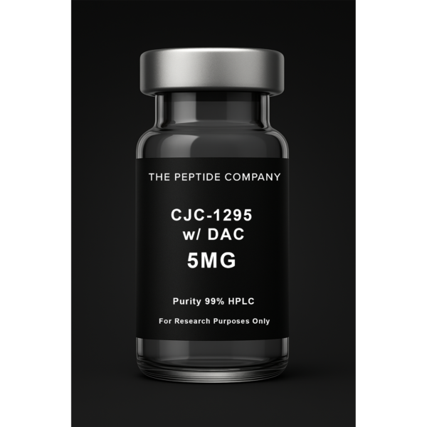
CJC w/ DAC – 5mg
CJC-1295 with DAC is a modified growth hormone–releasing hormone (GHRH) analog studied for prolonged stimulation of growth hormone signaling pathways and endocrine regulation. For research use only.

Introduction
Epithalon (Epitalon) is a synthetic tetrapeptide with the sequence Ala–Glu–Asp–Gly, modeled after endogenous pineal peptides. Research explores its roles in telomere regulation, cellular senescence, circadian rhythm biology, mitochondrial signaling, and oxidative-stress pathways. Its small molecular structure allows broad interaction across cellular regulatory networks.
Structural Biology of Epithalon
Epithalon’s tetrapeptide composition—Ala, Glu, Asp, Gly—confers high stability and efficient diffusion properties. It is structurally simpler and more defined than Epithalamin, the natural pineal extract from which its concept originates. Research focuses on its structural advantages, purity, and selective signaling behaviors.
Telomere Biology and Telomerase Research
Studies investigate Epithalon’s influence on telomerase reverse transcriptase (TERT) expression, telomerase activation, and telomere maintenance. Key areas include shelterin complex regulation (TRF1, TRF2, POT1, TIN2), DNA end-protection, and modulation of senescence markers such as p53, p21, and p16INK4a. Researchers examine how Epithalon affects genomic stability and replicative longevity.
Circadian Rhythm and Pineal Regulation
Epithalon is closely tied to circadian biology due to its pineal origins. Research explores its influence on melatonin cycles and transcription of circadian-clock genes including CLOCK, BMAL1, PER1/2, and CRY1/2. These pathways regulate sleep-wake cycles, endocrine rhythmicity, metabolic timing, and peripheral tissue transcriptional oscillations.
Mitochondrial Function and Oxidative-Stress Pathways
Mitochondrial resilience and oxidative balance are central themes in Epithalon research. Studies examine interactions with NRF2, SIRT1, FOXO transcription factors, and UPRmt (mitochondrial unfolded protein response). Epithalon is evaluated for influences on ROS handling, mitochondrial gene expression, and antioxidant signaling patterns.
Protein Homeostasis and Autophagy
Epithalon is studied for its impact on autophagic signaling (LC3-II, Beclin-1, ATG genes), proteasomal pathways, and protein-quality control systems. Research examines its potential role in maintaining proteostasis, mitigating misfolded protein accumulation, and supporting cellular cleanup mechanisms linked to aging biology.
Immune Signaling and Inflammatory Pathways
Research explores Epithalon’s modulation of cytokine networks including IL-6, TNF-α, IL-1β, and interferon-associated signaling. Studies also investigate its influence on neuroendocrine–immune communication, including hypothalamic–pituitary–immune axis dynamics.
Cellular Longevity and Aging Signatures
Epithalon research covers DNA-damage markers (γ-H2AX, oxidized guanine lesions), senescence-associated secretory phenotype (SASP) profiles, and transcriptional pathways associated with AMPK, PGC‑1α, SIRT family genes, and mitochondrial biogenesis. These studies help map Epithalon’s potential role in aging and cellular adaptation.
Summary
Epithalon is a structurally simple synthetic tetrapeptide studied for its involvement in telomere maintenance, circadian regulation, mitochondrial signaling, proteostasis, immune modulation, and longevity transcription networks. Its broad signaling interactions make it a critical compound in aging and mitochondrial research.
Educational & Research Disclaimer
This article is for educational and scientific research purposes only. No therapeutic claims or usage recommendations are provided. Compounds referenced are not approved for human use and are intended solely for controlled laboratory experimentation.
FAQ:
What is Epithalon in research?
Epithalon (Epitalon) is a synthetic tetrapeptide analog of epithalamin, studied for its potential roles in telomere biology, cellular senescence, and circadian-rhythm regulation under controlled laboratory conditions.
How does Epithalon function in laboratory studies?
In research models, Epithalon is explored for its influence on telomerase activity, chromatin structure, melatonin signaling, and age-associated gene expression. These findings remain experimental and limited to in-vitro or animal-model conditions.
Is Epithalon considered a therapeutic compound?
No. Epithalon supplied by The Peptide Company is for laboratory and in-vitro research only. It is not a therapy, supplement, drug, or product for human or clinical use.
What research applications involve Epithalon?
Researchers study Epithalon in models of aging biology, circadian-clock gene regulation, oxidative-stress responses, telomere maintenance, and experimental longevity pathways.
Does Epithalon affect telomere length in research?
Some experimental studies in cell cultures and animal models suggest telomerase-activation potential, though results are inconsistent and strictly preclinical. These observations do not imply any clinical effect.
How is Epithalon typically handled in research environments?
It is supplied as a lyophilized powder and stored away from heat and humidity. After reconstitution, it is refrigerated and used only within institutional laboratory workflows.
Can Epithalon be administered by consumers?
No. Epithalon is intended exclusively for controlled laboratory and in-vitro research studies.
2. Related Research Compounds
IGF-1 Analogues: LR3 and DES Structural Variations and Receptor Binding in Research Models
CJC-1295: GHRH Analog, DAC Conjugation, and Growth Hormone Pulsatility in Research
Sermorelin: GHRH Fragment Research and Growth Hormone Pulsatility Models
3. References
PMID: 11769766 — Peptide epithalon and telomerase activity regulation in aging models
PMID: 11217738 — Effects of epithalon on pineal peptides and circadian function
PMID: 11399890 — Telomere dynamics and peptide regulation in senescence research
PMID: 11708714 — Peptide-based modulation of chromatin and cellular aging markers
PMID: 11762917 — Experimental gerontology: pineal peptides and lifespan mechanisms
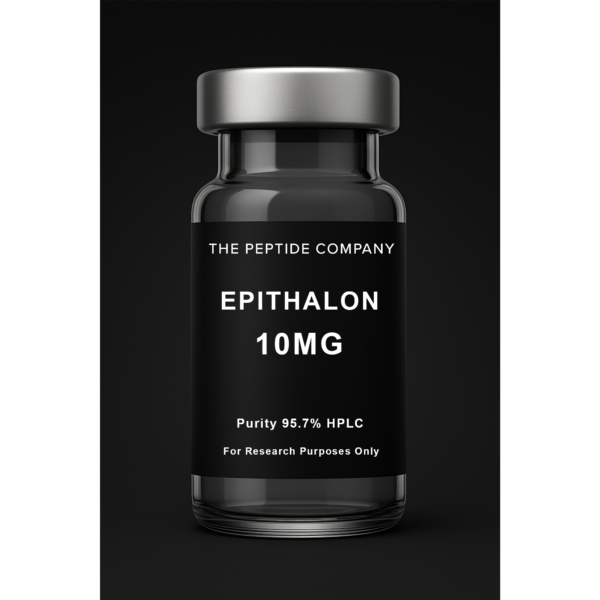
Epithalon 10mg
Epithalon 10mg is a research peptide studied for telomerase activation, cellular aging pathways, and circadian rhythm regulation in laboratory research models. For research use only.