Abstract & Overview
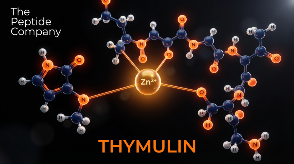
Thymulin — also designated facteur thymique sérique (FTS) or serum thymic factor (STF) — is an endogenous nonapeptide hormone produced by two distinct epithelial populations within the thymus. First isolated and biochemically characterised by Bach and colleagues in 1977, thymulin occupies a unique position in immunobiology as the only known thymic hormone whose biological activity is absolutely contingent upon coordination with a divalent zinc ion (Zn²⁺). In the absence of zinc, the peptide exists as an inactive apo-form; zinc binding induces a conformational transition that confers full receptor-binding competence and downstream signalling capacity [1][2].
Thymulin’s primary role is the orchestration of T-lymphocyte maturation — both within the thymic microenvironment and at extrathymic peripheral sites. Beyond this canonical immunological function, thymulin operates as a bidirectional communicator between the immune system and the hypothalamic-pituitary-adrenal (HPA) axis, modulating the secretion of multiple adenohypophyseal hormones including luteinizing hormone (LH), follicle-stimulating hormone (FSH), growth hormone (GH), prolactin (PRL), thyroid-stimulating hormone (TSH), and adrenocorticotropic hormone (ACTH) [3][4]. Circulating thymulin levels peak in the early postnatal period and decline progressively with age, establishing the peptide as a quantitative biomarker of immunosenescence [5].
“Thymulin is not toxic and one may foresee its clinical use as one of the major immunoregulatory agents.” — Bach JF, Medical Oncology & Tumor Pharmacotherapy (1989) [6].
Molecular Identity and Structural Architecture
Thymulin is a nonapeptide with the amino acid sequence H-Pyr-Ala-Lys-Ser-Gln-Gly-Gly-Ser-Asn-OH, where Pyr denotes a pyroglutamate residue at the N-terminus — a cyclised form of glutamine that confers resistance to aminopeptidase degradation. The molecular formula is C₃₃H₅₄N₁₂O₁₅ with a molar mass of 858.86 g/mol (CAS: 63958-90-7; PubChem CID: 3085284). The serum half-life of thymulin is approximately 10.3 minutes, reflecting rapid clearance that necessitates continuous thymic secretion for sustained biological activity [5].
The zinc-binding site is formed by coordination chemistry involving the N-terminal pyroglutamate, the ε-amino group of lysine at position 3, and the hydroxyl groups of the two serine residues at positions 4 and 8. This tetradentate coordination geometry creates a stable 1:1 Zn:peptide metallopeptide complex. Critically, monoclonal antibody studies by Dardenne et al. demonstrated that the zinc-bound conformation exposes a distinct epitope not present on the apo-peptide, confirming that zinc binding is not merely stabilising but structurally transformative [2]. Chelation of zinc with EDTA or similar agents abolishes biological activity, while re-introduction of Zn²⁺ ions reconstitutes activity within minutes [1].
Thymulin secretion is regulated by a network of endocrine and paracrine signals including prolactin, growth hormone, interleukins IL-1α and IL-1β, and opioid peptides (β-endorphins and β-enkephalins). The peptide follows a circadian secretory rhythm, and physiologically elevated ACTH levels correlate positively with plasma thymulin concentrations, reflecting the deep integration of thymic endocrinology with the HPA axis [3].
Mechanistic Rationale: Zinc Activation and Receptor Signalling
Zinc-Dependent Activation and T-Cell Maturation
The Zn²⁺-bound metallopeptide form of thymulin is the sole biologically active species. Upon zinc coordination, the peptide adopts a compact conformation that enables high-affinity binding to surface receptors expressed on immature lymphoid precursor cells within the thymic cortex and medulla. Binding of thymulin to these receptors initiates intracellular signalling cascades that prime T-cell precursors for progressive maturation steps, culminating in the expression of key surface phenotypic markers: CD90 (Thy-1), CD3, CD4, and CD8 [7].
Thymulin exerts both intra- and extrathymic effects on T-cell differentiation. Within the thymus, it acts in concert with thymic epithelial cells and their cytokine networks to orchestrate the sequential developmental programme from double-negative (CD4⁻CD8⁻) precursors through double-positive (CD4⁺CD8⁺) intermediates to mature single-positive (CD4⁺ or CD8⁺) T-cells. Extrathymically, thymulin can act on peripheral lymphoid precursors, partially restoring T-cell function in thymectomised animals — a property that distinguishes it from thymosin α₁ and thymopoietin, which lack significant extrathymic activity [7][8].
Thymulin also enhances natural killer (NK) cell cytotoxic activity, broadening its immunomodulatory profile beyond the T-cell lineage. Deficits in both Zn²⁺ and thymulin bioactivity have been documented in patients with Crohn’s disease and acute lymphoblastic leukaemia, suggesting that zinc-thymulin insufficiency may contribute to the immune dysregulation characteristic of these conditions [8].
Neuroendocrine Axis: Thymus-Pituitary Communication
Thymulin acts directly on anterior pituitary cells to modulate the secretion of multiple adenohypophyseal hormones. Research by Brown et al. demonstrated that thymulin stimulates LH release, and that co-incubation with gonadotropin-releasing hormone (GnRH) produces a synergistic effect on LH secretion and an additive effect on FSH release [3]. These interactions are mediated through second messenger pathways — specifically, accumulation of cyclic AMP (cAMP) and cyclic GMP (cGMP) following thymulin exposure in pituitary cell preparations — pointing to a receptor-mediated process whose molecular identity remains under investigation [3].
The neuroendocrine effects of thymulin are age-dependent: responsiveness of pituitary cells to thymulin declines in aged animals, paralleling the age-related fall in circulating thymulin levels. This bidirectional relationship — wherein thymulin modulates pituitary output while pituitary hormones (GH, PRL, ACTH) in turn regulate thymulin secretion — positions the peptide as a central node in the neuroendocrine-immune communication network [4][5].
Anti-Inflammatory and Cytokine Regulatory Mechanisms
A growing body of preclinical evidence positions thymulin as a potent negative regulator of inflammatory signalling. The metallopeptide suppresses the production of key pro-inflammatory cytokines — including interleukin-1β (IL-1β), interleukin-6 (IL-6), tumour necrosis factor-α (TNF-α), and interferon-γ (IFN-γ) — while concurrently elevating the counter-regulatory cytokine interleukin-10 (IL-10). This dual action shifts the immune microenvironment toward a controlled, anti-inflammatory state rather than simply suppressing immune activity [9].
At the intracellular signalling level, thymulin dampens the activity of nuclear factor kappa-B (NF-κB) and p38 mitogen-activated protein kinase (p38 MAPK) — two transcriptional regulators central to inflammatory gene expression. Additionally, thymulin reduces the production of heat shock proteins HSP70 and HSP72, which are typically upregulated during cellular stress and inflammation, suggesting interference with the broader stress-response axis [10].
Neuroprotective Effects and the Peptide Analog of Thymulin (PAT)
Thymulin and its synthetic analog PAT (Peptide Analog of Thymulin) have demonstrated significant neuroprotective and analgesic properties in preclinical models. Astrocytes appear to be the primary CNS target for thymulin’s anti-inflammatory action. In models of intracerebroventricular endotoxin injection, thymulin-related peptide attenuated brain inflammation, reduced endotoxin-induced hyperalgesia, and restored near-normal levels of IL-6 and IL-1β across specific brain tissue regions [11].
In chronic inflammatory pain models, thymulin attenuated spinal neuroinflammation through suppression of spinal microglial activation — evidenced by reduced Iba-1 expression — and inhibition of p38 MAPK phosphorylation and TNF-α production in the spinal cord. These findings suggest that thymulin may interfere with central sensitisation mechanisms, offering a potential avenue for the treatment of neuropathic pain and neuroinflammatory conditions including rheumatoid arthritis and neurodegenerative disease [11][12].
Research Applications and Experimental Evidence
Immunosenescence and Age-Related Immune Decline
The progressive decline of thymulin with age is one of the most reproducible findings in thymic endocrinology. Circulating thymulin peaks in the early postnatal period (approximately 2 pg/mL in umbilical cord blood) and falls to near-undetectable levels by the sixth decade of life. This decline correlates with the involution of the thymus and the contraction of the naive T-cell repertoire — hallmarks of immunosenescence. Research models indicate that thymulin supplementation can partially restore T-cell differentiation capacity and NK cell activity in aged subjects, suggesting potential utility as an immunorestorative agent in the context of ageing [5][8].
Zinc Deficiency and Immune Dysfunction
Because thymulin activity is absolutely dependent on zinc bioavailability, zinc deficiency states produce a functional thymulin insufficiency even when peptide synthesis is intact. Studies in zinc-deficient rodent models demonstrate impaired T-cell differentiation, reduced NK cell activity, and elevated susceptibility to infection — all of which can be partially reversed by zinc supplementation. Clinically relevant zinc deficiency states — including malnutrition, inflammatory bowel disease, and anorexia nervosa — are associated with significantly reduced thymulin bioactivity, providing a mechanistic link between nutritional zinc status and adaptive immune competence [1][13].
Tissue-Specific Protective Effects
Thymulin has demonstrated protective effects across multiple organ systems in preclinical research. In chemically induced diabetes models, thymulin suppressed hyperglycaemia and preserved pancreatic β-cell integrity by reducing the accumulation of pro-inflammatory cytokines that drive β-cell destruction. In nephrotoxicity models, thymulin mitigated renal damage via downregulation of inflammatory cascades and stress-response proteins. In colitis models, thymulin reduced colonic tissue inflammation by suppressing IL-1β, IL-6, TNF-α, and IFN-γ production. In pulmonary hypertension models, thymulin decreased IL-6 expression and reduced p38 MAPK activation, suggesting interference with cytokine-driven vascular remodelling [7].
Autoimmune and Oncological Research Models
Thymulin’s immunoregulatory properties have been investigated in models of autoimmune disease and haematological malignancy. In rheumatoid arthritis models, thymulin-related peptides attenuated joint inflammation and reduced pro-inflammatory cytokine burden. In models of acute lymphoblastic leukaemia, thymulin deficiency correlates with impaired immune surveillance, raising the possibility that thymulin restoration could support host anti-tumour immunity. Immunostimulatory effects have also been documented in animals infected with immunodeficiency virus and experimental encephalomyelitis [8][14].
Thymulin vs. Other Major Thymic Hormones: Comparative Profile
| Parameter | Thymulin (FTS) | Thymosin α₁ | Thymopoietin |
| Structure | Nonapeptide (9 AA) | 28-AA peptide | 49-AA peptide |
| Zinc Dependency | Absolute (metallopeptide) | None | None |
| Primary Action | T-cell differentiation | T-cell maturation/NK | T-cell differentiation |
| Extrathymic Activity | Yes (peripheral) | Yes | Limited |
| Neuroendocrine Effects | Extensive (HPA axis) | Limited | Not established |
| Anti-inflammatory | Yes (NF-κB, p38 MAPK) | Yes (moderate) | Limited data |
Pharmacological Considerations
Thymulin is a naturally occurring endogenous hormone available in synthetic form. Its small molecular size (858.86 Da) and nonapeptide structure render it amenable to solid-phase peptide synthesis with high purity. The serum half-life of approximately 10.3 minutes reflects rapid renal clearance, which has driven interest in developing more stable analogs such as PAT (Peptide Analog of Thymulin) with modified termini to resist peptidase degradation. The zinc-dependence of thymulin activity introduces an important pharmacological variable: the bioavailability of zinc at the site of action directly determines the proportion of peptide that exists in the active metallopeptide form [5][6].
Preclinical toxicology studies have not identified significant adverse effects associated with thymulin administration. Bach’s 1989 review noted the peptide’s favourable safety profile and proposed it as a candidate immunoregulatory therapeutic. Despite this, thymulin preparations have not advanced to formal clinical trials, a gap attributed in part to the complexity of zinc co-administration requirements and the availability of alternative immunomodulatory agents. The synthetic PAT analog, which retains the core immunomodulatory and neuroprotective properties while offering improved stability, represents the most clinically advanced thymulin-related compound currently under investigation [12][14].
Conclusion
Thymulin stands as one of the most structurally and functionally distinctive peptides in the thymic endocrine repertoire. Its absolute dependence on zinc for biological activity — a property unique among thymic hormones — creates a sophisticated regulatory checkpoint that couples immune function to systemic zinc homeostasis. As the primary orchestrator of T-lymphocyte maturation, thymulin governs the generation of the adaptive immune repertoire from early postnatal life through adulthood, with its progressive decline serving as a molecular signature of immunosenescence.
Beyond its canonical immunological role, thymulin’s extensive neuroendocrine interactions — modulating pituitary hormone secretion and responding to HPA axis signals — position it as a central integrator of immune-endocrine communication. Its anti-inflammatory properties, mediated through suppression of NF-κB and p38 MAPK pathways and elevation of IL-10, together with the neuroprotective analgesic effects demonstrated by PAT, open compelling research avenues in neuroinflammation, chronic pain, autoimmunity, and age-related immune decline. As synthetic analogs with improved pharmacokinetic profiles continue to be developed, thymulin’s translational potential as an immunorestorative and neuroprotective research compound remains substantial.
References
[1] Bach JF, Dardenne M, Pleau JM, Rosa J. Biochemical characterisation of a serum thymic factor. Nature. 1977;266(5597):55–7. doi:10.1038/266055a0. PMID: 300146.
[2] Dardenne M, Savino W, Berrih S, Bach JF. A zinc-dependent epitope on the molecule of thymulin, a thymic hormone. Proc Natl Acad Sci USA. 1985;82(20):7035–8. doi:10.1073/pnas.82.20.7035.
[3] Brown OA, Sosa YE, Dardenne M, Pléau JM, Goya RG. Studies on the gonadotropin-releasing activity of thymulin: changes with age. J Gerontol A Biol Sci Med Sci. 2000;55(4):B170–6. doi:10.1093/gerona/55.4.b170. PMID: 10811143.
[4] Brown OA, Sosa YE, Bolognani F, Goya RG. Thymulin stimulates prolactin and thyrotropin release in an age-related manner. Mech Ageing Dev. 1998;104(3):249–62. doi:10.1016/s0047-6374(98)00072-4. PMID: 9818729.
[5] Besman M, Zambrowicz A, Matwiejczyk M. Review of Thymic Peptides and Hormones: From Their Properties to Clinical Application. Int J Pept Res Ther. 2024;31:10. doi:10.1007/s10989-024-10666-y.
[6] Bach JF. Thymulin, a zinc-dependent hormone. Med Oncol Tumor Pharmacother. 1989;6(1):25–9. doi:10.1007/BF02985220. PMID: 2657247.
[7] Reggiani PC, Schwerdt JI, Console GM, Roggero EA, Dardenne M, Goya RG. Physiology and therapeutic potential of the thymic peptide thymulin. Curr Pharm Des. 2014;20(29):4690–6. doi:10.2174/1381612820666140130211157. PMID: 24588820.
[8] Dardenne M, Pléau JM, Nabarra B, et al. Contribution of zinc and other metals to the biological activity of the serum thymic factor. Proc Natl Acad Sci USA. 1982;79(17):5370–3. doi:10.1073/pnas.79.17.5370. PMID: 6957870.
[9] Haddad JJ, Saade NE, Safieh-Garabedian B. Thymulin: An emerging anti-inflammatory molecule. Curr Med Chem Anti-Inflamm Anti-Allergy Agents. 2005;4(3):333–8.
[10] Lunin SM, Khrenov MO, Novoselova TV, et al. Thymulin, a thymic peptide, prevents the overproduction of pro-inflammatory cytokines and heat shock protein Hsp70 in inflammation-bearing mice. Immunol Invest. 2008;37(8):858–70. doi:10.1080/08820130802447629. PMID: 18991101.
[11] Safieh-Garabedian B, Jabbur SJ, Dardenne M, Saadé NE. Thymulin related peptide attenuates inflammation in the brain induced by intracerebroventricular endotoxin injection. Neuropharmacology. 2011;60(2–3):496–504. doi:10.1016/j.neuropharm.2010.11.004. PMID: 21059360.
[12] Nasseri B, Zaringhalam J, Daniali S, et al. Thymulin treatment attenuates inflammatory pain by modulating spinal cellular and molecular signaling pathways. Int Immunopharmacol. 2019;70:225–234. doi:10.1016/j.intimp.2019.02.042. PMID: 30851702.
[13] Wade S, Bleiberg F, Mossé A, et al. Thymulin (Zn-facteur thymique sérique) activity in anorexia nervosa patients. Am J Clin Nutr. 1985;42(2):275–80. doi:10.1093/ajcn/42.2.275. PMID: 3927699.
[14] Dardenne M, Saade N, Safieh-Garabedian B. Role of thymulin or its analogue as a new analgesic molecule. Ann NY Acad Sci. 2006;1088:153–63. doi:10.1196/annals.1366.006. PMID: 17192563.
Disclaimer: This article is intended strictly for research and educational review purposes. Thymulin is an endogenous peptide hormone under preclinical investigation and has not been approved for human therapeutic use by any regulatory authority. All referenced studies were conducted in in vitro or preclinical (rodent) models unless otherwise stated. This document does not constitute medical advice and should not be used to guide clinical practice or personal health decisions.
thepeptidecompany.xyz | Research Division
What is Thymulin?
Thymulin is a thymic peptide studied for its role in immune system regulation, T-cell activity, and neuroendocrine signaling pathways.
What is Thymulin primarily researched for?
It is commonly studied for its involvement in thymus-derived immune signaling, inflammatory modulation, and cellular immune communication.
How does Thymulin interact with the immune system?
Research suggests Thymulin influences T-lymphocyte differentiation, immune signaling pathways, and cytokine-related activity in experimental models.
What biological pathways are associated with Thymulin?
Thymulin is studied in pathways related to immune regulation, neuroendocrine communication, inflammatory signaling, and thymic function.
Why is Thymulin important in thymus research?
It serves as a biomarker and signaling peptide associated with thymic activity and immune system maturation.
Is Thymulin studied outside of immune research?
Yes, experimental models also investigate its role in neuroendocrine interactions, stress signaling, and age-related thymic changes.
PMID:
6607412 — Thymulin structure and thymic hormone characterization
6140038 — Thymulin and T-cell differentiation research
6225511 — Immunoregulatory functions of thymulin
2951376 — Neuroendocrine interactions involving thymulin
8397102 — Thymulin and cytokine signaling pathways
10849509 — Thymic peptides and immune modulation
12745733 — Thymulin activity in inflammatory research
17026784 — Age-related thymic signaling and thymulin research
RELATED SEARCHES:
Thymosin Alpha-1 (Tα1): Immune Resilience and the Science of Thymic Restoration
Thymalin: Thymic Bioregulator Peptide, Immune Aging, and Epigenetic Control of Cellular Homeostasis
KPV: The Anti-Inflammatory Tripeptide and Cellular Repair Mechanism
LL-37: The Antimicrobial Peptide and Innate Immunity Blueprint
GHRP‑2 : Pituitary Axis Modulation, Ghrelin Receptor Activation, and Cellular Recovery Research
Abstract & Overview
Thymalin is a tissue-specific bioregulatory peptide derived from thymic tissue and classified within the broader family of cytomedins. It has been studied for its role in regulating immune cell differentiation, thymic function, and age-associated immune decline. Unlike thymosin peptides that act primarily through receptor-mediated immune signaling, Thymalin functions at the genomic and epigenetic level, influencing gene expression patterns involved in immune surveillance, cellular maturation, and systemic homeostasis. As a research compound, Thymalin serves as a central model for understanding peptide-based regulation of immune aging and thymic involution.
Background: Thymus Function and Immune Aging
The thymus is a primary lymphoid organ responsible for the maturation and selection of T lymphocytes. During early life, thymic activity is robust, ensuring effective immune surveillance and tolerance. With aging, the thymus undergoes involution, characterized by reduced epithelial tissue, diminished thymopoiesis, and impaired immune competence. This decline contributes to immunosenescence, increased susceptibility to infection, reduced vaccine responsiveness, and dysregulated inflammatory signaling. Research into thymic bioregulators such as Thymalin focuses on restoring or stabilizing thymic signaling pathways at the cellular and genomic level.
Molecular Classification and Cytomedin Biology
Thymalin belongs to the class of short regulatory peptides known as cytomedins, typically composed of two to four amino acids. These peptides exhibit pronounced tissue specificity and organotropism, allowing them to selectively influence gene expression within their target tissues. Thymalin’s peptide sequences were originally isolated from thymic extracts and later synthesized to enable controlled experimental investigation. Cytomedins differ fundamentally from classical hormones or cytokines, as their primary mode of action involves modulation of transcriptional and translational processes rather than receptor activation alone.
Mechanism of Action: Genomic and Epigenetic Regulation
The primary mechanism attributed to Thymalin involves peptide-mediated regulation of gene expression within immune and epithelial cells of thymic origin. Thymalin interacts with chromatin-associated proteins and nucleic acid structures, influencing transcriptional activity of genes responsible for lymphocyte differentiation, immune signaling balance, and cellular repair. Experimental data suggest that Thymalin modulates histone acetylation states and chromatin accessibility, thereby supporting stable gene expression patterns essential for immune competence. This epigenetic mode of action distinguishes Thymalin from short-acting immune peptides.
Effects on T-Cell Differentiation and Immune Balance
Research models indicate that Thymalin supports normalization of T-cell subpopulation ratios, including helper and cytotoxic T lymphocytes. By stabilizing thymic gene expression programs, Thymalin contributes to proper T-cell education and selection processes. This regulatory influence may help maintain immune tolerance while preserving effective pathogen response. In aging models, Thymalin has been associated with restoration of immune responsiveness and reduction of maladaptive inflammatory signaling.
Thymalin and Genomic Stability
An important aspect of Thymalin’s biological profile is its association with genomic stability. Studies have demonstrated increased expression of DNA repair enzymes and reduced markers of chromosomal instability following Thymalin exposure in experimental systems. These effects align with broader observations that tissue-specific bioregulators contribute to preservation of genomic integrity, particularly in rapidly renewing or immune-related tissues. Maintenance of genomic stability is central to preventing immune dysfunction and malignant transformation.
Comparative Analysis: Thymalin vs Thymosin Alpha-1
While both Thymalin and Thymosin Alpha-1 originate from thymic biology, their mechanisms and research applications differ significantly. Thymosin Alpha-1 primarily functions as an immune signaling peptide, enhancing innate and adaptive immune responses through receptor-mediated pathways. Thymalin, by contrast, operates at the level of gene regulation and epigenetic control, exerting longer-term modulatory effects on immune cell development and thymic function. This distinction positions Thymalin as a foundational bioregulator rather than an acute immune activator.
Role in Immune Aging and Systemic Homeostasis
Thymalin is frequently studied in the context of immune aging and systemic decline. By influencing thymic gene expression and lymphocyte maturation, Thymalin may counteract aspects of immunosenescence that contribute to chronic inflammation and impaired tissue repair. Its regulatory effects extend beyond the immune system, as balanced immune signaling is essential for maintaining systemic homeostasis and preventing age-associated pathologies.
Research Findings and Experimental Models
Experimental investigations involving Thymalin have demonstrated normalization of immune parameters in models of thymic dysfunction and aging. Observed outcomes include improved lymphocyte counts, enhanced immune responsiveness, and reduced inflammatory markers. In cellular studies, Thymalin has been shown to stimulate RNA synthesis and protein translation in immune cells, supporting its role as a genomic regulator. These findings underpin continued interest in Thymalin as a research tool for immune restoration studies.
Integration With Other Bioregulators
Within the bioregulator framework, Thymalin is often examined alongside peptides such as Vilon, Pancragen, Cardiogen, and Bronchogen. Each exhibits tissue-specific regulatory effects, while collectively contributing to systemic cellular balance. Thymalin’s role within this network highlights the cooperative nature of bioregulatory peptides in maintaining organism-wide homeostasis through targeted genomic modulation.
Limitations and Ongoing Research Questions
Despite extensive experimental study, important questions remain regarding Thymalin’s tissue specificity, long-term genomic effects, and interactions with other regulatory pathways. Further research is required to clarify the precise molecular targets of Thymalin and to delineate its role within complex immune and aging networks. As with all bioregulators, translation from experimental models to broader biological understanding remains an active area of investigation.
Summary
Thymalin represents a cornerstone thymic bioregulator peptide that provides critical insight into immune aging, thymic function, and epigenetic control of cellular homeostasis. Through genomic and chromatin-level regulation, Thymalin supports immune balance, genomic stability, and systemic resilience. Its study continues to inform broader research into peptide-based regulation of aging and immune competence.
Educational & Research Disclaimer
This document is provided for educational and scientific research purposes only. No medical, therapeutic, or usage claims are made. Thymalin and related compounds are not approved for human use and are intended solely for controlled laboratory and academic investigation.
FAQ:
What is Thymalin?
Thymalin is a thymus-derived bioregulatory peptide studied for its role in immune system signaling and cellular regulation.
What is Thymalin classified as?
Thymalin is classified as a cytomedin, a group of short peptides associated with tissue-specific cellular modulation.
How does Thymalin affect the immune system?
Research suggests Thymalin influences immune cell differentiation and signaling pathways related to thymic function.
Is Thymalin linked to aging research?
Thymalin is studied in aging models due to its association with immune system regulation and age-related cellular changes.
What role does Thymalin play in the thymus?
Thymalin is associated with thymic activity, particularly in processes related to immune cell development and maturation.
Does Thymalin influence gene expression?
Studies indicate Thymalin may interact with gene expression pathways, contributing to cellular homeostasis and regulation.
What biological processes is Thymalin studied for?
Thymalin is investigated in research involving immune signaling, epigenetic regulation, and cellular aging mechanisms.
How is Thymalin described in research contexts?
Thymalin is described as a tissue-specific regulatory peptide involved in immune modulation and cellular communication.
What makes Thymalin different from other peptides?
Its thymus-specific origin and focus on immune-related signaling pathways distinguish it from more generalized peptides.
Is Thymalin associated with epigenetic activity?
Research suggests Thymalin may play a role in epigenetic modulation, particularly in relation to immune system regulation and aging biology.
PMID:
PMID: 10378166 — Thymalin and its role in regulation of immune system function
PMID: 11241068 — Thymic peptides and their influence on T-cell differentiation and immune signaling
PMID: 12186792 — Cytomedins and their role in gene expression and cellular regulation
PMID: 12934759 — Thymalin effects on immune restoration and thymic activity
PMID: 14585163 — Bioregulatory peptides in aging and immune system modulation
PMID: 15798312 — Epigenetic regulation by short peptides in immune cells
PMID: 16804023 — Thymic peptides and mechanisms of immune homeostasis
PMID: 18261869 — Cytomedins and tissue-specific gene expression control
PMID: 21406990 — Role of peptide bioregulators in immune and endocrine signaling
PMID: 25900330 — Mechanisms of peptide regulation in aging and immune resilience
RELATED SEARCHES:
FOXO4-DRI : Targeting Cellular Senescence Through p53–FOXO4 Disruption and Senolytic Research
Thymosin Alpha-1 (Tα1): Immune Resilience and the Science of Thymic Restoration
KPV: The Anti-Inflammatory Tripeptide and Cellular Repair Mechanism
Introduction
Vasoactive Intestinal Peptide (VIP) is a 28–amino acid neuropeptide widely distributed throughout the central nervous system, peripheral nerves, and immune tissues. Originally identified in the gastrointestinal tract, VIP is now recognized as a multifunctional signaling molecule involved in neuroimmune communication, circadian rhythm regulation, vascular tone modulation, and anti-inflammatory transcriptional control in research models.
Molecular Structure and Biosynthesis
VIP is synthesized as part of a larger prepropeptide and processed through proteolytic cleavage into its active 28–amino acid form. It belongs to the secretin/glucagon peptide family and adopts an alpha-helical conformation critical for receptor binding. This structural arrangement enables high-affinity interactions with class B G‑protein–coupled receptors.
VIP Receptor Biology
VIP exerts its effects primarily through VPAC1 and VPAC2 receptors, with additional interactions involving PAC1 under certain conditions. These receptors are class B GPCRs coupled mainly to Gs proteins, leading to adenylate cyclase activation and increased intracellular cAMP. Downstream signaling cascades include PKA activation, CREB phosphorylation, and transcriptional regulation of immune and metabolic genes.
Neuroimmune Modulation
VIP is extensively studied for its role in immune regulation. Research models demonstrate VIP-mediated shifts toward anti-inflammatory cytokine profiles, including modulation of IL‑10, TNF‑α, IL‑6, and interferon-related pathways. VIP signaling influences T‑cell differentiation, macrophage polarization, and dendritic-cell activity, positioning it as a key neuroimmune regulator.
Circadian Rhythm and Suprachiasmatic Nucleus Signaling
VIP plays a critical role in circadian biology through its activity in the suprachiasmatic nucleus (SCN), the brain’s central circadian pacemaker. VIP–VPAC2 signaling synchronizes neuronal firing within the SCN and regulates clock-gene transcription, including CLOCK, BMAL1, PER, and CRY families. Research links VIP signaling to stability of circadian rhythms and temporal coordination of peripheral tissues.
Vascular and Smooth Muscle Research
VIP is a potent vasodilatory peptide studied for its effects on smooth muscle relaxation and vascular tone. These effects are mediated through cAMP-dependent pathways and nitric oxide interactions in endothelial research models. VIP signaling is also examined in pulmonary, cerebral, and gastrointestinal vascular systems.
Metabolic and Gastrointestinal Pathways
In metabolic research, VIP influences gastrointestinal secretion, motility, and epithelial barrier regulation. Studies examine its role in nutrient absorption, mucosal immune balance, and enteric nervous system signaling. VIP also appears in research on pancreatic function and metabolic homeostasis.
Neuroprotection and Cellular Stress Response
VIP is evaluated in models of neuronal stress for its influence on cell-survival pathways, antioxidant gene expression, and mitochondrial integrity. Research explores VIP-mediated activation of CREB-dependent survival programs and suppression of stress-induced apoptosis markers.
Summary
VIP is a multifunctional neuropeptide studied for its roles in neuroimmune regulation, circadian rhythm synchronization, vascular signaling, gastrointestinal physiology, and cellular stress adaptation. Its broad receptor distribution and cAMP-mediated signaling make VIP a central molecule in integrative neuroendocrine and immune research.
Educational & Research Disclaimer
This article is for educational and scientific research purposes only. No therapeutic claims or usage recommendations are provided. Compounds referenced are not approved for human use and are intended solely for controlled laboratory experimentation.
PMID:
- PMID: 10983204 – VIP as a neuroimmune signaling peptide
- PMID: 17666521 – VIP receptor biology (VPAC1 / VPAC2)
- PMID: 20363920 – VIP in immune and inflammatory regulation
- PMID: 21983043 – Circadian and autonomic effects of VIP
- PMID: 31570126 – Systemic regulatory roles of VIP in research models
FAQ:
What is VIP in research models?
Vasoactive Intestinal Peptide (VIP) is a 28-amino-acid neuropeptide studied for its roles in neuroimmune communication, circadian rhythm regulation, and systemic signaling.
How does VIP influence immune signaling?
VIP modulates cytokine production and immune cell activity through VPAC receptors, contributing to immune balance in experimental systems.
Is VIP involved in circadian regulation?
Yes. VIP signaling in the suprachiasmatic nucleus plays a key role in synchronizing circadian rhythms and neuronal timing.
What receptors does VIP act on?
VIP primarily activates VPAC1 and VPAC2 receptors, which are expressed across nervous, immune, and peripheral tissues.
Why is VIP considered a systemic regulatory peptide?
Research shows VIP integrates neural, immune, and autonomic signaling pathways, influencing multiple organ systems simultaneously.
RELATED SEARCHES:
Thymosin Alpha-1 (Tα1): Immune Resilience and the Science of Thymic Restoration
KPV: The Anti-Inflammatory Tripeptide and Cellular Repair Mechanism
LL-37: The Antimicrobial Peptide and Innate Immunity Blueprint
The Forgotten Immune Regulator
For decades, the thymus — the small gland behind the sternum — has been quietly training the immune system’s most specialized soldiers: T-cells. With age, chronic stress, or illness, this gland shrinks and loses efficiency. What follows is an immune system that’s reactive, fragmented, and often inflamed rather than balanced.
Among the most interesting molecules emerging from thymic research is Thymosin Alpha-1 (Tα1) — a naturally occurring 28-amino-acid peptide fragment first isolated in the 1970s. It has been studied for its ability to modulate immunity, rebalance cytokine signaling, and enhance the body’s intrinsic defense system rather than overstimulate it.
Today, Tα1 is recognized in research as a model for immune precision — restoring function where it’s weak and calming it where it’s overactive.
What Is Thymosin Alpha-1?
Tα1 originates from the thymus gland, a critical component of immune maturation. As an endogenous peptide, it’s part of the body’s natural regulatory framework — released to support T-cell differentiation, dendritic cell activation, and immune surveillance.
While discovered nearly half a century ago, its significance is now resurfacing with renewed attention from immunologists studying:
- Viral defense mechanisms
- Cancer immunotherapy adjuvants
- Age-related immune decline
- Immune “reset” models for chronic inflammation
Tα1 is not a stimulant. Instead, it appears to act as a modulator, tuning the immune system’s responsiveness — a concept known as immunorestoration.
Mechanism of Action: TLR Signaling and Immune Precision
Research suggests Thymosin Alpha-1 interacts with Toll-like Receptors (TLR-2 and TLR-9) located on dendritic cells and lymphocytes. This interaction sparks a cascade that reshapes cytokine balance:
- Upregulates Th1 cytokines → supports antiviral and antitumor defense
- Promotes interferon-α and interferon-γ → strengthens innate immunity
- Regulates IL-10 and TNF-α → reduces chronic, misdirected inflammation
- Enhances antigen presentation → improves immune “education” and tolerance
Rather than suppressing inflammation outright, Tα1 encourages the immune system to return to equilibrium — a key distinction from conventional immunosuppressants.
Research Highlights
1. Antiviral ActivityStudies have shown Tα1 to enhance host resistance in viral models, improving immune responsiveness without toxicity. It has been evaluated in hepatitis, influenza, and SARS-related contexts for its ability to optimize interferon signaling and T-cell activation.
2. Cancer ImmunomodulationTα1 has been explored as a co-adjuvant in several oncology studies. Its immune-modulating properties make it a candidate for enhancing checkpoint inhibitor efficacy and reducing treatment-induced immune fatigue.
3. Immune Aging (Immunosenescence)Aging leads to thymic involution — a gradual shrinking and functional decline. Research indicates that Tα1 may help re-establish naïve T-cell production and recalibrate immune responsiveness, positioning it as an intriguing molecule in longevity and immune rejuvenation research.
4. Cytokine Balance and Autoimmune ModulationBy fine-tuning IL-2, IL-6, and interferon pathways, Tα1 has demonstrated potential in restoring balance in hyperinflammatory or immunodeficient states. This is the foundation of its description as a bi-directional regulator.
Cellular Pathways Overview
At a cellular level, Thymosin Alpha-1 orchestrates immune recalibration through several mechanisms:
| Function | Molecular Target | Research Effect |
| Innate Immunity Activation | TLR-2 / TLR-9 | Enhances dendritic and NK cell activity |
| Cytokine Modulation | NF-κB pathway | Reduces chronic inflammation |
| T-Cell Maturation | Thymic microenvironment | Promotes balanced Th1/Th2 ratios |
| Antiviral Defense | Interferon induction | Strengthens host defense |
| Adaptive Tolerance | Antigen presentation | Supports immune precision |
Collectively, these effects represent a restoration of immune rhythm — not a blunt enhancement, but an optimization of communication between immune cells.
Synergistic Combinations (Research Context)
While each molecule has distinct properties, certain compounds show conceptual synergy with Tα1 in research models:
- KPV — complements Tα1’s immune regulation with localized anti-inflammatory effects (gut, skin, mucosa).
- LL-37 — enhances antimicrobial defense and innate immunity, pairing well in “programmable immunity” studies.
- GHK-Cu — may support tissue repair following inflammatory stress, aligning with the regenerative theme of Tα1.
- Glutathione — the antioxidant counterpart to Tα1’s immunomodulation, providing redox balance and cellular protection.
These associations help position Tα1 at the intersection of immune modulation, regeneration, and cellular protection — the three pillars of resilience biology.
Research Use and Safety
In research contexts, Tα1 has been explored across a wide range of concentrations.Typical study protocols involve 1.6–3.0 mg subcutaneous doses, though all data are drawn from controlled research environments.
Across published literature, reported tolerability is high, with mild injection-site reactions being the most common observation.It’s important to note that Thymosin Alpha-1 is not approved for consumer or clinical use in many regions, and is supplied strictly for in-vitro or laboratory research.
All references to Tα1 in this article are intended solely for educational and research purposes.
Summary: Restoring Balance, Not Forcing Response
Thymosin Alpha-1 stands out among bioactive molecules for one key reason — it doesn’t push, it calibrates.It represents a paradigm shift in immunology: moving from suppression and stimulation toward precision modulation.
As research continues, Tα1 may serve as a model for a new class of immune regulators — ones that restore homeostasis across inflammation, aging, and resilience.
Key Concepts
- Immune calibration over stimulation
- Thymic peptides and immune education
- Cytokine homeostasis and redox control
- Regeneration through modulation
References (Selection)
- Goldstein AL, et al. Proc Natl Acad Sci USA. (1977).
- Romani L, et al. Front Immunol. (2020).
- Garaci E, et al. Ann N Y Acad Sci. (2007).
- Di Cesare S, et al. Clin Exp Immunol. (2021).
- Khanna N, et al. Immunol Rev. (2018).
Educational & Research Disclaimer
This content is for educational and research purposes only.No medical advice or product claims are implied. All compounds discussed are not approved for human or clinical use and are intended for in-vitro laboratory research only.
—————————–
Selected References
PMID: 16224533 — Thymosin-α1 immune modulation and T-cell activation
PMID: 20114038 — Thymosin peptides and antiviral mechanisms
PMID: 11903012 — Thymic peptide regulation of cytokine balance
PMID: 28344541 — Immune restoration and inflammation control
Frontiers in Immunology — Thymic peptides and host-defense modulation
Journal of Peptide Science — Immunoregulatory bioactive peptides
FAQ:
What is Thymosin Alpha-1 (Tα1)?
Thymosin Alpha-1 is a synthetic peptide based on a naturally occurring thymic fragment, studied for its effects on immune modulation and T-cell function in research settings.
How does Thymosin Alpha-1 work in research?
In studies, Thymosin Alpha-1 has been shown to influence T-cell maturation, cytokine balance, and innate immune responses, helping researchers explore immune resilience and regulation.
Is Thymosin Alpha-1 approved for general consumer use?
No. Thymosin Alpha-1 discussed here is for research purposes only and is not approved for over-the-counter consumer use.
What are researchers investigating Thymosin Alpha-1 for?
Research explores Thymosin Alpha-1 in contexts such as immune support, viral response, vaccine adjuvancy, and immune recovery in experimental models.
How is Thymosin Alpha-1 typically used in studies?
Thymosin Alpha-1 is usually evaluated in controlled preclinical or clinical research protocols that monitor immune markers, safety, and response patterns.
Does Thymosin Alpha-1 have side effects in research?
Some studies report generally favorable tolerability, but any potential side effects are evaluated within structured research settings and are not established for general use.
Is Thymosin Alpha-1 the same as other “thymosin” products?
No. Thymosin Alpha-1 is a specific peptide sequence and should not be confused with other thymic extracts or thymosin formulations used in different research contexts.
Related Research Compounds:
KPV: The Anti-Inflammatory Tripeptide and Cellular Repair Mechanism
LL-37: The Antimicrobial Peptide and Innate Immunity Blueprint
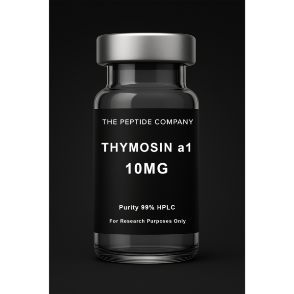
Thymosin Alpha-1 10mg
Thymosin Alpha-1 10mg is a research compound studied for immune signaling modulation, T-cell activity pathways, and inflammatory response regulation. For research use only.
From Inflammation to Regeneration
Inflammation is the body’s first response to damage or stress — an essential defense process that, when unresolved, becomes the foundation for chronic disease and tissue degeneration. The search for molecules that can resolve inflammation without suppressing immunity has led researchers to a naturally occurring tripeptide fragment known as KPV.KPV, short for Lysine-Proline-Valine, is a minimal yet powerful sequence derived from the larger melanocortin hormone α-melanocyte-stimulating hormone (α-MSH). Despite its simplicity, KPV demonstrates remarkable anti-inflammatory, wound-healing, and epithelial-protective activity across multiple biological systems.Following the immune-balancing precision of Thymosin Alpha-1, KPV represents the next logical step — focusing on inflammatory control and repair at the tissue interface.
What Is KPV?
KPV is a bioactive tripeptide naturally released during the breakdown of α-MSH, a hormone best known for its roles in pigmentation and immune modulation. Researchers first isolated the KPV fragment while studying α-MSH’s anti-inflammatory effects and discovered that this tiny segment alone retains potent biological activity.Unlike larger peptides, KPV’s short length provides:• High stability under physiological conditions• Strong receptor affinity for melanocortin-1 (MC1R)• Minimal immunogenicity and easy diffusion across epithelial barriersIt functions as a localized anti-inflammatory messenger, particularly within the skin, gastrointestinal tract, and mucosal linings.
Mechanism of Action
KPV’s activity is mediated primarily through the melanocortin-1 receptor (MC1R), a G-protein-coupled receptor expressed on keratinocytes, macrophages, and epithelial cells.Its mechanisms include:• Downregulation of pro-inflammatory cytokines such as IL-1β, TNF-α, and IL-6• Inhibition of NF-κB transcriptional activity, preventing chronic inflammatory gene expression• Upregulation of anti-inflammatory mediators including IL-10• Promotion of epithelial repair, cell migration, and collagen deposition• Stabilization of tight-junction proteins in gut and skin barriersIn contrast to corticosteroids or immunosuppressants, KPV doesn’t blunt the immune system. It promotes a resolution phase of inflammation — encouraging balance rather than suppression.
Research Highlights
1. Skin Barrier Restoration
Studies in dermatologic and wound-healing models demonstrate that topical or localized KPV reduces redness, edema, and pro-inflammatory cytokine expression, enhances keratinocyte proliferation and collagen synthesis, and accelerates wound closure and tissue remodeling. These findings have made KPV a molecule of interest in burn recovery, psoriasis, and cosmetic wound-healing research.
2. Gut Mucosa and Inflammatory Bowel Models
In gastrointestinal research, KPV has shown potential to reduce colonic inflammation in ulcerative colitis models, decrease infiltration of neutrophils and macrophages, and preserve intestinal barrier integrity through tight-junction reinforcement.
3. Systemic Anti-Inflammatory Potential
Pre-clinical work indicates that KPV can act beyond local tissues: reducing systemic inflammation markers in endotoxin-induced models and supporting metabolic and immune recovery under oxidative stress.
Cellular Pathways Overview
| Function | Primary Pathway | Observed Research Effect |
| Cytokine Suppression | NF-κB inhibition | Reduction in IL-1β, IL-6, TNF-α |
| Receptor Activation | MC1R signaling | Promotes anti-inflammatory gene expression |
| Barrier Protection | Tight-junction stabilization | Restores epithelial integrity |
| Wound Repair | MAPK / PI3K pathways | Accelerates healing and tissue regeneration |
| Oxidative Defense | Nrf2 activation (indirect) | Enhances redox balance |
Synergy and Comparison
KPV functions well as part of a multi-pathway research model for immune and tissue optimization.Conceptual pairings include:• GHK-Cu — synergistic effects on collagen repair and tissue remodeling• LL-37 — complementary antimicrobial and immune-balancing activity• Thymosin Alpha-1 — higher-order immune recalibration paired with inflammation resolution• Glutathione — redox and detoxification support during inflammatory recovery
Research Use and Safety
In the literature, KPV has been evaluated both topically and systemically. Typical concentrations in experimental models range from 100 µg to several milligrams per administration, depending on the study design.Across publications, no significant adverse events have been reported at research-level concentrations. However, KPV remains a research-only molecule and is not approved for clinical or consumer use.All mentions of KPV in this article are for educational and in-vitro research discussion only.
Summary: Precision Anti-Inflammation
KPV represents a new category of anti-inflammatory regulation — one that works with the immune system rather than against it. It demonstrates how targeted peptide fragments can deliver complex biological effects using elegant simplicity: three amino acids capable of redirecting inflammatory pathways, protecting tissues, and accelerating recovery.As research evolves, KPV continues to embody a central principle of modern bioactive design — resolve, don’t suppress.
Key Concepts
• Tripeptide fragment of α-MSH with independent biological activity• Modulates inflammation through MC1R and NF-κB signaling• Promotes epithelial repair and barrier stability• Serves as a bridge between immune modulation and regeneration research
References (Selection)
1. Brzoska T, et al. J Invest Dermatol. (2008).2. Catania A, et al. Ann N Y Acad Sci. (2010).3. Getting SJ, et al. Br J Pharmacol. (2001).4. Zhao H, et al. Peptides. (2021).5. Wang L, et al. J Inflamm Res. (2022).
Educational & Research Disclaimer
This content is for educational and research purposes only. No medical advice or product claims are implied. All compounds discussed are not approved for human or clinical use and are intended for in-vitro laboratory research only.
——————————
Selected References
- PMID: 19765659 — α-MSH peptide signaling and MC1R modulation
- PMID: 19406120 — Anti-inflammatory actions of melanocortin peptides
- PMID: 16239978 — NF-κB suppression via melanocortin pathways
- PMID: 21610182 — Peptide-induced epithelial repair and barrier protection
- Frontiers in Immunology — Melanocortin system and inflammation resolution
- Journal of Peptide Science — Bioactive tripeptides and immunomodulation
FAQ:
What is KPV?
KPV is a tripeptide fragment of α-MSH studied for its anti-inflammatory and tissue-protective properties. It is used exclusively in research settings.
How does KPV work in research?
KPV modulates MC1R and NF-κB signaling pathways, which help regulate inflammation and support epithelial repair in experimental models.
Is KPV approved for human use?
No. KPV is a research-only molecule and is not approved for clinical or consumer use.
What are researchers studying KPV for?
Current research includes inflammation control, epithelial barrier support, tissue protection, and immune modulation.
How is KPV typically studied?
Studies evaluate KPV using in-vitro assays or experimental models to examine its effects on inflammatory pathway responses.
Does KPV have known side effects?
Published research reports minimal adverse effects at research-level concentrations, but no clinical safety data exists.
Related Research Compounds
Thymosin Alpha-1 (Tα1): Immune Resilience and the Science of Thymic Restoration
LL-37: The Antimicrobial Peptide and Innate Immunity Blueprint
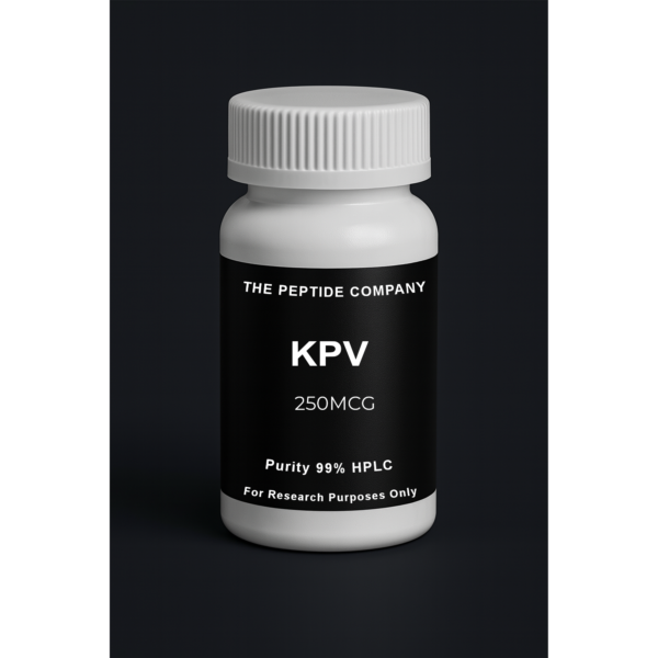
KPV Capsules 250mcg (60 ct)
KPV 250mcg capsules are a research compound studied for anti-inflammatory signaling, NF-κB pathway modulation, and epithelial barrier integrity research. For research use only.
The Body’s Built-In Defense Signal
Long before adaptive immunity learns to recognize specific pathogens, the body relies on a fast, powerful system known as innate immunity. At the center of this system is a family of antimicrobial peptides — small molecules capable of destroying pathogens, guiding immune cell responses, and initiating tissue repair.Among these, LL-37 is the most extensively studied. Derived from the human cathelicidin precursor hCAP18, LL-37 is a 37-amino-acid peptide with broad-spectrum antimicrobial activity and potent immunomodulatory effects. It serves as both a first-response defender and a coordinator of tissue regeneration, making it one of the most versatile molecules in innate immune biology.As the next step in your “Regeneration & Immunity” series, LL-37 builds on the themes established by Thymosin Alpha-1 (immune calibration) and KPV (anti-inflammation and repair) — completing the triad of immune balance, inflammation control, and direct pathogen defense.
What Is LL-37?
LL-37 is the only known human cathelicidin-derived peptide, produced primarily by neutrophils, epithelial cells (skin, lungs, gut), macrophages, and dendritic cells.It becomes active when the precursor protein hCAP18 is cleaved into its functional form, LL-37. This peptide is present at infection sites, mucosal surfaces, and damaged tissues — essentially anywhere the body requires rapid protection.Its biological spectrum includes antibacterial activity, antiviral and antifungal effects, immune signaling, chemotaxis, and wound healing.
Mechanism of Action
LL-37 functions through a multi-layered set of biological mechanisms.1. Direct Antimicrobial Activity:LL-37 disrupts microbial membranes by binding to negatively charged lipid bilayers, forming pores or destabilizing membrane integrity, and leading to rapid pathogen lysis.2. Immune Cell Activation and Chemotaxis:It interacts with receptors such as FPR2, TLR2, TLR4, and P2X7, helping recruit immune cells and enhance antigen presentation.3. Modulation of Inflammation:LL-37 downregulates excessive cytokine release, balances NF-κB activity, and enhances appropriate acute inflammation.4. Wound Healing, Repair, and Angiogenesis:LL-37 promotes keratinocyte migration, fibroblast activity, collagen formation, and VEGF signaling — playing a significant role in tissue regeneration.
Research Highlights
1. Broad-Spectrum Antimicrobial Defense:LL-37 has demonstrated strong activity across bacterial, viral, and fungal pathogens, with rapid membrane disruption as its primary mechanism.2. Lung and Respiratory Immunity:In airway research, LL-37 supports mucosal defense, viral clearance, and controlled inflammation.3. Skin Regeneration:LL-37 accelerates epithelial closure, enhances angiogenesis, reduces biofilm formation, and supports regulated repair mechanisms.4. Antiviral and Immunomodulatory Behavior:LL-37 can disrupt viral envelopes, enhance interferon signaling, and strengthen innate antiviral defense.
Cellular Pathways Overview
| Function | Target Pathway | Research Effect |
| Antimicrobial Defense | Membrane permeabilization | Direct pathogen lysis |
| Immune Activation | FPR2, TLR2, TLR4 | Increased innate immune responsiveness |
| Chemotaxis | GPCR signaling | Recruitment of leukocytes |
| Tissue Repair | EGFR, MAPK, VEGF | Accelerated healing and angiogenesis |
| Inflammation Modulation | NF-κB regulation | Controlled cytokine output |
Synergy and Research Context
LL-37 integrates naturally into multi-pathway research models involving:• KPV — inflammation control and epithelial repair support• Thymosin Alpha-1 — immune modulation and balance• GHK-Cu — regenerative effects and collagen remodeling• Glutathione — oxidative balance during immune activation and tissue repairTogether, these molecules form a conceptual network around immune activation, inflammation resolution, and tissue regeneration.
Research Use and Safety
LL-37 has been studied across many biological systems, but its effects are highly concentration-dependent. Low to moderate concentrations support defense and repair, while high concentrations may induce excess inflammation or cytotoxicity in vitro.No significant toxicity has been observed in regulated research dosing ranges. LL-37 remains a research-only compound, not approved for human or consumer use.All mentions of LL-37 in this article are for educational and in-vitro research discussion only.
Summary
LL-37 represents one of nature’s most efficient biological designs — a molecule capable of killing pathogens, guiding immune cells, regulating inflammation, and accelerating tissue repair.As interest in host defense peptides grows, LL-37 stands out as a blueprint for next-generation antimicrobial and regenerative research.
References (Selection)
1. Sørensen OE, et al. Nat Rev Immunol. (2006).2. Vandamme D, et al. Cell Mol Life Sci. (2012).3. Dürr UHN, et al. Biochim Biophys Acta. (2006).4. Nell MJ, et al. J Leukoc Biol. (2006).5. Heilborn JD, et al. J Invest Dermatol. (2003).
Educational & Research Disclaimer
This content is for educational and research purposes only. No medical advice or product claims are implied. All compounds discussed are not approved for human or clinical use and are intended for in-vitro laboratory research only.
—————–
FAQ
What is LL-37 in research?
LL-37 is a human-derived antimicrobial peptide studied for its roles in innate immunity, host defense, epithelial barrier function, and pathogen response in preclinical models.
How does LL-37 function in laboratory studies?
Research shows LL-37 interacts with microbial membranes, modulates cytokine signaling, and influences immune cell activation, making it a key peptide in host-defense exploration.
Is LL-37 considered a therapeutic product?
No. LL-37 from The Peptide Company is provided strictly for laboratory and in-vitro research use. It is not a therapy, drug, supplement, or product for human use.
What research applications involve LL-37?
LL-37 is explored in models of infection defense, wound repair, microbiome regulation, epithelial integrity, inflammation modulation, and innate immune signaling.
Does LL-37 have antimicrobial activity in studies?
Yes. LL-37 has been shown in preclinical research to disrupt bacterial membranes and modulate pathogen-related immune responses. These findings are experimental only.
How is LL-37 typically stored in research settings?
Researchers store LL-37 lyophilized in cool, dry, stable environments away from light and reconstitute it under institutional laboratory protocols.
Can LL-37 be applied or administered by consumers?
No. LL-37 is not intended for any form of self-administration. It is for controlled laboratory and in-vitro research environments only.
Related Research Compounds:
Thymosin Alpha-1 (Tα1): Immune Resilience and the Science of Thymic Restoration
KPV: The Anti-Inflammatory Tripeptide and Cellular Repair Mechanism
References
PMID: 12711666 — Human cathelicidin LL-37 antimicrobial activity
PMID: 17034334 — LL-37 in innate immunity and host defense
PMID: 19348957 — Epithelial barrier modulation by LL-37
PMID: 32503546 — LL-37 and immune-cell signaling interactions
PMID: 25485019 — Antimicrobial peptides and pathogen membrane disruption
Frontiers in Immunology — Host-defense peptides in innate immune pathways
Nature Reviews Microbiology — Human antimicrobial peptide mechanisms
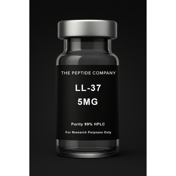
LL-37 – 5MG | High-Purity Research Compound
LL-37 is a synthetic antimicrobial peptide studied for its role in innate immune signaling, antimicrobial defense mechanisms, and inflammatory pathway modulation in research models. For research use only.