
Introduction
SLU‑PP‑332 is a synthetic small‑molecule agonist of estrogen‑related receptors (ERRs), primarily ERRα and ERRγ, developed as a research tool to study mitochondrial oxidative metabolism and exercise‑mimetic transcriptional programs. Unlike compounds that activate AMPK directly, SLU‑PP‑332 operates at the nuclear receptor level, driving coordinated expression of genes involved in oxidative phosphorylation, fatty‑acid oxidation, and mitochondrial biogenesis.
Estrogen‑Related Receptors: ERRα and ERRγ
ERRα and ERRγ are orphan nuclear receptors that regulate cellular energy metabolism. They are highly expressed in tissues with high oxidative demand, including skeletal muscle, cardiac muscle, and brown adipose tissue. ERRs function as transcriptional regulators by binding to estrogen‑related response elements (ERREs) in the promoters of metabolic genes.
Mechanism of Action of SLU‑PP‑332
SLU‑PP‑332 binds to ERRα and ERRγ, stabilizing their active conformations and enhancing transcriptional activity. This activation promotes expression of mitochondrial genes involved in electron transport chain function, fatty‑acid β‑oxidation, and tricarboxylic acid cycle flux. Research distinguishes this mechanism from energy‑stress‑based activation pathways such as AMPK.
Mitochondrial Oxidative Metabolism
ERR activation by SLU‑PP‑332 leads to upregulation of genes encoding components of oxidative phosphorylation, including complexes I–V of the electron transport chain. Studies examine increases in mitochondrial respiration, ATP generation efficiency, and oxidative capacity in skeletal muscle and cardiac research models.
Exercise‑Mimetic Transcriptional Programs
SLU‑PP‑332 is widely studied as an exercise mimetic due to its ability to induce transcriptional programs associated with endurance training. Research explores overlaps with exercise‑induced gene expression, including pathways regulated by PGC‑1α, NRF1, and mitochondrial transcription factor A (TFAM).
Fatty‑Acid Oxidation and Metabolic Flexibility
Activation of ERRs enhances expression of enzymes involved in fatty‑acid transport and oxidation. Research examines how SLU‑PP‑332 influences metabolic substrate selection, favoring lipid utilization over glycolysis in oxidative tissues.
Skeletal Muscle and Cardiac Research
In skeletal muscle models, SLU‑PP‑332 is used to study endurance capacity, oxidative fiber programming, and mitochondrial density. Cardiac research focuses on its effects on myocardial energy metabolism, contractile efficiency, and resistance to metabolic stress.
Comparative Signaling Context
SLU‑PP‑332‑mediated ERR activation is mechanistically distinct from AMPK‑dependent pathways triggered by AICAR or energy depletion. Research highlights how ERR agonism can drive oxidative programs without inducing catabolic stress responses associated with ATP depletion.
Research Limitations and Specificity
As a synthetic ERR agonist, SLU‑PP‑332 is used exclusively in controlled experimental systems. Studies emphasize tissue specificity, receptor subtype selectivity, and dose‑dependent transcriptional outcomes when interpreting results. Off‑target nuclear receptor interactions remain an area of active investigation.
Summary
SLU‑PP‑332 is a powerful research compound for studying estrogen‑related receptor biology, mitochondrial oxidative metabolism, and exercise‑mimetic transcriptional programs. By acting at the level of nuclear receptor signaling, it provides unique insights into metabolic remodeling and endurance‑associated gene regulation.
Educational & Research Disclaimer
This article is for educational and scientific research purposes only. No therapeutic claims or usage recommendations are provided. Compounds referenced are not approved for human use and are intended solely for controlled laboratory experimentation.
FAQ:
What is SLU-PP-332?
SLU-PP-332 is a synthetic small-molecule agonist of estrogen-related receptors (ERRs), primarily ERRα and ERRγ. It is used in research to study mitochondrial oxidative metabolism and transcriptional programs associated with energy expenditure.
How does SLU-PP-332 work?
SLU-PP-332 activates estrogen-related receptors, which are nuclear transcription factors that regulate genes involved in mitochondrial biogenesis, oxidative phosphorylation, and fatty-acid oxidation. This activation shifts cellular metabolism toward increased oxidative capacity.
Why is SLU-PP-332 described as exercise-mimetic in research?
ERR activation by SLU-PP-332 induces transcriptional profiles similar to those observed during endurance exercise, including enhanced mitochondrial function and oxidative metabolism, without requiring mechanical muscle contraction.
What pathways are commonly studied using SLU-PP-332?
Research using SLU-PP-332 often focuses on ERRα/ERRγ signaling, mitochondrial oxidative phosphorylation, fatty-acid utilization, and coordination with downstream metabolic regulators such as PGC-1α.
Is SLU-PP-332 related to estrogen signaling?
No. Despite the name, estrogen-related receptors do not bind estrogen. ERRs are orphan nuclear receptors that regulate energy metabolism independently of classical estrogen signaling.
Is SLU-PP-332 a peptide?
No. SLU-PP-332 is not a peptide. It is a small-molecule compound designed to selectively activate estrogen-related receptors.
What research models use SLU-PP-332?
SLU-PP-332 has been studied in cellular systems and animal models to examine mitochondrial metabolism, exercise-mimetic gene expression, and systemic energy regulation.
Is SLU-PP-332 approved for clinical use?
SLU-PP-332 is a research compound and is not approved for clinical or therapeutic use. Its applications remain experimental.
Selected References (PMIDs)
- PMID: 37258673 – Identification of SLU-PP-332 as an ERR agonist inducing exercise-like metabolic programs
- PMID: 16007134 – Estrogen-related receptors as regulators of mitochondrial function
- PMID: 18316377 – ERRα control of oxidative metabolism and mitochondrial gene expression
- PMID: 20603086 – ERRγ regulation of skeletal muscle oxidative capacity
- PMID: 24011593 – Nuclear receptor control of mitochondrial biogenesis and energy metabolism
- PMID: 33208945 – Transcriptional networks linking ERR signaling and energy expenditure
RELATED SEARCHES:
Mazdutide : Dual GLP‑1 and Glucagon Receptor Activation in Metabolic and Lipid Regulation Research
AICAR : AMPK Activation, Cellular Energy Sensing, and Exercise‑Mimetic Signaling in Research Models
Apelin : APJ Receptor Signaling, Cardiovascular Regulation, and Metabolic Research Pathways
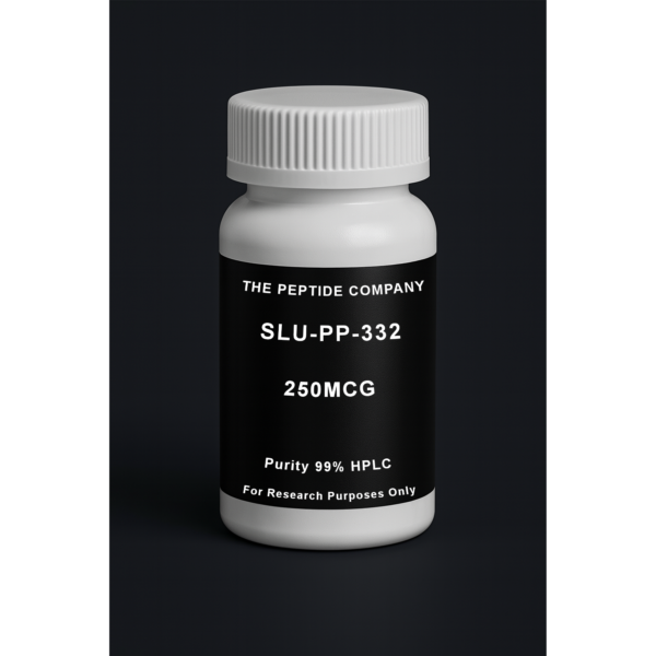
SLU-PP-332 250mcg (100 ct)
SLU-PP-332 250mcg is a research compound studied for ERR signaling, mitochondrial biogenesis pathways, and exercise-mimetic metabolic mechanisms. For research use only.
Introduction
5‑Aminoimidazole‑4‑carboxamide ribonucleotide (AICAR) is a cell‑permeable adenosine analog widely used in research to study cellular energy sensing and metabolic regulation. Once inside cells, AICAR is phosphorylated to ZMP, an AMP mimetic that activates AMP‑activated protein kinase (AMPK). Through this mechanism, AICAR serves as a foundational tool for investigating energy stress responses, mitochondrial biogenesis, and exercise‑mimetic signaling pathways.
Chemical Properties and Intracellular Conversion
AICAR enters cells via nucleoside transporters and is rapidly converted to ZMP by adenosine kinase. ZMP structurally and functionally resembles AMP, allowing it to interact with AMPK regulatory subunits. This conversion bypasses upstream energetic stress, enabling controlled activation of energy‑sensing pathways in experimental models.
AMPK Structure and Activation Mechanism
AMPK is a heterotrimeric kinase composed of catalytic α and regulatory β and γ subunits. ZMP binding to the γ subunit promotes AMPK activation by enhancing phosphorylation of the α subunit at Thr172 and protecting it from dephosphorylation. Research examines how AICAR‑induced AMPK activation differs from physiological activation driven by ATP depletion.
Metabolic Reprogramming and Substrate Utilization
Activation of AMPK by AICAR shifts cellular metabolism toward catabolic processes that generate ATP. Research models demonstrate increased fatty acid oxidation through inhibition of acetyl‑CoA carboxylase (ACC), enhanced glucose uptake via GLUT4 translocation, and suppression of anabolic pathways such as lipid and protein synthesis.
Mitochondrial Biogenesis and Oxidative Capacity
AICAR‑mediated AMPK activation influences mitochondrial biogenesis through regulation of PGC‑1α and related transcriptional networks. Studies examine changes in mitochondrial DNA content, respiratory enzyme expression, and oxidative phosphorylation capacity following AMPK activation in skeletal muscle and other tissues.
Exercise‑Mimetic Signaling Research
AICAR is frequently described in research as an exercise mimetic due to its ability to activate AMPK‑dependent pathways associated with endurance adaptation. Experimental models explore overlaps and distinctions between AICAR‑induced signaling and mechanical exercise, including effects on muscle fiber composition and metabolic efficiency.
Cardiac and Skeletal Muscle Research
In muscle research, AICAR is used to study energy stress responses, contractile efficiency, and metabolic flexibility. Cardiac models investigate its influence on substrate utilization, ischemic tolerance, and mitochondrial resilience under energetic challenge.
Signal Specificity and Research Limitations
While AICAR is a powerful research tool, ZMP can interact with AMP‑sensitive enzymes beyond AMPK. Studies emphasize the importance of distinguishing AMPK‑dependent and AMPK‑independent effects when interpreting results. Dose, exposure time, and tissue context significantly influence signaling outcomes in experimental systems.
Summary
AICAR is a widely used experimental compound for probing AMPK activation, energy‑sensing mechanisms, mitochondrial adaptation, and exercise‑related metabolic signaling. Its ability to selectively engage cellular energy pathways makes it a central molecule in metabolic and mitochondrial research.
Educational & Research Disclaimer
This article is for educational and scientific research purposes only. No therapeutic claims or usage recommendations are provided. Compounds referenced are not approved for human use and are intended solely for controlled laboratory experimentation.
FAQ:
What is AICAR?
AICAR (5-Aminoimidazole-4-carboxamide ribonucleotide) is a synthetic adenosine analog commonly used in research to study cellular energy sensing and AMP-activated protein kinase (AMPK) signaling pathways.
How does AICAR activate AMPK?
Inside cells, AICAR is converted to ZMP, an AMP mimetic that binds to AMPK and promotes its activation, allowing researchers to study downstream effects of energy stress signaling without altering ATP directly.
Why is AICAR described as an exercise-mimetic in research?
AICAR activates many of the same intracellular pathways stimulated by endurance exercise—particularly AMPK-dependent pathways involved in mitochondrial biogenesis and substrate utilization—making it useful in exercise physiology models.
What pathways are commonly studied using AICAR?
AICAR is frequently used to investigate AMPK-related pathways such as mitochondrial biogenesis (PGC-1α), glucose uptake, fatty-acid oxidation, and metabolic adaptation under energetic stress.
Is AICAR a peptide?
No. AICAR is not a peptide. It is a small-molecule nucleoside analog, though it is often discussed alongside peptides due to its use in metabolic and signaling research.
What research models use AICAR?
AICAR has been studied in cell culture, animal models, and controlled laboratory settings to explore metabolic regulation, mitochondrial function, and energy-sensing mechanisms.
Is AICAR used clinically?
AICAR is primarily a research compound and is not approved for general clinical use. Its role is largely limited to experimental and mechanistic studies.
Selected References (PMIDs)
- PMID: 11340166 – AICAR activation of AMPK and effects on cellular metabolism
- PMID: 15987730 – AMPK regulation of mitochondrial biogenesis via PGC-1α
- PMID: 15075399 – AICAR-induced glucose uptake independent of insulin
- PMID: 17106097 – Exercise-like metabolic adaptations induced by AICAR
- PMID: 17434812 – AMPK as a central energy sensor in metabolic regulation
- PMID: 20071205 – AMPK signaling and mitochondrial function in skeletal muscle
Related Searches:
Apelin : APJ Receptor Signaling, Cardiovascular Regulation, and Metabolic Research Pathways
Cardiogen: Short Peptide Bioregulator for Cardiac and Myocardial Tissue Research
Introduction
Nicotinamide adenine dinucleotide (NAD⁺) is a ubiquitous redox cofactor essential for cellular energy metabolism, mitochondrial function, and regulatory signaling. Beyond its classical role in oxidation–reduction reactions, NAD⁺ serves as a substrate for multiple enzyme families that govern DNA repair, chromatin remodeling, stress responses, and metabolic adaptation. As a result, NAD⁺ occupies a central position in modern cellular and mitochondrial research.
Chemical Structure and Redox Function
NAD⁺ consists of two nucleotides joined through their phosphate groups: one containing an adenine base and the other nicotinamide. The nicotinamide moiety undergoes reversible reduction to NADH, enabling electron transfer reactions. This NAD⁺/NADH redox couple is fundamental to glycolysis, the tricarboxylic acid cycle, and oxidative phosphorylation.
Role in Mitochondrial Energy Metabolism
Within mitochondria, NAD⁺ accepts electrons generated during the TCA cycle and delivers them to complex I of the electron transport chain via NADH. This process drives proton pumping, establishes the electrochemical gradient, and ultimately supports ATP synthesis. Research examines how compartmentalized NAD⁺ pools influence mitochondrial efficiency, redox balance, and adaptive responses to energetic stress.
NAD⁺-Consuming Enzymes
NAD⁺ functions not only as a redox cofactor but also as a consumable substrate for several enzyme families. Sirtuins (SIRT1–SIRT7) utilize NAD⁺ for deacetylation and ADP-ribosylation reactions that regulate gene expression, mitochondrial protein function, and stress resistance. Poly(ADP-ribose) polymerases (PARPs) consume NAD⁺ during DNA repair, linking NAD⁺ availability to genomic maintenance.
NAD⁺ and Chromatin Regulation
Through sirtuin activity, NAD⁺ levels influence chromatin structure and transcriptional programs. Research models show that NAD⁺-dependent deacetylation affects histones, transcription factors, and co-regulators, thereby coordinating metabolic state with gene expression. This positions NAD⁺ as a molecular bridge between metabolism and epigenetic control.
Cellular Stress, DNA Repair, and Redox Homeostasis
During oxidative or genotoxic stress, NAD⁺ consumption by PARPs increases to facilitate DNA repair. Excessive activation can deplete cellular NAD⁺ pools, disrupting energy metabolism. Research explores how cells balance NAD⁺ regeneration, redox homeostasis, and repair processes to maintain viability under stress.
NAD⁺ Salvage and Biosynthetic Pathways
Cells maintain NAD⁺ levels through de novo synthesis and salvage pathways. The salvage pathway recycles nicotinamide into NAD⁺ via intermediates such as NMN, coordinated by enzymes including NAMPT and NMNATs. Research focuses on how these pathways regulate intracellular NAD⁺ availability across nuclear, cytosolic, and mitochondrial compartments.
Systemic and Intercellular Signaling Roles
Beyond individual cells, NAD⁺ metabolism influences intercellular communication and systemic physiology. Studies examine extracellular NAD⁺ turnover, ectoenzyme activity, and the role of NAD⁺-derived metabolites in immune and inflammatory signaling. These findings expand the relevance of NAD⁺ beyond classical metabolism.
Summary
NAD⁺ is a central molecular hub integrating redox chemistry, mitochondrial energy production, DNA repair, chromatin regulation, and stress-response signaling. Its dual role as both a cofactor and a consumable substrate makes NAD⁺ a key determinant of cellular resilience and metabolic adaptation in advanced biological research.
Educational & Research Disclaimer
This article is for educational and scientific research purposes only. No therapeutic claims or usage recommendations are provided. Compounds referenced are not approved for human use and are intended solely for controlled laboratory experimentation.
PMID:
- PMID: 33353981 (NAD⁺ metabolism in ageing) PMC
- PMID: 24786309 (NAD⁺ + sirtuins in aging/disease) PubMed
- PMID: 29883761 (NAD metabolism in aging/longevity) PubMed
- PMID: 29482842 (CD38 biology; NAD⁺ consumption) PubMed
- PMID: 30355082 (Sirtuins/NAD⁺ in cardio-metabolic disease models)
FAQ:
What is NAD⁺ in research models?
NAD⁺ (nicotinamide adenine dinucleotide) is an essential redox cofactor that supports cellular energy metabolism and acts as a substrate for enzymes involved in DNA repair and gene regulation.
How does NAD⁺ support cellular energy production?
NAD⁺ shuttles electrons in glycolysis and the TCA cycle, enabling oxidative phosphorylation and ATP generation through mitochondrial respiration.
What’s the difference between NAD⁺ and NADH?
NAD⁺ is the oxidized form and NADH is the reduced form. The NAD⁺/NADH ratio is a core indicator of cellular redox state and metabolic flux.
Why is NAD⁺ linked to sirtuins and longevity pathways?
Sirtuins use NAD⁺ to regulate protein deacetylation, influencing mitochondrial biogenesis, stress responses, and metabolic adaptation in research systems.
How is NAD⁺ regulated inside cells?
Cells maintain NAD⁺ through biosynthesis and salvage pathways (notably from nicotinamide), while enzymes like CD38 and PARPs consume NAD⁺ during signaling and repair processes.
How is NAD⁺ typically measured in lab studies?
Common approaches include LC–MS/MS quantification, enzymatic cycling assays, and paired measurement of NAD⁺/NADH to assess redox balance.
Is NAD⁺ itself a peptide?
No. NAD⁺ is a nucleotide-derived coenzyme, not a peptide—though it’s often discussed alongside bioactive molecules studied for cellular optimization.
Related Searches:
MOTS-c: The Mitochondrial-Encoded Peptide for Metabolic Regulation and Cellular Resilience
NAD+ 1000mg
NAD+ 1000mg is a research compound studied for cellular energy metabolism, redox balance, mitochondrial function, and sirtuin-associated signaling pathways. For research use only.

Introduction
Humanin is a small mitochondrial-derived peptide (MDP) encoded within the mitochondrial genome and translated in the cytoplasm. It was originally identified in studies of neuronal survival and has since become a central molecule in research on mitochondrial stress signaling, apoptosis resistance, metabolic regulation, and neuroprotection. Humanin represents a class of peptides that function as retrograde signals, allowing mitochondria to communicate cellular stress states to the nucleus.
Mitochondrial Origin and Structure
Humanin consists of 24 amino acids and is encoded within the 16S rRNA region of mitochondrial DNA. Despite its small size, Humanin exhibits a conserved sequence across species, suggesting evolutionary importance. Structural studies indicate that Humanin adopts conformations compatible with receptor binding and intracellular protein–protein interactions.
Humanin Receptor and Signaling Complexes
Humanin interacts with both intracellular targets and cell-surface receptor complexes. One well-studied signaling route involves a trimeric receptor complex composed of CNTFRα, WSX‑1, and gp130, leading to activation of STAT3-dependent transcriptional pathways. Research also explores Humanin’s receptor-independent intracellular actions.
Anti-Apoptotic Mechanisms
Humanin is extensively studied for its ability to suppress apoptotic signaling. It directly interacts with pro-apoptotic BCL‑2 family members, including BAX, tBID, and BimEL, preventing mitochondrial outer membrane permeabilization and cytochrome c release. These mechanisms position Humanin as a key modulator of intrinsic apoptosis pathways in research models.
Mitochondrial Stress and Retrograde Signaling
As a mitochondrial-derived peptide, Humanin participates in retrograde signaling networks that link mitochondrial function to nuclear gene expression. Research investigates its role in modulating stress-response transcription, mitochondrial quality control, and cellular adaptation to oxidative and metabolic challenges.
Neuroprotective Research Pathways
Humanin is widely studied in neuronal models for its influence on cell survival, synaptic integrity, and resistance to neurotoxic stressors. Research examines its interactions with amyloid-associated stress, excitotoxic signaling, and mitochondrial dysfunction in neural tissues.
Metabolic and Endocrine Research
Beyond the nervous system, Humanin is explored for its effects on metabolic regulation. Studies examine its influence on insulin signaling pathways, glucose metabolism, lipid handling, and cellular energy balance. These findings position Humanin within a broader network of mitochondrial peptides involved in systemic metabolic communication.
Inflammatory and Immune Signaling
Humanin signaling intersects with inflammatory pathways through modulation of cytokine expression and stress-responsive immune transcription. Research explores its potential role in balancing pro‑ and anti‑inflammatory signaling during cellular stress conditions.
Summary
Humanin is a mitochondrial-derived peptide studied for its roles in apoptosis suppression, mitochondrial stress signaling, neuroprotection, metabolic regulation, and immune-modulatory pathways. Its ability to function as a retrograde signal highlights the mitochondrion as an active regulator of cellular fate and resilience in advanced biological research.
Educational & Research Disclaimer
This article is for educational and scientific research purposes only. No therapeutic claims or usage recommendations are provided. Compounds referenced are not approved for human use and are intended solely for controlled laboratory experimentation.
PMID:
- PMID: 11416100 – Discovery of humanin and neuroprotective effects
- PMID: 14593182 – Humanin and mitochondrial-mediated apoptosis
- PMID: 18755891 – Cytoprotective signaling pathways of humanin
- PMID: 23336290 – Humanin analogs and stress resistance
- PMID: 30559258 – Humanin as a mitochondrial-derived peptide in aging research
FAQ:
What is humanin in research models?
Humanin is a mitochondrial-derived peptide studied for its role in cellular protection, stress resistance, and survival signaling under pathological conditions.
How does humanin protect cells?
Humanin interferes with pro-apoptotic signaling pathways and supports mitochondrial integrity, reducing oxidative and metabolic stress–induced cell death.
Is humanin linked to mitochondrial function?
Yes. Humanin originates from mitochondrial DNA and is closely associated with mitochondrial communication and bioenergetic regulation.
What systems are studied with humanin?
Research models explore humanin in neuroprotection, metabolic regulation, aging, and cellular stress response pathways.
How is humanin investigated in laboratory settings?
Humanin is examined through receptor binding studies, apoptosis assays, mitochondrial stress models, and signaling pathway analyses.
Related Searches:
MOTS-c: The Mitochondrial-Encoded Peptide for Metabolic Regulation and Cellular Resilience
NMN: NAD⁺ Precursor Biology, Cellular Metabolism, and Mitochondrial Research
Introduction
Glutathione (GSH) is a tripeptide composed of glutamate, cysteine, and glycine and is one of the most abundant intracellular antioxidants. It plays a central role in maintaining cellular redox balance, detoxification processes, mitochondrial function, and regulation of cellular signaling pathways. Due to its broad involvement in oxidative stress management, glutathione is a foundational molecule in biochemical and cellular research.
Molecular Structure and Biosynthesis
Glutathione’s unique γ-glutamyl bond between glutamate and cysteine confers stability and functional specificity. Biosynthesis occurs in two ATP-dependent steps catalyzed by glutamate–cysteine ligase (GCL) and glutathione synthetase (GS). Cellular availability of cysteine is rate-limiting, linking glutathione synthesis to amino acid metabolism and redox demand.
Redox Cycling and Antioxidant Function
Glutathione exists in reduced (GSH) and oxidized (GSSG) forms, enabling dynamic redox cycling. Glutathione peroxidases (GPxs) utilize GSH to neutralize hydrogen peroxide and lipid peroxides, while glutathione reductase regenerates GSH from GSSG using NADPH. The GSH:GSSG ratio is a key indicator of cellular redox status in research models.
Detoxification and Conjugation Pathways
Glutathione participates in phase II detoxification through conjugation reactions catalyzed by glutathione S-transferases (GSTs). These reactions increase solubility of electrophilic compounds, facilitating their removal. Research explores glutathione’s role in xenobiotic metabolism, environmental toxin handling, and cellular defense against reactive intermediates.
Mitochondrial Function and Oxidative Stress
Mitochondrial glutathione pools are essential for maintaining oxidative phosphorylation efficiency and preventing oxidative damage to mitochondrial DNA, lipids, and proteins. Studies examine glutathione transport into mitochondria and its role in preserving mitochondrial membrane integrity and respiratory chain function under stress conditions.
Regulation of Cellular Signaling
Beyond antioxidant activity, glutathione modulates redox-sensitive signaling pathways. Protein S-glutathionylation serves as a reversible post-translational modification that influences enzyme activity, transcription factor binding, and signal transduction. This positions glutathione as a regulator of cellular communication in research settings.
Immune and Inflammatory Research
Glutathione status influences immune cell function, cytokine production, and inflammatory signaling. Research investigates how glutathione levels affect lymphocyte proliferation, macrophage activity, and the balance between pro- and anti-inflammatory transcriptional programs.
Summary
Glutathione is a central tripeptide antioxidant studied for its roles in redox homeostasis, detoxification, mitochondrial protection, and regulation of cellular signaling. Its integration into metabolic, immune, and stress-response pathways makes glutathione a cornerstone molecule in cellular and biochemical research.
Educational & Research Disclaimer
This article is for educational and scientific research purposes only. No therapeutic claims or usage recommendations are provided. Compounds referenced are not approved for human use and are intended solely for controlled laboratory experimentation.
- PMID: 16492803 – Meister & Anderson, glutathione metabolism overview
- PMID: 17021671 – Redox regulation and cellular signaling
- PMID: 18973918 – Role of glutathione in detoxification pathways
- PMID: 23766848 – Mitochondrial glutathione and oxidative stress
- PMID: 33208454 – Glutathione depletion and redox imbalance in disease models
FAQ:
What is glutathione and why is it important in research models?
Glutathione (GSH) is a tripeptide composed of glutamate, cysteine, and glycine that functions as the primary intracellular antioxidant, maintaining redox balance and protecting cells from oxidative damage.
How does glutathione regulate cellular redox homeostasis?
Glutathione cycles between reduced (GSH) and oxidized (GSSG) states, buffering reactive oxygen species and preserving redox-sensitive signaling pathways.
What role does glutathione play in detoxification pathways?
Glutathione conjugates xenobiotics and endogenous toxins via glutathione S-transferases, facilitating cellular detoxification and excretion.
Is glutathione involved in mitochondrial function?
Yes. Mitochondrial glutathione protects respiratory chain components from oxidative stress and supports ATP production and mitochondrial integrity.
How is glutathione studied in laboratory research?
Research models examine glutathione synthesis, depletion, redox ratios (GSH:GSSG), and enzyme activity to assess oxidative stress, detoxification capacity, and cellular resilience.
Related Searches:
MOTS-c: The Mitochondrial-Encoded Peptide for Metabolic Regulation and Cellular Resilience
NMN: NAD⁺ Precursor Biology, Cellular Metabolism, and Mitochondrial Research
Glutathione 1500mg
Glutathione 1500mg is a research compound studied for cellular redox balance, oxidative stress modulation, and detoxification pathway mechanisms. For research use only.

Overview
BAM15 (N5,N6-bis(2-fluorophenyl)[1,2,5]oxadiazolo[3,4-b]pyrazine-5,6-diamine) is a synthetic small molecule identified as a mitochondrial protonophore.
Originally discovered through high-throughput screening for mitochondrial modulators, BAM15 functions as a selective mitochondrial uncoupler, separating electron transport from ATP synthesis.
In research settings, this compound has been used to explore energy expenditure, fat oxidation, metabolic flexibility, and mitochondrial efficiency, offering a controlled method to increase caloric utilization without increasing food intake.
Preclinical models suggest BAM15 increases resting energy expenditure, decreases hepatic steatosis, improves insulin sensitivity, and alters lipid metabolism — making it a potent tool for studying obesity, type 2 diabetes, and aging biology.
Mechanism of Action (Research Context)
BAM15 acts as a protonophore, meaning it facilitates proton (H⁺) transport across the mitochondrial inner membrane independent of ATP synthase.
Under normal conditions, mitochondria create a proton gradient (Δψ) to power oxidative phosphorylation and ATP production.
By dissipating this gradient, BAM15 uncouples substrate oxidation from ATP generation, forcing the mitochondria to oxidize more fuel substrates (fatty acids and glucose) to maintain energy balance.
This increased proton leak leads to higher oxygen consumption and heat generation, a process known as mitochondrial uncoupling–induced thermogenesis.
Unlike earlier uncouplers such as DNP (2,4-dinitrophenol), BAM15 demonstrates selectivity and safety advantages in preclinical models — showing no significant rise in body temperature or systemic toxicity at experimental doses.
On a molecular level:
- BAM15 integrates within the mitochondrial inner membrane lipid bilayer.
- It transports protons via reversible ion-shuttling, collapsing the proton motive force.
- This elevates substrate oxidation, increases NADH turnover, and enhances mitochondrial respiration.
These properties make BAM15 an important research probe for studying bioenergetic efficiency, mitochondrial stress adaptation, and metabolic signaling pathways such as AMPK and PGC-1α.
Potential Research Benefits (Reported in Literature)
• Increases whole-body energy expenditure and fat oxidation without hyperthermia
• Improves insulin sensitivity and glucose tolerance in diet-induced obese models
• Reduces hepatic steatosis and adiposity in rodent studies
• Enhances mitochondrial respiratory capacity and turnover of defective mitochondria
• Lowers reactive oxygen species (ROS) accumulation by optimizing electron transport efficiency
• Demonstrates lifespan-extension potential in some cellular aging models
• Offers a tool to study non-hormonal thermogenic mechanisms independent of adrenergic signaling
Selected Research Highlights
• Energy Expenditure: BAM15 increased oxygen consumption rate (OCR) and resting metabolic rate in murine studies without altering food intake or causing febrile responses.
• Liver Fat Reduction: Rodents fed high-fat diets and treated with BAM15 displayed reduced hepatic triglyceride accumulation and improved hepatic insulin signaling.
• Insulin Sensitivity: HOMA-IR and glucose-tolerance testing improved significantly in BAM15-treated groups, correlating with enhanced mitochondrial oxidation.
• Mitochondrial Health: Research indicates improved mitochondrial dynamics, with increased biogenesis markers (PGC-1α, NRF-1) and reduced fission protein expression (DRP1).
• Safety Profile: Unlike older uncouplers, BAM15 did not significantly elevate body temperature, demonstrating a more favorable margin between efficacy and thermogenic toxicity.
Chemical / Physical Information
• Chemical Name: N5,N6-bis(2-fluorophenyl)[1,2,5]oxadiazolo[3,4-b]pyrazine-5,6-diamine
• Molecular Formula: C₁₄H₁₀F₂N₆O₂
• Molecular Weight: 332.27 Da
• Appearance: Pale yellow crystalline solid
• Solubility: Soluble in DMSO and ethanol; sparingly soluble in water
• Storage: Store at −20 °C protected from light and moisture; for extended storage, maintain under inert gas.
Reconstituted solutions should be aliquoted and frozen to prevent repeated freeze–thaw cycles.
Metabolic Implications (Research Context)
BAM15 has provided researchers with a model to explore controlled mitochondrial inefficiency as a therapeutic strategy.
By modestly reducing ATP yield per molecule of substrate oxidized, BAM15 increases caloric dissipation as heat — effectively “wasting” energy to rebalance metabolic homeostasis.
This mechanism allows investigation into:
- Adipose tissue thermogenesis independent of β-adrenergic stimulation
- Hepatic lipid oxidation and non-alcoholic fatty liver disease (NAFLD) models
- Muscle mitochondrial turnover and metabolic flexibility
- Mitochondrial hormesis — the adaptive benefits of mild mitochondrial stress
The uncoupling process also activates AMP-activated protein kinase (AMPK) and downstream mitochondrial biogenesis pathways, offering a valuable tool for studying exercise-mimetic or calorie-restriction-mimetic mechanisms.
Regulatory & Compliance Notes
BAM15 is an experimental compound and is not approved for therapeutic or dietary use by any regulatory authority.
All handling and application must be confined to controlled laboratory environments under institutional safety oversight.
Documentation such as Certificates of Analysis (COA) and Material Safety Data Sheets (MSDS) should accompany all research-grade material.
References (Selection)
- Alexopoulos SJ et al. (2020). Mitochondrial uncoupler BAM15 reverses obesity and insulin resistance in mice. Nature Communications.
- Gao AW, et al. (2021). Mitochondrial uncoupling as a strategy for metabolic disease: lessons from BAM15. Cell Metabolism.
- Mills EL, et al. (2020). Uncoupling mitochondrial respiration in metabolic research: mechanistic insights from BAM15. J Biol Chem.
- Krahmer N, et al. (2022). Mitochondrial efficiency and thermogenesis under BAM15-induced proton leak. EMBO Reports.
- Wallace DC. (2023). Mitochondrial bioenergetics in aging and disease: uncouplers and beyond. Trends Endocrinol Metab.
Disclaimer
This publication is intended for educational and research purposes only.
BAM15 is not approved for human or veterinary use.
All research must adhere to institutional biosafety and ethical standards governing chemical handling, metabolic studies, and mitochondrial research.
——————————
Selected References
PMID: 32395069 — Mitochondrial uncouplers and metabolic modulation
PMID: 31594992 — BAM15 mechanisms in energy expenditure
PMID: 33093089 — Uncoupling agents and obesity/metabolic regulation
PMID: 30728358 — Mitochondrial-targeted therapeutics in metabolism
Frontiers in Endocrinology — Mitochondrial bioenergetics and metabolic control
Journal of Biological Chemistry — Uncoupling-driven energy metabolism
FAQ:
What is BAM15?
BAM15 is a mitochondrial protonophore studied for its ability to uncouple oxidative phosphorylation and influence metabolic energy expenditure in research models.
How does BAM15 work in research?
BAM15 disrupts the mitochondrial proton gradient, increasing energy expenditure without directly impacting appetite or food intake in experimental systems.
Is BAM15 approved for human use?
No. BAM15 is strictly a research compound and is not approved for clinical, therapeutic, or consumer use.
What are researchers studying BAM15 for?
Research explores BAM15 in contexts such as metabolic rate modulation, obesity models, thermogenesis, and mitochondrial function.
Does BAM15 affect muscle tissue or lean mass?
Studies suggest that BAM15 may increase metabolic activity without significantly reducing lean mass, though findings are limited to preclinical models.
How is BAM15 typically evaluated in research?
BAM15 is assessed using controlled laboratory experiments that monitor mitochondrial activity, metabolic rate, and energy balance.
Does BAM15 have known side effects in studies?
Some experimental data show tolerability within certain dosage ranges, but comprehensive safety profiles are not established.
Related Research Compounds
MOTS-c: The Mitochondrial-Encoded Peptide for Metabolic Regulation and Cellular Resilience
NMN: NAD⁺ Precursor Biology, Cellular Metabolism, and Mitochondrial Research
A New Class of Bioactive Molecules
Mitochondria are traditionally described as the “powerhouses” of the cell, responsible for ATP production and energy balance. In recent years, a new perspective has emerged: mitochondria also behave as endocrine signaling hubs, releasing peptides that regulate systemic metabolism, stress responses, and cellular adaptation.
Among these peptides, MOTS-c has generated significant scientific interest. Unlike most bioactive peptides encoded in the cell nucleus, MOTS-c is encoded directly within mitochondrial DNA (mtDNA) — a rare feature that places it at the intersection of metabolism, exercise physiology, and cellular resilience.
MOTS-c connects longevity science, metabolic regulation, and mitochondrial stress response research — making it the ideal entry point into the “Mitochondrial & Metabolic Optimization” phase of your content series.
What Is MOTS-c?
MOTS-c (Mitochondrial Open Reading Frame of the 12S rRNA type-c) is a 16-amino-acid peptide encoded by the mitochondrial genome. First identified in 2015, MOTS-c challenges the traditional belief that peptide-coding sequences exist only in the nucleus.
MOTS-c has been observed to:
• Enhance metabolic flexibility
• Increase glucose utilization
• Improve insulin sensitivity
• Activate stress-response pathways
• Support mitochondrial homeostasis
Because mitochondria respond dynamically to nutrients, exercise, and oxidative stress, MOTS-c is increasingly viewed as a key coordinator of adaptive metabolism.
Mechanism of Action
MOTS-c regulates nutrient sensing and cellular stress pathways through several core mechanisms.
1. AMPK Activation
MOTS-c activates AMPK, the body’s primary energy sensor. This leads to:
• Increased glucose uptake
• Enhanced fatty acid oxidation
• Improved insulin sensitivity
• Suppressed energy-consuming anabolic pathways
2. Exercise-Mimetic Effects
MOTS-c is released during physical stress and enhances:
• Muscular glucose transport
• Endurance capacity
• Stress tolerance
3. Nuclear Translocation
Under metabolic stress, MOTS-c can translocate into the nucleus and regulate gene transcription related to:
• Stress resistance
• Antioxidant defense
• Metabolic efficiency
4. Folate & Methionine Pathways
MOTS-c influences one-carbon metabolism and redox balance — essential for DNA repair, methylation, and detoxification.
Research Highlights
1. Metabolic Health
MOTS-c enhances glucose uptake, increases insulin sensitivity, and improves metabolic flexibility.
2. Weight Regulation
Supports fatty acid oxidation and counters metabolic slowdown during caloric surplus.
3. Exercise Performance
Improves endurance, fatigue resistance, and mitochondrial biogenesis signals in research models.
4. Stress Resilience
Activates antioxidant genes and stress-response pathways through AMPK and nuclear signaling.
5. Age-Related Decline
MOTS-c levels decrease with age; restoring them in research settings improves metabolic and physical performance.
Synergistic Combinations (Research Context)
MOTS-c integrates naturally with other metabolic or mitochondrial research molecules:
• 5-Amino-1MQ — complements MOTS-c by enhancing NAD+ availability
• SS-31 — supports mitochondrial membrane stability
• NAD+ precursors — synergize with AMPK and redox pathways
• Glutathione — supports antioxidant demands during mitochondrial output
These molecules form the basis of the “Mitochondrial Optimization” research cluster.
Research Use and Safety
MOTS-c has been evaluated in cellular, animal, and metabolic studies.
Key points:
• No significant toxicity at research levels
• Dose-dependent metabolic effects
• Increasingly studied for metabolic syndrome and aging research
• Not approved for medical, clinical, or consumer use
All discussions refer strictly to research-only contexts.
Summary
MOTS-c represents a new era of mitochondrial biology and metabolic regulation. Its ability to improve metabolic flexibility, enhance stress responses, and support cellular energy balance makes it central to longevity and performance research.
As interest in mitochondrial peptides expands, MOTS-c stands out as a key regulator of cellular adaptation and resilience.
References (Selection)
1. Lee C, et al. Cell Metabolism. (2015).
2. Reynolds JC, et al. Aging Cell. (2021).
3. Zarse K, et al. Aging. (2019).
4. Kim KH, et al. Nat Commun. (2018).
5. Cobb LJ, et al. J Cachexia Sarcopenia Muscle. (2016).
Educational & Research Disclaimer
This content is for educational and research purposes only. No medical advice or product claims are implied. Compounds discussed are not approved for human or clinical use and are intended for in-vitro laboratory research only.
————————
FAQ:
What is MOTS-c in research?
MOTS-c is a mitochondrial-encoded peptide studied for its roles in metabolic regulation, stress adaptation, and cellular resilience in preclinical models.
What pathways does MOTS-c influence in studies?
Research suggests MOTS-c interacts with AMPK activation, mitochondrial signaling, and cellular energy regulation, especially under metabolic or oxidative stress.
Is MOTS-c considered a therapeutic compound?
No. MOTS-c is a research molecule used exclusively in laboratory and in-vitro models. It is not approved for medical, dietary, or clinical use.
How is MOTS-c typically used in research environments?
Researchers investigate MOTS-c in cell assays and animal models to study energy balance, metabolic signaling, and stress responses. All use must follow lab protocols.
Does MOTS-c impact exercise or performance in research studies?
Some preclinical studies explore MOTS-c’s effect on exercise tolerance and metabolic flexibility, but these findings are experimental and not applicable to human use.
How should MOTS-c be stored in a laboratory?
Labs typically store MOTS-c lyophilized in a cool, dry environment protected from light, following their institutional handling and stability procedures.
Is MOTS-c safe for human consumption or self-administration?
No. MOTS-c sold by The Peptide Company is for laboratory and in-vitro use only. It is not for human use, self-administration, or clinical application.
Selected References
PMID: 26562337 — Mitochondrial-encoded peptides and metabolic regulation
PMID: 25738465 — MOTS-c activation of AMPK and cellular energy pathways
PMID: 31447076 — Exercise-induced MOTS-c and metabolic homeostasis
PMID: 33414491 — Mitochondrial-derived peptides in stress adaptation
PMID: 35408335 — MOTS-c signaling in metabolic flexibility and resilience
Frontiers in Endocrinology — Mitochondrial peptides in energy regulation
Nature Communications — MOTS-c and nuclear signaling under metabolic stress
Related Research Compounds:
NMN: NAD⁺ Precursor Biology, Cellular Metabolism, and Mitochondrial Research
A New Frontier in Mitochondrial Medicine
Mitochondria regulate cellular energy, stress responses, redox balance, and metabolic flexibility. As these systems decline with age or metabolic stress, mitochondrial dysfunction becomes a driver of fatigue, impaired tissue repair, and systemic metabolic decline.
SS-31, also known as Elamipretide or MTP-131, is a mitochondria-targeted tetrapeptide designed to selectively target the inner mitochondrial membrane and interact with cardiolipin. By stabilizing mitochondrial structure and reducing oxidative stress, SS-31 has become a leading research tool for studying mitochondrial restoration.
What Is SS-31?
SS-31 is a synthetic mitochondria-targeted tetrapeptide with the sequence D-Arg-Dmt-Lys-Phe-NH₂.
It can:
• Cross mitochondrial membranes
• Concentrate at the inner mitochondrial membrane
• Bind electrostatically to cardiolipin
• Support mitochondrial architecture and ATP production
Unlike general antioxidants, SS-31 modulates ROS production at the source while preserving physiological signaling.
Mechanism of Action
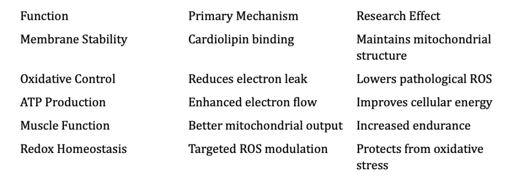
1. Cardiolipin Stabilization
SS-31 binds directly to cardiolipin, helping maintain membrane curvature, respiratory chain structure, and ATP synthesis efficiency.
2. Reduced ROS Production
By stabilizing membrane architecture, SS-31 reduces “electron leak” that generates excess ROS. This improves redox balance while maintaining physiological signaling.
3. Enhanced ATP Generation
With more efficient electron flow, SS-31 supports improved mitochondrial energy output and muscular performance.
4. Protection Against Mitochondrial Stress
SS-31 has been evaluated in models of ischemia-reperfusion injury, age-related decline, metabolic syndrome, and mitochondrial diseases.
Research Highlights
1. Muscle Function and Fatigue
Improves skeletal muscle endurance, mitochondrial respiration, and ATP turnover.
2. Cardiac Performance
Supports healthier mitochondrial morphology, reduces oxidative stress, and improves cardiac output in research settings.
3. Age-Related Mitochondrial Decline
Reverses cardiolipin oxidation, restores respiratory chain stability, and enhances endurance in aging models.
4. Mitochondrial Disorders
Improves mitochondrial structure and oxygen utilization in studies of mitochondrial pathology.
Cellular Pathways Overview
Synergistic Combinations (Research Context)
• MOTS-c — enhances mitochondrial signaling and AMPK activation
• 5-Amino-1MQ — improves NAD⁺ availability and metabolic pathways
• NAD⁺ precursors — support sirtuin activity and mitochondrial energy
• Glutathione — reinforces redox balance during mitochondrial output
Research Use and Safety
SS-31 has undergone extensive preclinical and early clinical evaluation.
Key points:
• Generally well-tolerated in research environments
• Effects are most pronounced under mitochondrial stress
• Not approved for clinical or consumer use
All descriptions refer strictly to laboratory and research-only contexts.
Summary
SS-31 represents a shift in mitochondrial science toward targeted membrane stabilization. By binding cardiolipin, reducing electron leak, and supporting ATP generation, SS-31 restores a fundamental layer of cellular energy production.
Its effects on muscle function, cardiac resilience, and age-related mitochondrial decline position SS-31 as a central molecule in metabolic and longevity research.
References (Selection)
1. Birk AV, et al. J Am Soc Nephrol. (2013).
2. Szeto HH. Pharmacology & Therapeutics. (2014).
3. Zhao K, et al. J Biol Chem. (2004).
4. Kloner RA, et al. Circulation. (2015).
5. Dai DF, et al. Aging Cell. (2017).
Educational & Research Disclaimer
This content is for educational and research purposes only. No medical advice or product claims are implied. Compounds discussed are not approved for human or clinical use and are intended for in-vitro laboratory research only.
———————–
FAQ:
What is SS-31 (Elamipretide) in research?
SS-31 is a mitochondria-targeted tetrapeptide studied for promoting mitochondrial protection, stabilizing cardiolipin, and supporting cellular energy output in preclinical models.
How does SS-31 interact with mitochondria in studies?
Research shows SS-31 localizes to the inner mitochondrial membrane, where it binds cardiolipin and may help reduce oxidative stress and improve electron transport efficiency.
Is SS-31 considered a therapeutic compound?
No. SS-31 from The Peptide Company is intended strictly for laboratory and in-vitro research use and is not approved for human, medical, or clinical use.
What research applications involve SS-31?
SS-31 is explored in cell and animal models examining mitochondrial dysfunction, bioenergetics, oxidative stress, membrane stability, and aging-related metabolic decline.
Does SS-31 improve ATP production in studies?
Some preclinical work suggests SS-31 may improve mitochondrial coupling efficiency and ATP output, though these findings remain research-only and non-medical.
How is SS-31 typically handled in the lab?
Researchers store SS-31 lyophilized in cool, light-protected conditions and reconstitute it following standard laboratory procedures and institutional guidelines.
Can SS-31 be self-administered?
No. SS-31 is not for human use, self-administration, or consumption of any kind. It is for controlled research environments only.
Related Research Compounds:
MOTS-c: The Mitochondrial-Encoded Peptide for Metabolic Regulation and Cellular Resilience
NMN: NAD⁺ Precursor Biology, Cellular Metabolism, and Mitochondrial Research
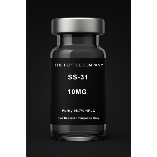
SS-31 10mg
SS-31 10mg is a research compound studied for mitochondrial targeting, bioenergetic efficiency, and cardiolipin stabilization pathway mechanisms. For research use only.
Introduction
NMN (nicotinamide mononucleotide) is a central intermediate in the NAD⁺ salvage pathway and is widely studied for its role in cellular metabolism, mitochondrial redox cycles, genomic maintenance, and energy signaling. As a precursor to NAD⁺, NMN significantly influences sirtuin activity, DNA repair processes, metabolic adaptation, and mitochondrial function.
NAD⁺ Metabolism and the Salvage Pathway
NMN is generated from nicotinamide via NAMPT, the rate‑limiting enzyme of the salvage pathway. NMN is then converted into NAD⁺ through NMNAT enzymes (NMNAT1, NMNAT2, NMNAT3), distributed across the nucleus, cytosol, and mitochondria. Research explores how NMN availability affects intracellular NAD⁺ pools, sirtuin consumption, PARP‑mediated DNA repair, redox homeostasis, and metabolic resilience.
Mitochondrial NAD⁺ Biology
Mitochondria require NAD⁺ for electron transfer chain (ETC) activity. NAD⁺ accepts electrons in the TCA cycle, feeds complex I through NADH, and enables ATP production. Research shows NMN-supported NAD⁺ levels influence mitochondrial biogenesis, mitophagy, redox balance, UPRmt (mitochondrial unfolded protein response), and maintenance of mitochondrial membrane potential.
Energy Metabolism and Redox Cycling
NAD⁺/NADH ratios are crucial for glycolysis, beta‑oxidation, oxidative phosphorylation, and metabolic flux. NMN research investigates how restored NAD⁺ pools regulate AMPK activity, influence metabolic efficiency, and maintain healthy redox cycling across different tissues.
Sirtuin Pathways
Sirtuins (SIRT1, SIRT2, SIRT3, SIRT6) are NAD⁺‑dependent deacetylases critical for gene expression, mitochondrial stability, stress adaptation, and chromatin regulation. Studies show that NMN availability can modulate sirtuin activity, impacting metabolic transcription, antioxidant defenses, genomic maintenance, and mitochondrial protein acetylation patterns.
DNA Repair and PARP Activity
PARP enzymes consume NAD⁺ in response to DNA damage. NMN-supported NAD⁺ pools may influence PARP activation, DNA strand repair efficiency, and cellular responses to oxidative stress. Excessive PARP activation depletes NAD⁺, making NAD⁺replenishment and salvage pathway dynamics essential to genomic maintenance.
Comparative Mechanistic Notes: NMN vs NR vs NAD⁺
NMN converts directly to NAD⁺ via NMNAT. NR (nicotinamide riboside) must first convert to NMN before entering this step. Exogenous NAD⁺ cannot directly cross membranes efficiently and must be broken down into NMN or NR precursors. Mechanistic studies evaluate differences in transporters, enzymatic conversion rates, and intracellular uptake among these compounds.
Cellular Stress, Autophagy, and AMPK
Research explores NMN’s involvement in AMPK activation, autophagic flux, mitochondrial quality control, antioxidant transcription, and metabolic stress adaptation. These pathways integrate NMN into mitochondrial resilience and cell‑protection models.
Summary
NMN is a critical NAD⁺ precursor examined for its roles in mitochondrial energy metabolism, redox cycling, genomic stability, sirtuin activity, and metabolic adaptation. Its central position in the salvage pathway makes it a key subject of modern mitochondrial, metabolic, and cellular‑repair research.
Educational & Research Disclaimer
This article is for educational and scientific research purposes only. No therapeutic claims or usage recommendations are provided. Compounds referenced are not approved for human use and are intended solely for controlled laboratory experimentation.
FAQ:
What is NMN in research?
Nicotinamide mononucleotide (NMN) is a biochemical NAD⁺ precursor studied in laboratory models for its role in cellular metabolism, oxidative stress pathways, and mitochondrial function.
How does NMN support NAD⁺ levels in research settings?
NMN is enzymatically converted into NAD⁺ through the salvage pathway. Researchers use it to explore how NAD⁺ fluctuations affect metabolism, DNA repair, mitochondrial respiration, and cellular stress responses.
Is NMN considered a therapeutic agent?
No. NMN from The Peptide Company is strictly for laboratory and in-vitro research. It is not a drug, supplement, or consumer product.
What areas of study involve NMN?
Research includes mitochondrial bioenergetics, sirtuin activation, metabolic regulation, redox balance, cellular aging models, and oxidative stress biology.
Can NMN influence mitochondrial performance in studies?
In controlled laboratory environments, NMN is examined for its relationship with ATP production, mitochondrial efficiency, and NAD⁺-dependent enzymatic activity.
How is NMN handled in research workflows?
NMN is stored cool and protected from light. After reconstitution, it is handled under validated institutional laboratory protocols only.
Is NMN intended for human use?
No. NMN is not for human consumption or clinical application of any kind.
Related Research Compounds
MOTS-c: The Mitochondrial-Encoded Peptide for Metabolic Regulation and Cellular Resilience
References
PMID: 25642957 — NAD⁺ metabolism, aging, and mitochondrial homeostasis
PMID: 29127247 — NMN and NAD⁺ salvage pathway in metabolic research
PMID: 26730458 — NAD⁺ precursors and mitochondrial function in experimental models
PMID: 31395777 — Redox regulation and sirtuin activity driven by NAD⁺ modulation
PMID: 23434792 — Cellular energy metabolism and NAD⁺ biosynthesis dynamics