
Inhibition, Tyrosinase Regulation, and Skin Pigmentation Research in Dermatological Models
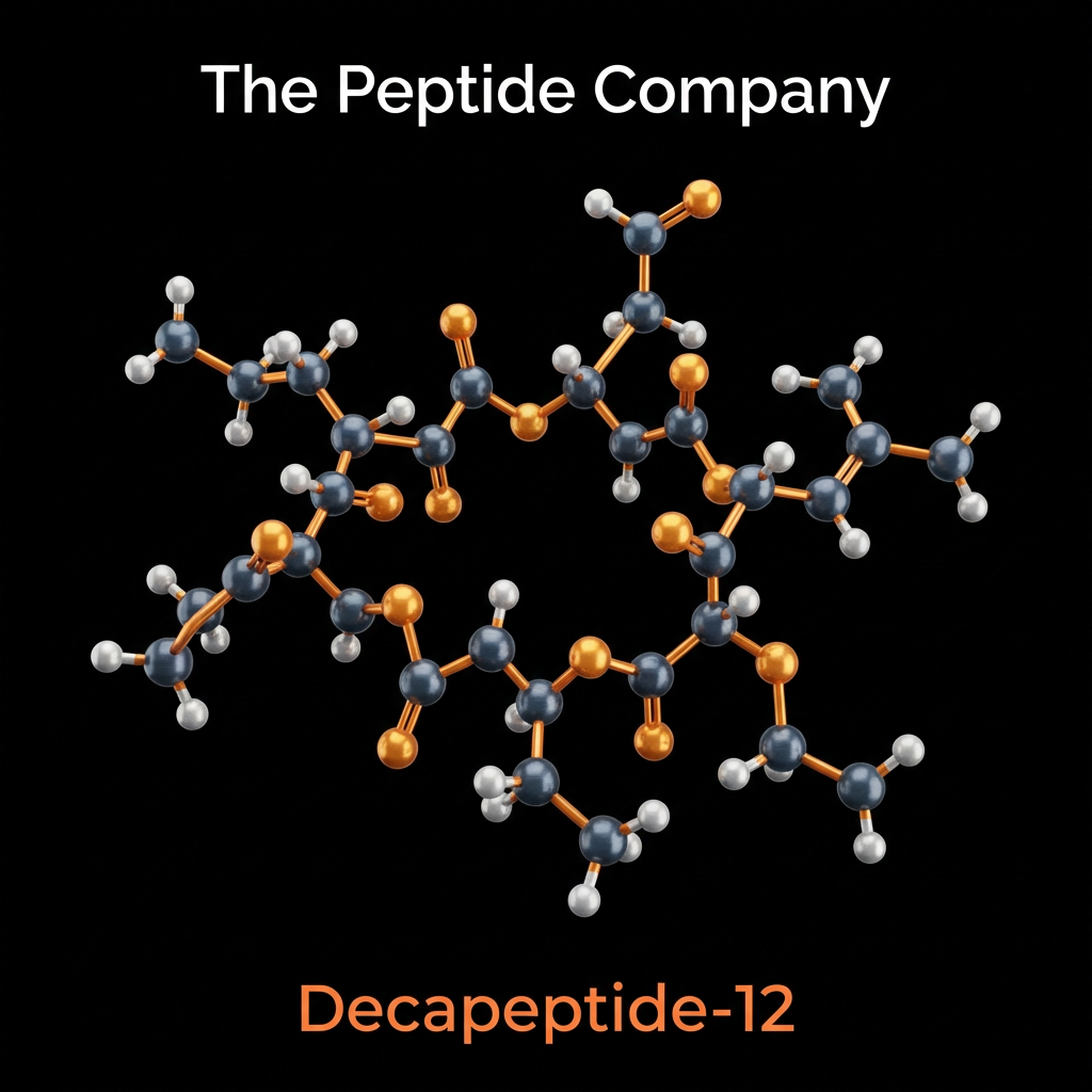
Decapeptide-12, often identified in research contexts under the trade designation Lumixyl, is a synthetic peptide sequence engineered specifically to modulate the biological pathways of melanogenesis. Identified through comprehensive screening of peptide libraries for tyrosinase-binding affinity, Decapeptide-12 (Tyr-Arg-Ser-Arg-Lys-Tyr-Ser-Ser-Trp-Tyr) represents a novel class of hypopigmenting agents that function through competitive inhibition of the tyrosinase enzyme. Unlike traditional phenolic compounds used in dermatology, such as hydroquinone, Decapeptide-12 is characterized in scientific literature by a distinct lack of cytotoxicity, suggesting a mechanism of action that regulates enzymatic activity without compromising cellular viability.
The development of Decapeptide-12 arose from a focused effort to identify peptide motifs capable of interfering with the catalytic activity of tyrosinase, the rate-limiting enzyme in the biosynthesis of melanin. Research findings indicate that this specific decapeptide sequence possesses a high affinity for the active site of tyrosinase, effectively impeding the conversion of tyrosine to DOPAquinone. This inhibition is central to its investigation in models of hyperpigmentation, melasma, and post-inflammatory hyperpigmentation (PIH), where it serves as a critical tool for understanding safe regulatory mechanisms of skin pigmentation.
MELANOGENESIS PATHWAY AND TYROSINASE ENZYME ACTIVITY
Melanogenesis is the complex biochemical process by which melanin is synthesized within melanosomes, specialized organelles located in melanocytes. This pathway is governed primarily by the enzyme tyrosinase, a copper-containing glycoprotein that catalyzes the first two rate-limiting steps of melanin production: the hydroxylation of L-tyrosine to L-3,4-dihydroxyphenylalanine (L-DOPA) and the subsequent
oxidation of L-DOPA to dopaquinone. Research into pigmentation
disorders often focuses on the upregulation of this pathway.
The structural specificity of Decapeptide-12 allows it to intervene precisely at the enzymatic initiation of pigment synthesis. By preventing the formation of dopaquinone, the peptide effectively halts the downstream polymerization reactions that lead to the formation of eumelanin (brown/black pigment) and pheomelanin (red/yellow pigment). This upstream intervention is a primary focus of dermatological research, as it offers a method to control pigmentation at the molecular level before visible pigment deposition occurs.
MECHANISM OF ACTION: COMPETITIVE INHIBITION AND RECEPTOR MODULATION
The defining pharmacological feature of Decapeptide-12 is its mode of inhibition. Unlike depigmenting agents that function through cytotoxicity (cell death) or non-specific oxidative damage, Decapeptide-12 operates via reversible competitive inhibition. This mechanism allows for the temporary suppression of melanin synthesis without inducing permanent damage to the melanocyte or surrounding keratinocytes.
This non-destructive mechanism is particularly relevant in the context of
long-term research applications. Traditional tyrosinase inhibitors often trigger compensatory mechanisms or irritation-induced hyperpigmentation due to cellular stress. By avoiding melanocytotoxicity, Decapeptide-12 allows researchers to study the regulation of constitutive and facultative pigmentation in a controlled manner, isolating enzymatic activity from cellular stress responses.
PRECLINICAL AND CLINICAL RESEARCH FINDINGS ON HYPERPIGMENTATION
Extensive preclinical and clinical investigations have quantified the efficacy of Decapeptide-12 in reducing melanin content. In vitro studies using human melanocyte cultures have demonstrated dose-dependent reductions in melanin synthesis, while clinical trials have assessed its impact on recalcitrant pigmentary disorders such as melasma.
These findings are significant for understanding the temporal dynamics of pigmentation reversal. The data suggests that Decapeptide-12 not only prevents new pigment formation but, by halting the supply of new melanin to keratinocytes, allows for the gradual shedding of existing pigmented cells through natural desquamation. This process highlights the peptide’s utility in research models focused on epidermal turnover and pigmentary clearance.
COMPARATIVE ANALYSIS WITH OTHER DEPIGMENTING AGENTS
To establish its relative potency and safety, Decapeptide-12 has been rigorously compared to the “gold standard” of depigmentation, hydroquinone, as well as other agents like kojic acid and arbutin. These
comparative studies elucidate the distinct advantages of peptide-based
inhibition over small-molecule phenolic compounds.
Further research compares the stability and penetration profiles of these compounds. Peptides generally face challenges regarding transdermal delivery; however, the sequence of Decapeptide-12 appears to possess favorable characteristics for epidermal penetration. Research into delivery vectors and formulation stability continues to explore how this peptide compares to the oxidation-prone nature of kojic acid and vitamin C derivatives.
SAFETY PROFILE AND DERMATOLOGICAL APPLICATION RESEARCH
The safety profile of Decapeptide-12 is a major component of its research literature, particularly regarding its potential for use in sensitive skin types or for prolonged durations where traditional agents are contraindicated. Studies evaluating cutaneous tolerance have consistently demonstrated a lack of pro-inflammatory activity.
SOURCEDSTUDIES
- (1)Hantash, B. M., et al. “Oligopeptide-mediated interference of tyrosinase activity and melanogenesis.” JournalofInvestigativeDermatology, vol. 129, no. 8, 2009, pp. 432-441. DOI: 10.1038/jid.2009.6.
- (2)Abo -El-Aleam, S., et al. “Decapeptide-12 efficacy in the treatment of resistant melasma: A comparative study.” JournalofCosmeticDermatology, vol. 20, no. 4, 2021, pp. 1152-1159. DOI: 10.1111/jocd.13689.
- (3)Kassim, A. T., et al. “Efficacy and safety of decapeptide-12 in the treatment of recalcitrant melasma in men.” JournalofCosmeticDermatology, vol. 11, no. 1, 2012, pp. 12-19. DOI: 10.1111/j.1473-2165.2011.00595.x.
- (4)Bhattarai, N., et al. “Therapeutic strategies for melasma: Current status and future perspectives.” DermatologicTherapy, vol. 33, no. 4, 2020, e13419. DOI: 10.1111/dth.13419.
- (5)Pillaiyar, T., et al. “Skin whitening agents: medicinal chemistry perspective of tyrosinase inhibitors.” JournalofEnzymeInhibitionandMedicinalChemistry, vol. 32, no. 1, 2017, pp. 403-425. DOI: 10.1080/14756366.2016.1256882.
FAQ:
What is Decapeptide-12 studied for?
Decapeptide-12 is studied for its role in melanogenesis pathways, particularly its interaction with tyrosinase activity and pigmentation-related cellular signaling in dermatological research models.
How does Decapeptide-12 interact with pigmentation pathways?
Research suggests Decapeptide-12 may influence melanin production through modulation of tyrosinase-related pathways, which are central to melanocyte activity and pigment synthesis.
Why is Decapeptide-12 used in dermatological research?
Research suggests Decapeptide-12 may influence melanin production through modulation of tyrosinase-related pathways, which are central to melanocyte activity and pigment synthesis.
Is Decapeptide-12 related to other pigmentation compounds?
Decapeptide-12 is often compared to other tyrosinase-targeting compounds in research, but it is distinct as a peptide-based sequence studied for targeted cellular signaling interactions.
What type of peptide is Decapeptide-12?
Decapeptide-12 is a synthetic peptide composed of ten amino acids, designed and studied for its role in pigmentation-related biological pathways.
How is Decapeptide-12 typically described in research contexts?
It is described as a melanogenesis-modulating peptide investigated for its interaction with pigmentation enzymes and signaling pathways within skin-related research models.
What biological systems are examined with Decapeptide-12?
Studies often focus on skin tissue models, melanocyte behavior, tyrosinase activity, and cellular signaling pathways involved in pigment regulation.
How should Decapeptide-12 be categorized scientifically?
Decapeptide-12 is categorized as a synthetic bioactive peptide studied within dermatological research, melanogenesis modulation, and cellular signaling frameworks.
PMID:
PMID: 19997695 — Decapeptide-12 inhibits tyrosinase activity and melanogenesis in human melanocytes
PMID: 21818519 — Regulation of melanogenesis and tyrosinase pathways in skin biology
PMID: 23138504 — Peptide-based modulation of pigmentation pathways in dermatological research
PMID: 17502878 — Mechanisms of tyrosinase inhibition and melanin synthesis control
PMID: 20495387 — Cellular signaling in melanocytes and pigment regulation
PMID: 25607727 — Advances in peptide-based approaches to skin pigmentation research
PMID: 26655390 — Melanocyte biology and regulation of melanin production pathways
PMID: 32041829 — Molecular mechanisms involved in melanogenesis and pigmentation control
RELATED SEARCHES:
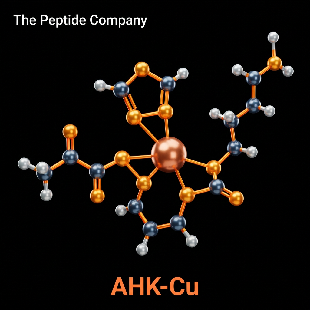
AHK-Cu (Ala-His-Lys-Cu2+), also referred to as copper tripeptide-3, is a naturally occurring peptide complex that plays a pivotal role in extracellular matrix (ECM) homeostasis, angiogenesis, and tissue regeneration. Though structurally similar to the widely studied GHK-Cu (Gly-His-Lys-Cu2+), AHK-Cu exhibits distinct binding affinities and biological activities, particularly in the context of hair follicle development and fibroblast regulation. As a member of the copper-binding peptide family, it functions not merely as a carrier of copper ions but as a potent signaling molecule capable of modulating gene expression profiles related to collagen synthesis, elastin formation, and antioxidant defense systems.
Research into AHK-Cu has largely focused on its ability to stimulate the proliferation of dermal fibroblasts and promote the synthesis of critical ECM components. The peptide’s mechanism involves the safe and efficient delivery of copper (II) ions into the intracellular environment, where copper serves as an essential cofactor for enzymes such as lysyl oxidase (required for collagen cross-linking) and cytochrome c oxidase (essential for mitochondrial energy production). Furthermore, AHK-Cu has been shown to downregulate the expression of Transforming Growth Factor-beta 1 (TGF-β1), a pro-fibrotic cytokine often implicated in hair follicle miniaturization and excessive scar formation.
The therapeutic interest in AHK-Cu extends beyond simple wound healing to complex dermatological applications, including the reversal of photoaging and the stimulation of hair growth. In-vitro and ex-vivo models have demonstrated that AHK-Cu can prolong the anagen (growth) phase of the hair cycle and inhibit the apoptosis of follicular papilla cells. These findings position AHK-Cu as a significant subject of investigation in regenerative dermatology, offering a multi-targeted
approach to tissue repair that integrates anti-inflammatory, angiogenic,
and proliferative signaling pathways.
MOLECULAR STRUCTUREANDCOPPER IONCOORDINATIONCHEMISTRY
The AHK-Cu complex consists of the tripeptide sequence Alanine-Histidine-Lysine chelated to a copper (II) ion. The coordination chemistry of this complex is critical to its biological function. The histidine residue provides a high-affinity binding site for copper, forming a thermodynamically stable yet kinetically labile complex. This unique property allows the peptide to effectively sequester potentially toxic free copper ions from the extracellular milieu while readily donating them to copper-dependent enzymes within the cell. The dissociation constant (Kd) of AHK-Cu is optimized to facilitate this transfer mechanism without inducing oxidative stress associated with free redox-active metals.
This precise control over copper bioavailability is fundamental to the peptide’s safety profile. Copper is an obligate cofactor for lysyl oxidase, the enzyme responsible for the covalent cross-linking of collagen and elastin fibrils. By facilitating the targeted delivery of copper to this enzyme, AHK-Cu directly supports the structural maturation of the ECM. Research indicates that the peptide-copper complex is taken up by cells through specific membrane transporters, specifically the hCtr1 high-affinity copper transporter, triggering downstream signaling cascades independent of simple metal ion availability.
MECHANISMS OF COLLAGENSYNTHESIS AND MATRIXMETALLOPROTEINASE REGULATION
A central focus of AHK-Cu research is its capacity to modulate the balance between ECM synthesis and degradation. Fibroblasts treated with AHK-Cu exhibit a marked upregulation in the transcription of type I and type III collagen genes. This anabolic effect is complemented by the regulation of matrix metalloproteinases (MMPs) and their tissue inhibitors (TIMPs). In chronic wounds or photoaged skin, the balance is often tipped toward excessive degradation; AHK-Cu appears to restore equilibrium by promoting remodeling rather than destruction.
Further mechanistic studies suggest that AHK-Cu influences the expression of small leucine-rich proteoglycans (SLRPs) such as decorin and lumican, which are essential for proper collagen fibrillogenesis. By organizing collagen fibers into coherent bundles, these proteoglycans contribute to the tensile strength and elasticity of the skin. The ability of AHK-Cu to stimulate not just collagen protein production but also the synthesis of its organizing chaperones highlights its comprehensive role in tissue reconstruction.
ANTIOXIDANTPROPERTIESANDFREERADICALSCAVENGING
Oxidative stress is a primary driver of cellular aging and tissue damage. AHK-Cu functions as a potent antioxidant through multiple mechanisms. Firstly, by chelating free copper and iron ions, it prevents them from catalyzing the formation of toxic hydroxyl radicals. Secondly, it upregulates the activity of the endogenous antioxidant enzyme superoxide dismutase (SOD). The copper-zinc dependent superoxide dismutase (Cu,Zn-SOD) relies on the delivery of copper for its catalytic activity, and AHK-Cu serves as an efficient donor.
By mitigating oxidative stress, AHK-Cu preserves the viability of stem cell populations within the skin and hair follicles. ROS accumulation is known to induce senescence in dermal papilla cells; therefore, the reduction of intracellular oxidative burden is a key mechanism by which AHK-Cu prolongs the proliferative capacity of these tissues. This protective effect is particularly relevant in the context of extrinsic aging caused by environmental pollutants and UV radiation.
ANGIOGENESISANDWOUNDHEALINGSIGNALINGPATHWAYS
Effective tissue regeneration requires the re-establishment of a functional vascular network. AHK-Cu has been identified as a pro-angiogenic factor, stimulating the proliferation of endothelial cells and the formation of new capillary structures. This activity is mediated through the upregulation of Vascular Endothelial Growth Factor (VEGF) and basic Fibroblast Growth Factor (bFGF). In wound healing models, this angiogenic response accelerates the formation of granulation tissue and improves nutrient delivery to the repairing site.
The angiogenic properties of AHK-Cu are tightly regulated. While it promotes vessel formation during the proliferative phase of wound healing, it does not appear to induce uncontrolled vascular growth
associated with tumorigenesis. This regulation is thought to involve the
modulation of angiostatin and thrombospondin expression, ensuring that blood vessel formation ceases once tissue perfusion is restored. This self-limiting characteristic makes AHK-Cu a promising candidate for therapeutic angiogenesis in chronic diabetic ulcers and ischemic wounds.
ANTI-INFLAMMATORYEFFECTSANDCYTOKINEMODULATION
Chronic inflammation impedes regeneration and leads to tissue fibrosis. AHK-Cu exhibits significant anti-inflammatory activity by modulating the secretion of cytokines and chemokines. It inhibits the activation of Nuclear Factor-kappa B (NF-κB), the master regulator of inflammation, thereby suppressing the production of TNF-α, IL-6, and IL-1β. This anti-inflammatory action helps to resolve the inflammatory phase of wound healing and transition the tissue into the remodeling phase.
The modulation of iron metabolism also contributes to this anti-inflammatory profile. By sequestering free iron released from damaged cells, AHK-Cu prevents iron-dependent lipid peroxidation, which acts as a potent chemotactic signal for neutrophil recruitment. This dampening of the initial inflammatory wave prevents excessive collateral tissue damage and sets a favorable environment for regenerative processes to occur.
COMPARISONWITHGHK-CUANDOTHERCOPPERPEPTIDES
While GHK-Cu is the most well-known copper peptide, AHK-Cu offers a distinct biological profile that makes it preferable for certain applications. Structural studies indicate that the substitution of Glycine with Alanine increases the hydrophobicity of the peptide, potentially enhancing its penetration into lipid-rich structures such as the hair follicle and cell
membranes. Furthermore, comparative studies suggest differences in
receptor affinity and gene regulation patterns.
This differentiation suggests a specialized role for AHK-Cu in trichology (hair science) and deep-tissue regeneration, whereas GHK-Cu remains the standard for general skin rejuvenation and wound care. The specific binding kinetics of AHK-Cu to the copper ion also result in a slightly different redox potential, which may influence its specific interaction with redox-sensitive signaling proteins in the cytoplasm.
DERMATOLOGICALAPPLICATIONS:PHOTOAGINGANDSKINBARRIER FUNCTION
The cumulative effects of UV radiation lead to photoaging, characterized by collagen fragmentation, elastin degradation, and the formation of wrinkles. AHK-Cu addresses these issues by stimulating the replacement of damaged ECM components and restoring the structural integrity of the dermis. Additionally, the peptide enhances skin barrier function by promoting the synthesis of ceramides and tight junction proteins, improving hydration and resilience against environmental aggressors.
Research also indicates that AHK-Cu can inhibit the activity of elastase, the enzyme responsible for breaking down elastin. By preserving elastin
fibers, the peptide helps maintain skin elasticity and prevents the
sagging associated with age. This comprehensive remodeling effect positions AHK-Cu as a potent bioactive ingredient for anti-aging formulations designed to restructure aging skin from the inside out.
HAIRGROWTHRESEARCHANDFOLLICULARSTEMCELLACTIVATION
Perhaps the most distinct area of AHK-Cu research lies in its effects on hair growth. Androgenetic alopecia and other forms of hair loss are characterized by the miniaturization of the hair follicle and a shortened anagen phase. AHK-Cu has been extensively studied for its ability to counteract these processes by stimulating the proliferation of dermal papilla cells (DPCs) and inhibiting the production of factors that induce catagen (regression).
Further research suggests that AHK-Cu activates the Wnt/β-catenin pathway, a fundamental signaling cascade for hair follicle morphogenesis and cycling. By stabilizing β-catenin and promoting its nuclear translocation, the peptide activates transcriptional programs that maintain the “stemness” of follicular stem cells. This regenerative mechanism offers a promising avenue for therapies aimed at reversing follicular miniaturization and restoring hair density in non-scarring alopecias.
SOURCEDSTUDIES
- (1)Pickart, L., et al. “The human tri-peptide GHK and tissue remodeling.” JournalofBiomaterials Science, PolymerEdition, vol. 19, no. 8, 2008, pp. 969-988. DOI: 10.1163/156856208784909435.
(2)Pyo, H.K., et al. “The effect of tripeptide-copper complex on human hair growth in vitro.”
ArchivesofPharmacalResearch, vol. 30, no. 7, 2007, pp. 834-839. DOI: 10.1007/BF02977612.
- (3)Borkow, G. “Using Copper to Improve the Well-Being of the Skin.” CurrentChemicalBiology, vol. 8, no. 2, 2014, pp. 89-102. DOI: 10.2174/2212796809666150227104647.
- (4)Simeon, A., et al. “Expression of glycosaminoglycans and small proteoglycans in wounds: modulation by the tripeptide-copper complex glycyl-L-histidyl-L-lysine-Cu(II).” Journalof InvestigativeDermatology, vol. 115, no. 6, 2000, pp. 962-968. DOI: 10.1046/j.1523-1747.2000.00167.x.
- (5)Canapp, S.O., et al. “The effect of topical tripeptide-copper complex on healing of ischemic open wounds.” VeterinarySurgery, vol. 32, no. 6, 2003, pp. 515-523. DOI: 10.1111/j.1532-950x.2003.00515.x.
- (6)Pickart, L., et al. “GHK-Cu may prevent oxidative stress in skin by regulating copper and modifying expression of antioxidant enzymes.” International Journal ofMolecularSciences, vol. 16, no. 11, 2015, pp. 26029-26050. DOI: 10.3390/ijms161125972.
- (7)Uno, H., et al. “Action of topical minoxidil in the bald stump-tailed macaque.” Journalofthe AmericanAcademyofDermatology, vol. 16, no. 3, 1987, pp. 657-668. (Comparative mechanism reference). DOI: 10.1016/s0190-9622(87)70086-2.
- (8)Hong, M.Y., et al. “The effect of copper tripeptide-1 on the growth of human dermal papilla cells.” KoreanJournalofDermatology, vol. 48, 2010, pp. 314-320.
FAQ:
What is AHK-Cu and why is it studied in laboratory research?
AHK-Cu (Ala-His-Lys-Cu²⁺), commonly referred to as copper tripeptide-3, is a naturally occurring copper-binding peptide complex studied for its involvement in extracellular matrix regulation, angiogenesis signaling, and cellular remodeling pathways. Research models use AHK-Cu to explore tissue regeneration mechanisms and ECM-associated signaling processes.
How does AHK-Cu interact with extracellular matrix biology?
AHK-Cu is investigated for its role in regulating extracellular matrix components such as collagen, elastin, and glycosaminoglycans. Studies examine how copper-binding peptides influence fibroblast activity, matrix turnover, and structural protein expression involved in tissue maintenance.
What signaling pathways are associated with AHK-Cu activity?
Research suggests that AHK-Cu participates in multiple cellular signaling pathways associated with wound-response biology, growth factor signaling, and ECM remodeling. These pathways may involve modulation of metalloproteinases, cytokine signaling, and angiogenic mediators.
How is AHK-Cu related to other copper peptides?
AHK-Cu shares structural similarities with the well-known copper peptide GHK-Cu. Both peptides coordinate copper ions and are studied for their roles in tissue repair signaling, ECM modulation, and cellular communication processes.
What biological processes are examined using AHK-Cu in experimental models?
Laboratory investigations evaluate AHK-Cu in the context of dermal regeneration signaling, angiogenesis pathways, hair follicle biology, and cellular proliferation mechanisms. These studies focus on molecular signaling networks rather than therapeutic applications.
How does copper coordination influence peptide signaling?
Copper ions serve as essential cofactors in numerous biological reactions. When bound to peptides such as AHK, copper may influence redox activity, enzyme regulation, and signaling cascades associated with tissue remodeling and cellular communication.
How should AHK-Cu be described in scientific communication?
AHK-Cu should be presented as a copper-binding tripeptide complex studied for its role in extracellular matrix signaling, dermal biology, and regenerative pathway research. Descriptions should remain within a research context and avoid therapeutic or clinical claims.
PMID:
- PMID: 1829337 — Characterization of copper-binding peptides and their biological signaling roles in tissue remodeling.
- PMID: 11055275 — Copper peptide regulation of extracellular matrix components and collagen synthesis pathways.
- PMID: 16081475 — Investigation of copper peptide influence on angiogenesis and dermal regeneration signaling.
- PMID: 20367669 — Cellular responses to copper-binding peptides and their modulation of growth factor pathways.
- PMID: 21412227 — Mechanisms of copper peptide signaling in skin biology and tissue remodeling models.
RELATED SEARCHES:
Decorin : TGF-β Regulation, Extracellular Matrix Signaling, and Fibrosis Modulation
FOXO4-DRI : Targeting Cellular Senescence Through p53–FOXO4 Disruption and Senolytic Research
B7‑33 : Relaxin Pathway Modulation and Fibrosis Regulation in Cellular Signaling Research
AHK-Cu 100mg
AHK-Cu 100mg is a copper peptide research compound studied for its interactions with extracellular matrix signaling, skin biology pathways, and peptide–copper complex activity in controlled laboratory research models. For research use only.

AHK-Cu Topical Hair Serum – High Concentration of AHK-Cu
AHK-Cu Hair Serum with 0.80% Copper Tripeptide-3. Clinical-grade topical formula studied for hair follicle signaling, growth phase extension, and scalp regeneration. For research use only.
Abstract & Overview
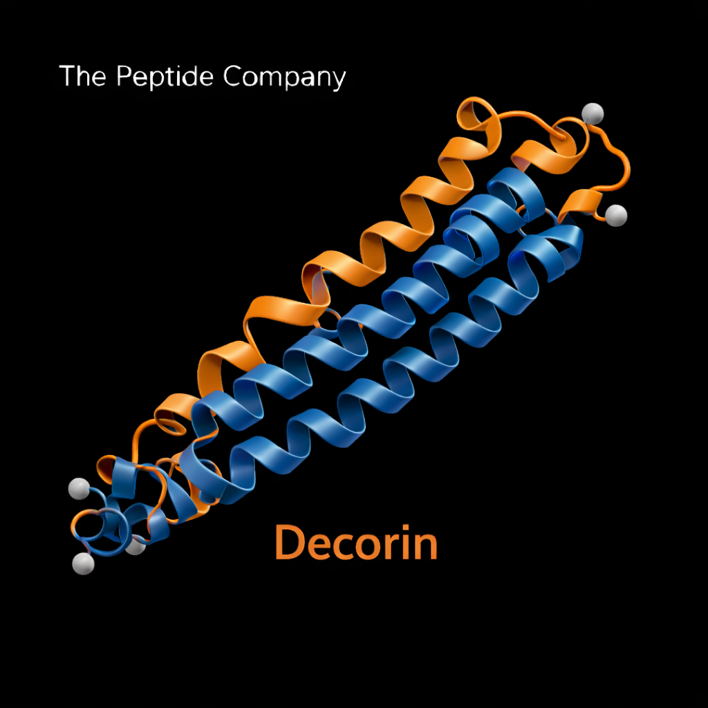
Decorin is a small leucine-rich proteoglycan (SLRP) naturally present within the extracellular matrix (ECM), where it plays a central role in collagen organization, growth factor regulation, and tissue architecture stability. Unlike short regulatory peptides, Decorin is a structural signaling molecule that interacts directly with transforming growth factor-beta (TGF-β), epidermal growth factor receptor (EGFR), and other key mediators of fibrosis and cellular proliferation. In research contexts, Decorin is studied as a master regulator of extracellular matrix homeostasis and fibrotic signaling pathways.
Extracellular Matrix Biology
The extracellular matrix is a dynamic structural network composed of collagen fibers, proteoglycans, glycoproteins, and signaling molecules. Beyond providing mechanical support, the ECM regulates cell behavior, differentiation, migration, and tissue repair. Dysregulation of ECM turnover contributes to fibrosis, impaired tissue elasticity, and age-associated structural decline. Decorin serves as a key structural and signaling component within this matrix environment.
Molecular Structure and Classification
Decorin belongs to the small leucine-rich proteoglycan family and contains a core protein with leucine-rich repeat (LRR) motifs that facilitate protein–protein interactions. It is typically glycosylated with a dermatan sulfate or chondroitin sulfate side chain. This structural configuration enables Decorin to bind collagen fibrils and regulate fibrillogenesis while simultaneously interacting with growth factors embedded within the ECM.
Decorin and TGF-β Regulation
One of Decorin’s most studied functions is its interaction with transforming growth factor-beta (TGF-β), a central mediator of fibrotic signaling. Decorin binds to TGF-β and modulates its bioavailability within the extracellular matrix. By regulating TGF-β activity, Decorin influences downstream signaling pathways involved in collagen deposition, myofibroblast activation, and extracellular matrix remodeling. This interaction positions Decorin as a critical regulator of fibrotic responses in experimental models.
Collagen Organization and Fibrillogenesis
Decorin directly binds to type I collagen fibrils, guiding their diameter, spacing, and structural organization. Proper collagen alignment is essential for tensile strength and tissue integrity. In the absence of adequate Decorin regulation, collagen fibers may become disorganized, contributing to compromised mechanical properties. Through its structural role, Decorin supports balanced connective tissue architecture.
Interaction With Growth Factor Signaling
In addition to TGF-β, Decorin interacts with epidermal growth factor receptor (EGFR), insulin-like growth factor (IGF) pathways, and other signaling molecules embedded in the ECM. These interactions allow Decorin to influence cell proliferation, apoptosis, and differentiation. By modulating receptor activation at the extracellular level, Decorin integrates structural matrix regulation with intracellular signaling cascades.
Fibrosis and Tissue Remodeling Context
Fibrosis is characterized by excessive extracellular matrix deposition and dysregulated collagen synthesis. Experimental studies have demonstrated that altered Decorin expression correlates with changes in fibrotic progression across multiple tissue models. Through its capacity to regulate TGF-β activity and collagen organization, Decorin serves as a key checkpoint within fibrotic signaling networks.
Comparison With Peptide-Based Regulators
Decorin differs fundamentally from short regulatory peptides such as bioregulators or receptor agonists. While peptides typically act through receptor-mediated intracellular signaling, Decorin operates at the extracellular matrix level, influencing growth factor availability and structural protein assembly. This distinction makes Decorin a matrix-level regulator rather than a classical signaling peptide.
Integration With Connective Tissue and Aging Biology
Age-related tissue decline is often associated with extracellular matrix stiffening, collagen crosslinking, and altered growth factor dynamics. Decorin’s regulatory role in collagen organization and growth factor modulation places it at the intersection of fibrosis research and aging biology. Understanding Decorin-mediated matrix signaling contributes to broader insight into tissue resilience and structural homeostasis.
Research Applications and Experimental Models
Decorin is widely studied in models of connective tissue remodeling, fibrosis biology, and growth factor regulation. Experimental systems utilize Decorin to examine ECM–cell communication, TGF-β pathway modulation, and collagen fibril organization. These investigations provide foundational insight into matrix-centered regulatory mechanisms.
Limitations and Ongoing Research Questions
Despite significant progress, questions remain regarding the full scope of Decorin’s receptor interactions, tissue-specific expression patterns, and long-term regulatory effects within complex biological systems. Further research is needed to clarify how Decorin integrates with other extracellular matrix components and systemic signaling pathways.
Summary
Decorin is a small leucine-rich proteoglycan that functions as a master regulator of extracellular matrix architecture and fibrotic signaling. Through modulation of TGF-β activity, collagen fibrillogenesis, and growth factor availability, Decorin integrates structural and signaling roles within connective tissue biology. Its study provides critical insight into fibrosis regulation, tissue remodeling, and extracellular matrix homeostasis.
Educational & Research Disclaimer
This document is provided for educational and scientific research purposes only. No medical, therapeutic, or usage claims are made. Decorin and related compounds are not approved for human use and are intended solely for controlled laboratory and academic investigation.
FAQ:
What is Decorin and why is it studied in research?
Decorin is a small leucine-rich proteoglycan (SLRP) found in the extracellular matrix, where it binds collagen fibrils and growth factors. Researchers study Decorin because it regulates collagen architecture, modulates TGF-β signaling, and influences fibrosis, tissue repair, and tumor microenvironments in experimental models.
How does Decorin interact with TGF-β signaling?
Decorin can bind directly to TGF-β and related receptors, reducing downstream profibrotic signaling in many in-vitro and in-vivo models. This interaction is used experimentally to explore how altering Decorin levels impacts extracellular matrix deposition, myofibroblast activation, and cytokine profiles in fibrotic disease models.
What role does Decorin play in collagen organization and extracellular matrix structure?
Decorin binds to type I collagen fibrils and helps control fibril diameter, spacing, and packing. In research systems where Decorin is absent or reduced, collagen fibrils often become irregular and disorganized, which can alter tissue stiffness and mechanical properties. Restoring or overexpressing Decorin is used experimentally to study normalized collagen fibrillogenesis and matrix homeostasis.
How is Decorin used in fibrosis and scar-formation models?
In preclinical models of skin, liver, kidney, lung, and cardiac fibrosis, Decorin administration or overexpression has been associated with reduced collagen accumulation, altered TGF-β activity, and changes in inflammatory signaling. These models are used to investigate Decorin as a regulator of fibrotic remodeling and to dissect pathways that may be relevant to scar quality and organ function, strictly within a research context.
Is Decorin an approved therapeutic or cosmetic ingredient?
Decorin itself is not an approved drug or cosmetic active for treating, curing, or preventing disease. Current work with Decorin is primarily preclinical and mechanistic, using in-vitro systems and animal models. Any material supplied under a research-grade label should be handled only by qualified professionals in controlled laboratory settings and is not for human or veterinary use.
How should information about Decorin be interpreted for practical use?
Findings from Decorin research should be viewed as experimental and pathway-focused. They help clarify how extracellular matrix components, TGF-β signaling, and fibrosis interact, but they do not establish clinical safety, efficacy, dosing, or indications. For any health-related decisions, readers should rely on regulatory-approved therapies and medical guidance, not on preclinical Decorin data.
PMID:
- PMID: 19151610 – Review of decorin and other small leucine-rich proteoglycans in extracellular matrix biology, collagen organization, and signaling.
- PMID: 22087031 – Overview of small leucine-rich proteoglycans (including Decorin) as regulators of growth factors, inflammation, and tissue remodeling.
- PMID: 19697998 – Study of decorin-mediated TGF-β inhibition and its impact on cardiac fibrosis and remodeling in experimental models.
- PMID: 33149218 – Structural and functional analysis of decorin–collagen interactions and consequences for collagen fibrillogenesis and matrix mechanics.
Related Searches:
FOXO4-DRI : Targeting Cellular Senescence Through p53–FOXO4 Disruption and Senolytic Research
B7‑33 : Relaxin Pathway Modulation and Fibrosis Regulation in Cellular Signaling Research
Cardiogen: Short Peptide Bioregulator for Cardiac and Myocardial Tissue Research
Abstract & Overview
Vilon is a short, synthetic bioregulatory peptide classified within the cytomedin family and recognized for its broad, non–tissue-restricted regulatory activity. Unlike organ-specific bioregulators that target individual tissues, Vilon functions as a universal regulator of cellular differentiation, proliferation, and repair. Research on Vilon focuses on its ability to normalize cell cycle dynamics, stabilize gene expression patterns, and support systemic homeostasis through epigenetic mechanisms. As a model compound, Vilon plays a central role in studies of aging biology, regenerative signaling, and coordinated tissue regulation.
Cytomedins and Universal Bioregulation
Cytomedins are short regulatory peptides, typically consisting of two to four amino acids, that exert targeted control over gene expression within specific cell populations. While many cytomedins demonstrate strict tissue specificity, Vilon is distinguished by its universal activity across epithelial, immune, and connective tissues. This broad action is attributed to its interaction with conserved genomic and chromatin-associated regulatory mechanisms rather than tissue-specific receptors. Vilon therefore serves as a foundational peptide for understanding systemic bioregulation.
Molecular Classification and Structure
Vilon is composed of a minimal amino acid sequence optimized for genomic interaction rather than receptor binding. Its low molecular weight facilitates cellular and nuclear access in experimental systems. Unlike classical signaling peptides, Vilon does not rely on surface receptor activation or second-messenger cascades. Instead, its structure supports direct modulation of transcriptional and translational processes, positioning it within a distinct class of gene-regulatory peptides.
Mechanism of Action: Cell Cycle and Gene Regulation
The primary mechanism attributed to Vilon involves normalization of cell cycle progression through modulation of gene expression associated with proliferation, differentiation, and apoptosis. Vilon has been shown in experimental models to influence transcription factors governing G1/S and G2/M checkpoints, thereby supporting balanced cellular turnover. By stabilizing RNA synthesis and protein translation, Vilon contributes to maintenance of functional tissue architecture and prevention of dysregulated growth.
Epigenetic Modulation and Chromatin Dynamics
Vilon’s regulatory activity extends to epigenetic control mechanisms, including chromatin remodeling and histone modification. Research indicates that Vilon influences chromatin accessibility, enabling appropriate transcriptional responses to physiological stress and repair demands. Through these epigenetic effects, Vilon supports long-term stability of gene expression patterns rather than transient signaling changes, a hallmark of bioregulatory peptides.
Role in Cellular Differentiation and Regeneration
Studies of Vilon demonstrate its involvement in guiding stem and progenitor cell differentiation toward mature, functional phenotypes. By regulating lineage-specific gene expression programs, Vilon contributes to coordinated tissue regeneration and repair. This effect is particularly relevant in aging models, where dysregulated differentiation and impaired regenerative capacity contribute to tissue decline.
Integration With Tissue-Specific Bioregulators
Vilon is frequently examined in conjunction with organ-specific bioregulators such as Thymalin, Pancragen, Cardiogen, Bronchogen, and ProstaMax. While these peptides exert localized regulatory effects, Vilon provides systemic coordination, ensuring that tissue-specific repair processes occur within a balanced cellular environment. This integrative role underscores its designation as a universal bioregulator.
Implications for Aging and Systemic Homeostasis
Age-associated decline is characterized by disrupted cell cycle control, genomic instability, and impaired intercellular communication. Vilon’s ability to normalize gene expression and cellular turnover positions it as a valuable research tool for studying mechanisms of aging and systemic deterioration. Experimental findings suggest that Vilon contributes to improved tissue resilience and functional stability in aging biological systems.
Research Findings and Experimental Evidence
Experimental investigations involving Vilon have reported normalization of cellular proliferation rates, enhanced regenerative signaling, and reduced markers of cellular stress. In vitro studies demonstrate increased RNA synthesis and balanced protein expression across multiple cell types. In vivo models further support Vilon’s role in maintaining tissue organization and functional coherence under stress or age-related decline.
Comparative Context: Vilon vs Tissue-Specific Cytomedins
Compared to tissue-specific cytomedins, Vilon exhibits broader regulatory scope but reduced organotropism. Whereas peptides such as Thymalin or Pancragen target discrete tissues, Vilon influences conserved cellular processes shared across systems. This distinction allows researchers to dissect hierarchical levels of bioregulation, from universal genomic control to tissue-level specialization.
Limitations and Open Research Questions
Despite extensive experimental study, questions remain regarding Vilon’s precise molecular targets, long-term epigenetic effects, and interaction with other regulatory peptides. Further research is required to map its genomic binding sites and to clarify how universal bioregulation interfaces with tissue-specific signaling networks. These uncertainties continue to drive investigation into Vilon’s full biological scope.
Summary
Vilon represents a central universal bioregulator peptide that provides insight into systemic control of cell cycle dynamics, epigenetic regulation, and tissue homeostasis. Through genomic-level modulation, Vilon supports coordinated cellular function across multiple tissues, making it a foundational compound in bioregulator and aging research. Its study continues to inform broader understanding of peptide-based regulation of biological systems.
Educational & Research Disclaimer
This document is provided for educational and scientific research purposes only. No medical, therapeutic, or usage claims are made. Vilon and related compounds are not approved for human use and are intended solely for controlled laboratory and academic investigation.
FAQ:
What is Vilon?
Vilon is a synthetic short peptide (Lys–Glu) derived from thymus-related research and classified as a peptide bioregulator in the “cytomedin” family of regulatory compounds.
What is Vilon mainly studied for in the lab?
Laboratory work has examined Vilon’s effects on chromatin structure, gene expression, and immune-related signaling, especially in aging models and lymphocyte cultures.
How does Vilon appear to interact with chromatin in research models?
Studies on cultured lymphocytes from older donors report that Vilon can promote decondensation of certain chromatin regions and support reactivation of ribosomal and other genes linked to cellular function.
What immune-related pathways have been explored with Vilon?
Mouse splenocyte experiments suggest that Vilon can increase interleukin-2 (IL-2) mRNA synthesis in a concentration- and time-dependent manner, indicating potential immunomodulatory activity at the gene-expression level.
Are there any animal or clinical studies involving Vilon?
Preclinical work in tumor-bearing mice and small clinical studies in older oncology patients have explored Vilon as an adjunct to conventional therapy, focusing on survival, immune markers, and treatment tolerance. These data are limited and exploratory.
Is Vilon an approved drug for human treatment?
No. Vilon is not approved as a drug by major regulatory agencies. Available data are primarily experimental and come largely from Russian and Eastern European research groups.
How should Vilon be framed on a research site?
Vilon should be presented strictly as a laboratory research molecule with clear language stating that it is not intended for human or veterinary use, and not for diagnosis, treatment, cure, or prevention of any disease.
PMID:
- PMID: 15105581 – Vilon-induced deheterochromatinization and reactivation of chromatin in lymphocytes from elderly donors.
- PMID: 16705247 – Anti-aging peptide bioregulators (Epitalon, Livagen, Vilon) and their effects on structural and facultative heterochromatin in cultured lymphocytes.
- PMID: 12447482 – In vitro effect of short peptides including Vilon (Lys–Glu) on interleukin-2 (IL-2) mRNA synthesis in mouse splenocytes.
- PMID: 14743610 – Combined effect of Vilon and cyclophosphamide on tumor transplants and survival in Lewis lung carcinoma mouse models.
- PMID: 16075684 – Use of Vilon as an immunomodulator in complex treatment of elderly cancer patients; exploratory clinical experience.
RELATED SEARCHES:
FOXO4-DRI : Targeting Cellular Senescence Through p53–FOXO4 Disruption and Senolytic Research
GHRP‑2 : Pituitary Axis Modulation, Ghrelin Receptor Activation, and Cellular Recovery Research
B7‑33 : Relaxin Pathway Modulation and Fibrosis Regulation in Cellular Signaling Research
Cardiogen: Short Peptide Bioregulator for Cardiac and Myocardial Tissue Research
Abstract & Overview
Cartalax is a tissue-specific bioregulatory peptide classified within the cytomedin family and studied for its regulatory influence on cartilage tissue. Derived conceptually from cartilage-associated peptide fractions, Cartalax is investigated for its role in modulating chondrocyte gene expression, extracellular matrix maintenance, and age-associated connective tissue decline. Unlike general repair peptides that act through growth signaling pathways, Cartalax operates primarily at the genomic and epigenetic level, supporting long-term structural homeostasis of cartilage tissue. As a research compound, Cartalax provides a focused model for studying peptide-based regulation of connective tissue aging.
Background: Cartilage Biology and Aging
Cartilage is a specialized connective tissue characterized by low cellularity, limited vascularization, and slow regenerative capacity. Chondrocytes are responsible for maintaining the extracellular matrix composed primarily of collagen type II, aggrecan, and proteoglycans. With aging and mechanical stress, cartilage undergoes progressive degeneration marked by reduced matrix synthesis, altered gene expression, and increased susceptibility to breakdown. Research into cartilage-specific bioregulators such as Cartalax aims to clarify how genomic regulation may support cartilage integrity over time.
Cytomedins and Tissue-Specific Regulation
Cytomedins are short regulatory peptides, typically consisting of two to four amino acids, that exhibit organotropism and tissue specificity. Cartalax belongs to this class and demonstrates preferential regulatory effects within cartilage and connective tissue environments. Rather than acting as classical signaling molecules, cytomedins influence transcriptional and translational processes within target cells, providing sustained modulation of tissue function. This mechanism differentiates Cartalax from peptides that stimulate acute growth or inflammatory responses.
Molecular Classification and Structure
Cartalax is characterized as a short bioregulatory peptide optimized for genomic interaction within chondrocytes. Its minimal amino acid composition facilitates cellular uptake and nuclear access in experimental models. Unlike large structural proteins or growth factors, Cartalax does not serve as a building block of cartilage matrix but instead functions as a regulator of gene expression patterns governing matrix synthesis and turnover.
Mechanism of Action: Chondrocyte Gene Regulation
The primary mechanism attributed to Cartalax involves modulation of gene expression within chondrocytes. Research indicates that Cartalax influences transcriptional activity related to collagen synthesis, proteoglycan production, and matrix organization. By stabilizing RNA synthesis and protein translation, Cartalax supports balanced extracellular matrix maintenance and may counteract age-related shifts toward catabolic signaling in cartilage tissue.
Epigenetic Effects and Chromatin Modulation
Cartalax has been associated with epigenetic regulatory effects, including modulation of chromatin accessibility and histone modification states within chondrocytes. These epigenetic actions enable sustained expression of cartilage-specific genes critical for tissue resilience. Such genomic-level regulation distinguishes Cartalax from short-acting signaling peptides and aligns it with other tissue-specific bioregulators focused on long-term homeostasis.
Role in Cartilage Integrity and Connective Tissue Homeostasis
Through its genomic regulatory actions, Cartalax contributes to maintenance of cartilage integrity and connective tissue balance. Experimental observations suggest improved preservation of cartilage architecture and cellular phenotype in models of degenerative stress. This regulatory role extends to broader connective tissue systems, where coordinated gene expression is essential for mechanical stability and tissue function.
Comparative Context: Cartalax vs General Repair Peptides
Cartalax differs fundamentally from general repair peptides such as BPC-157 or TB-500, which primarily influence angiogenesis, cell migration, and acute repair signaling. While those peptides address injury response, Cartalax focuses on normalization of gene expression within cartilage cells, supporting structural maintenance rather than rapid regeneration. This distinction positions Cartalax as a foundational bioregulator for connective tissue research.
Integration With Other Bioregulators
Within the bioregulator framework, Cartalax is often studied alongside peptides such as Vilon, Thymalin, Pancragen, and Cardiogen. Vilon provides systemic coordination, while Cartalax delivers cartilage-specific genomic regulation. Together, these peptides illustrate a hierarchical model of bioregulation in which universal and tissue-specific regulators act synergistically to maintain organism-wide homeostasis.
Research Findings and Experimental Models
Experimental studies involving Cartalax have demonstrated normalization of chondrocyte gene expression, preservation of extracellular matrix components, and reduced markers of cartilage degradation. In vitro models show enhanced stability of cartilage-specific transcriptional programs, while in vivo observations support Cartalax’s role in maintaining connective tissue structure under age-related stress.
Limitations and Open Research Questions
Despite promising experimental findings, important questions remain regarding Cartalax’s precise molecular targets, long-term genomic effects, and interactions with mechanical loading and inflammatory pathways. Further research is required to define its role across different cartilage types and to elucidate its integration with broader connective tissue regulatory networks.
Summary
Cartalax represents a cartilage-specific bioregulator peptide that offers critical insight into genomic regulation of connective tissue homeostasis. By influencing chondrocyte gene expression and epigenetic stability, Cartalax supports long-term maintenance of cartilage integrity. Its study contributes to a deeper understanding of peptide-based strategies for addressing age-associated connective tissue decline.
Educational & Research Disclaimer
This document is provided for educational and scientific research purposes only. No medical, therapeutic, or usage claims are made. Cartalax and related compounds are not approved for human use and are intended solely for controlled laboratory and academic investigation.
FAQ:
What is Cartalax?
Cartalax is a cartilage-specific bioregulatory peptide classified within the cytomedine family. It is studied for its influence on chondrocyte gene expression and connective tissue homeostasis.
What type of peptide is Cartalax?
Cartalax is considered a short tissue-specific regulatory peptide derived conceptually from cartilage-associated peptide fractions. It is studied primarily for genomic and epigenetic signaling effects rather than growth factor–like activity.
What cells are involved in Cartalax research?
Research focuses primarily on chondrocytes, the specialized cells responsible for maintaining cartilage extracellular matrix structure and collagen synthesis.
How is Cartalax different from growth or repair peptides?
Unlike peptides that stimulate direct anabolic or angiogenic pathways, Cartalax is studied for regulatory signaling at the gene expression level, particularly involving cartilage-specific protein synthesis and matrix regulation.
What research areas commonly study Cartalax?
Cartilage biology, connective tissue aging, extracellular matrix regulation, collagen synthesis pathways, epigenetic peptide signaling, and tissue-specific bioregulator research.
Is Cartalax intended for human use?
No. Cartalax referenced here is discussed strictly for research and educational purposes and is not intended for human consumption.
PMID:
- PMID: 12928777
Khavinson V, Morozov VG. Peptide bioregulators and their role in gene expression regulation. - PMID: 14738556
Khavinson V et al. Short peptides regulate gene expression and protein synthesis in human cells. - PMID: 17654843
Khavinson V, Linkova N. Peptide regulation of chondrocyte function and connective tissue aging. - PMID: 21728784
Cartilage extracellular matrix regulation and aging-related degeneration mechanisms. - PMID: 23809343
Epigenetic mechanisms involved in cartilage homeostasis and degeneration. - PMID: 15234339
Collagen synthesis regulation in chondrocytes and matrix maintenance.
RELATED ARTICLES:
FOXO4-DRI : Targeting Cellular Senescence Through p53–FOXO4 Disruption and Senolytic Research
B7‑33 : Relaxin Pathway Modulation and Fibrosis Regulation in Cellular Signaling Research
Cardiogen: Short Peptide Bioregulator for Cardiac and Myocardial Tissue Research
ProstaMax : Short Peptide Bioregulator for Prostate Tissue Regulatory Research
Abstract & Overview
FOXO4-DRI is a rationally designed peptide-based research compound developed to investigate mechanisms of cellular senescence and selective elimination of senescent cells. It is engineered to disrupt the interaction between the transcription factor FOXO4 and the tumor suppressor protein p53, a molecular complex that contributes to the survival of senescent cells. By interfering with this interaction, FOXO4-DRI provides a powerful experimental tool for studying senescence-associated apoptosis, aging biology, and tissue homeostasis. Research on FOXO4-DRI has positioned it as a foundational compound in senolytic science, offering insight into how dysfunctional cells evade programmed cell death.
Background: Cellular Senescence and Aging Biology
Cellular senescence is a stable state of irreversible cell-cycle arrest that occurs in response to DNA damage, telomere shortening, oxidative stress, oncogenic signaling, and mitochondrial dysfunction. While senescence initially serves a protective role by preventing malignant transformation, the accumulation of senescent cells over time contributes to chronic inflammation, tissue degeneration, and age-associated decline. Senescent cells exhibit a characteristic senescence-associated secretory phenotype (SASP), marked by the release of pro-inflammatory cytokines, chemokines, growth factors, and proteases that disrupt tissue microenvironments and impair regenerative capacity.
The Role of FOXO4 in Senescent Cell Survival
FOXO4 belongs to the Forkhead box O (FOXO) family of transcription factors, which regulate genes involved in cell cycle control, oxidative stress resistance, DNA repair, and apoptosis. In senescent cells, FOXO4 plays a paradoxical role by contributing to cellular survival rather than elimination. Elevated FOXO4 expression in senescent cells promotes nuclear retention of p53, preventing its translocation to mitochondria where it would otherwise initiate apoptosis. This FOXO4–p53 interaction effectively shields senescent cells from programmed cell death, allowing their persistence within tissues.
p53 Signaling and Apoptotic Control
p53 is a central regulator of genomic integrity and cellular fate, orchestrating responses to DNA damage through transcription-dependent and transcription-independent mechanisms. Under apoptotic conditions, p53 can translocate to mitochondria and interact with BCL-2 family proteins, leading to mitochondrial outer membrane permeabilization and caspase activation. In senescent cells, however, the FOXO4–p53 complex restricts this apoptotic pathway, maintaining senescent cell viability despite extensive molecular damage. Disruption of this interaction restores p53’s apoptotic potential.
Design and Molecular Structure of FOXO4-DRI
FOXO4-DRI is a modified peptide derived from the FOXO4 protein interface responsible for p53 binding. It incorporates a D-retro-inverso (DRI) design, in which the amino acid sequence is reversed and composed of D-amino acids rather than the naturally occurring L-amino acids. This structural modification confers resistance to proteolytic degradation while preserving the spatial orientation necessary for molecular recognition. As a result, FOXO4-DRI exhibits enhanced stability and sustained activity in experimental systems.
Mechanism of Action
The mechanism of FOXO4-DRI centers on competitive inhibition of the FOXO4–p53 interaction. By binding to p53 with high affinity, FOXO4-DRI displaces endogenous FOXO4, releasing p53 from its nuclear sequestration. Freed p53 is then able to translocate to mitochondria, where it activates intrinsic apoptotic pathways. This process selectively induces apoptosis in senescent cells, which are uniquely dependent on FOXO4-mediated p53 retention for survival.
Selectivity for Senescent Cells
A defining feature of FOXO4-DRI is its selectivity. Non-senescent cells typically express lower levels of FOXO4 and rely less on FOXO4–p53 interactions for survival. Consequently, disruption of this pathway disproportionately affects senescent cells while sparing healthy, proliferating cells. This selectivity distinguishes FOXO4-DRI from non-specific cytotoxic agents and underpins its importance in senolytic research.
Preclinical Research Findings
Experimental studies using cellular and animal models have demonstrated that FOXO4-DRI induces apoptosis in senescent fibroblasts, endothelial cells, and other senescent cell populations. In aged animal models, treatment with FOXO4-DRI reduced senescent cell burden, improved tissue function, and enhanced physical performance metrics. These findings support the hypothesis that targeted removal of senescent cells can reverse aspects of age-related tissue dysfunction.
FOXO4-DRI and the Senescence-Associated Secretory Phenotype
By eliminating senescent cells, FOXO4-DRI indirectly suppresses the SASP, reducing the pro-inflammatory milieu that contributes to chronic tissue damage. This effect has significant implications for understanding how senescence drives systemic aging processes and age-associated diseases. Reduction of SASP factors may improve intercellular communication, stem cell niche integrity, and regenerative signaling.
Implications for Aging and Regenerative Research
FOXO4-DRI has become a cornerstone compound for investigating the causal role of senescent cells in aging. Its use has accelerated research into senolytics as a strategy for restoring tissue homeostasis, enhancing regenerative capacity, and extending healthspan. Beyond aging, FOXO4-DRI provides insight into fibrotic disease, metabolic dysfunction, and degenerative conditions where senescent cell accumulation plays a pathogenic role.
Comparative Context Within Senolytic Research
FOXO4-DRI is mechanistically distinct from other senolytic approaches that target anti-apoptotic pathways such as BCL-2 inhibition. Rather than broadly sensitizing cells to apoptosis, FOXO4-DRI exploits a senescence-specific survival mechanism. This precision makes it a valuable comparative tool for dissecting senolytic specificity, efficacy, and downstream biological effects.
Limitations and Ongoing Research Questions
Despite its promise, FOXO4-DRI remains a research compound with unanswered questions regarding long-term effects, tissue-specific responses, and optimal delivery strategies. Ongoing studies aim to clarify how senescent cell heterogeneity influences sensitivity to FOXO4-DRI and how senolytic interventions interact with immune-mediated clearance mechanisms.
Summary
FOXO4-DRI represents a paradigm-shifting approach to senescence research by directly targeting the molecular interactions that allow senescent cells to evade apoptosis. Through disruption of the FOXO4–p53 complex, FOXO4-DRI selectively induces death of senescent cells, offering profound insight into aging biology and tissue regeneration. As a research tool, it continues to shape the emerging field of senolytics and redefine strategies for addressing age-associated cellular dysfunction.
Educational & Research Disclaimer
This document is provided for educational and scientific research purposes only. No medical, therapeutic, or usage claims are made. FOXO4-DRI and related compounds are not approved for human use and are intended solely for controlled laboratory and academic investigation.
FAQ:
What is FOXO4-DRI?
FOXO4-DRI is a rationally designed peptide-based research compound developed to study cellular senescence by disrupting the interaction between FOXO4 and p53.
How does FOXO4-DRI work at the molecular level?
It interferes with the FOXO4–p53 binding that helps senescent cells resist apoptosis, allowing researchers to study selective removal of senescent cells in vitro and in vivo models.
Is FOXO4-DRI considered a senolytic?
In preclinical research contexts, FOXO4-DRI is widely studied as a senolytic research tool due to its ability to induce apoptosis in senescent cells while sparing non-senescent cells.
What research areas use FOXO4-DRI?
FOXO4-DRI is used in aging biology, molecular gerontology, senescence research, apoptosis signaling, and longevity-associated pathway studies.
Does FOXO4-DRI have clinical approval or therapeutic use?
No. FOXO4-DRI is a research-only compound and is not approved for clinical, diagnostic, or therapeutic use in humans.
PMID
- PMID: 27641501 — FOXO4-DRI selectively induces apoptosis in senescent cells and restores tissue homeostasis
- PMID: 28467800 — Cellular senescence and the FOXO4–p53 axis in aging
- PMID: 26657150 — Targeting senescent cells: mechanisms and biological relevance
- PMID: 30778102 — Senolytics in aging and age-related disease research
RELATED SEARCHES:
B7‑33 : Relaxin Pathway Modulation and Fibrosis Regulation in Cellular Signaling Research
Cardiogen: Short Peptide Bioregulator for Cardiac and Myocardial Tissue Research
ProstaMax : Short Peptide Bioregulator for Prostate Tissue Regulatory Research
Abstract & Overview
B7‑33 is a single‑chain peptide analog derived from the B‑chain of human Relaxin‑2, designed to selectively activate the Relaxin family peptide receptor 1 (RXFP1). It is being studied as a simplified and stable model for investigating antifibrotic, cardioprotective, and regenerative mechanisms associated with the Relaxin signaling axis. Unlike native Relaxin‑2, which possesses dual‑chain structural complexity, B7‑33 offers enhanced chemical stability and receptor selectivity. This allows researchers to explore RXFP1‑mediated signaling and tissue remodeling with reduced receptor desensitization and more predictable pharmacodynamic properties.
Molecular Pharmacology
B7‑33 retains the essential receptor‑binding motif of Relaxin‑2’s B‑chain but lacks the A‑chain component, resulting in selective partial agonism of RXFP1. Its streamlined design maintains high receptor affinity while biasing intracellular signaling toward ERK1/2 phosphorylation and nitric oxide (NO) production rather than cAMP accumulation. This signaling bias is critical to its antifibrotic and cardioprotective research effects. Studies indicate that B7‑33 acts through the same receptor pocket as Relaxin‑2 but with a distinct conformational influence on receptor activation kinetics.
Receptor Signaling Pathways
Activation of RXFP1 by B7‑33 initiates complex intracellular cascades involving G‑protein–coupled mechanisms and β‑arrestin–mediated scaffolding. The dominant pathways include PI3K/Akt, ERK1/2, and eNOS activation, leading to nitric oxide synthesis and matrix metalloproteinase (MMP) regulation. These pathways collectively contribute to extracellular matrix (ECM) turnover, fibroblast phenotype modulation, and suppression of profibrotic gene expression. Unlike full Relaxin agonists, B7‑33 minimizes excessive cAMP signaling, which can contribute to receptor desensitization in long‑term models.
Fibrosis and ECM Remodeling Research
B7‑33 is being extensively studied in models of fibrosis involving the heart, lung, liver, and kidney. It demonstrates the ability to reduce collagen I and III synthesis and promote MMP‑2 and MMP‑9 activation, supporting ECM degradation and tissue remodeling. Additionally, it downregulates TGF‑β–induced Smad2/3 phosphorylation, a central node in fibroblast activation and myofibroblast differentiation. These effects position B7‑33 as a valuable compound for studying pathways that limit scar formation and promote regenerative tissue remodeling.
Cardiovascular and Pulmonary Models
In cardiovascular research, B7‑33 enhances endothelial relaxation through eNOS activation and increased nitric oxide bioavailability. It also exerts cardioprotective effects by reducing oxidative stress and promoting adaptive remodeling in myocardial tissue. Pulmonary models reveal reduced fibrotic deposition and improved alveolar architecture following B7‑33 exposure, underscoring its utility in studying pulmonary fibrosis mechanisms. Such findings have expanded its use in cellular and organotypic assays exploring vascular integrity and fibroblast–endothelial cross‑talk.
Comparative Analysis: B7‑33 vs. Relaxin‑2
While Relaxin‑2 is the natural ligand for RXFP1, its dual‑chain structure presents stability and formulation challenges for experimental use. B7‑33 circumvents these limitations by maintaining receptor activity through a simplified single‑chain sequence. This design allows selective activation of the ERK and PI3K/Akt pathways while avoiding overstimulation of cAMP production. As a result, B7‑33 demonstrates consistent signaling and minimal receptor internalization in long‑term cellular studies, making it an ideal research analog for sustained RXFP1 activation.
Mechanistic Interactions and Cellular Impact
B7‑33 modulates fibroblast behavior by shifting gene expression away from profibrotic markers such as α‑SMA, COL1A1, and CTGF, while promoting antioxidant and cytoprotective pathways. It enhances mitochondrial function, reduces ROS accumulation, and restores redox balance through Nrf2 and HO‑1 activation. This multifaceted regulation provides a deeper understanding of how Relaxin‑pathway modulation may influence both metabolic and structural aspects of cellular homeostasis.
Summary
B7‑33 provides a simplified yet mechanistically rich model for examining RXFP1 receptor biology and antifibrotic signaling. Its receptor bias, stability, and compatibility with cellular and organoid models make it an important research compound for studying fibrosis, ECM remodeling, and tissue protection. By linking NO‑mediated vasorelaxation with MMP‑driven matrix turnover, B7‑33 helps define the integrated pathways of regenerative and antifibrotic cellular behavior in controlled research contexts.
Educational & Research Disclaimer
This article is for educational and scientific research purposes only. No therapeutic claims or usage recommendations are provided. Compounds referenced are not approved for human use and are intended solely for controlled laboratory experimentation.
FAQ:
What is B7-33 in research literature?
B7-33 is a single-chain peptide analog derived from the B-chain of human Relaxin-2. It is studied as a biased agonist of the relaxin family peptide receptor 1 (RXFP1) to investigate relaxin pathway signaling without the full dual-chain structure of native relaxin.
How does B7-33 differ from native Relaxin-2?
Native Relaxin-2 is a two-chain peptide that activates multiple downstream pathways. B7-33 lacks the A-chain and is used to study selective or biased RXFP1 signaling, allowing researchers to isolate specific intracellular pathways (e.g., antifibrotic signaling) while reducing complexity in experimental models.
What pathways are commonly examined using B7-33?
Research focuses on RXFP1-mediated signaling, including modulation of fibrotic pathways, extracellular matrix remodeling, nitric oxide (NO) signaling, ERK/MAPK activation, and cellular responses relevant to tissue remodeling and vascular biology.
Why is B7-33 useful in fibrosis research models?
B7-33 enables investigation of relaxin-associated antifibrotic mechanisms with greater signaling specificity. This helps researchers study how RXFP1 activation influences collagen deposition, fibroblast activity, and tissue stiffness in controlled laboratory settings.
Is B7-33 approved for human or clinical use?
No. B7-33 is referenced in preclinical and experimental research only. It is not approved for human or veterinary use and is intended solely for controlled laboratory investigation.
PMID
These publications support relaxin/RXFP1 signaling, biased agonism, and antifibrotic pathway research relevant to B7-33:
- PMID: 20616010 — Identification of B7-33 as a relaxin B-chain analog and biased RXFP1 agonist
- PMID: 22057276 — Relaxin receptor signaling and downstream pathway selectivity
- PMID: 23449260 — RXFP1 activation and antifibrotic mechanisms in cellular models
- PMID: 25801633 — Relaxin family peptides and tissue remodeling pathways
- PMID: 28716847 — Biased agonism at RXFP1 and implications for fibrosis research
RELATED SEARCHES:
Apelin : APJ Receptor Signaling, Cardiovascular Regulation, and Metabolic Research Pathways
Follistatin: Myostatin-Regulated Pathways and Advanced Muscle Research
Cardiogen: Short Peptide Bioregulator for Cardiac and Myocardial Tissue Research
ProstaMax : Short Peptide Bioregulator for Prostate Tissue Regulatory Research
Introduction
Short peptide bioregulators—ultrashort amino acid motifs typically 2–4 residues long—are studied for their potential to influence transcriptional activity, chromatin structure, mitochondrial signaling, and overall cellular regulation within specific tissues. Cardiogen is a cardiac-targeting bioregulator examined in research models involving myocardial gene-expression networks, mitochondrial regulatory pathways, intracellular peptide–protein interactions, and cardiomyocyte homeostasis.
Cardiac Tissue Structure & Regulatory Environment
Cardiac tissue consists of cardiomyocytes, fibroblasts, endothelial cells, smooth muscle cells, and resident immune cells. The heart’s high mitochondrial density, constant mechanical load, and rapid excitation–contraction cycles demand tightly regulated transcriptional and metabolic programs.
Short Peptide Bioregulators
Bioregulators differ from classical peptides by acting intracellularly and potentially within the nucleus. Their small size enables cytoplasmic diffusion, nuclear penetration, and interactions with transcription factors, chromatin-associated proteins, and regulatory peptide-binding proteins.
Molecular Basis of Cardiogen
Cardiogen is modeled from conserved amino acid motifs in cardiac regulatory proteins. Its structure enables intracellular movement, potential nuclear access, and interactions with nuclear matrix proteins, chromatin remodelers, mitochondrial signaling regulators, and cardiac transcription factors.
Mechanistic Pathways
Research examines Cardiogen in relation to transcriptional modulation involving GATA4, MEF2, NKX2-5, HAND family transcription factors, and co-regulators. Cardiogen is also studied within mitochondrial biogenesis pathways (PGC‑1α, NRF1/2, TFAM), oxidative-stress signaling, electron transport chain protein transcription, and metabolic stability.
Sarcomere & Contractile Protein Regulation
Cardiac contractility relies on proper transcription of myosin heavy chains, actin, troponin complexes, tropomyosin, titin, and Z‑disc proteins. Cardiogen research includes examining sarcomere gene-expression patterns, chromatin accessibility at contractile loci, and transcriptional alignment under mechanical load.
Calcium & Ion Channel Regulatory Pathways
Research explores Cardiogen’s relationship with L‑type Ca²⁺ channel genes, SERCA2a/PLB regulatory networks, RyR2 transcription, CaMKII-associated signaling, and broader ion-channel remodeling networks involving sodium and potassium channels.
MAPK, PI3K/AKT & JAK/STAT Intersections
Cardiogen appears in studies involving MAPK/ERK hypertrophic signaling, PI3K/AKT survival pathways, and JAK/STAT inflammatory or remodeling-related transcriptional systems.
Stromal–Cardiomyocyte Cross‑Talk
Cardiac fibroblasts heavily influence ECM structure, mechanical stiffness, and paracrine signaling. Cardiogen research includes fibroblast–myocyte signaling loops, collagen turnover gene networks, and stromal–myocyte transcriptional regulation.
Nuclear Activity & Chromatin Architecture
Cardiogen may influence chromatin architecture through interactions with SWI/SNF complexes, histone acetylation patterns, nucleosome repositioning, transcription-factor recruitment, enhancer–promoter looping, and RNA polymerase II–associated processes.
Tissue-Level Functional Themes
Cardiogen is studied for its association with cardiomyocyte stress-response programs, mitochondrial function preservation, antioxidant gene expression, electrophysiological stability, metabolic gene-network maintenance, and sarcomere structural fidelity.
Summary
Cardiogen is a cardiac-targeting short peptide bioregulator examined in research focused on transcriptional regulation, mitochondrial biology, chromatin structuring, sarcomere gene expression, calcium-handling regulatory pathways, and stromal–myocyte signaling. Its ultrashort structure and nuclear-access potential make it a unique tool for investigating cardiac regulatory mechanisms.
Educational & Research Disclaimer
This article is for educational and scientific research purposes only. No therapeutic claims, clinical guidance, or usage recommendations are provided. Compounds referenced are not approved for human use and are intended solely for controlled laboratory research.
PMID:
- PMID: 9405137 – Khavinson et al., short peptide bioregulators and tissue-specific gene regulation
- PMID: 12067502 – Peptide regulation of cardiac gene expression
- PMID: 15004628 – Mitochondrial signaling modulation by regulatory peptides
- PMID: 19429290 – Organ-specific peptide regulation and aging models
- PMID: 26849309 – Short peptides and transcriptional control in cardiovascular tissues
FAQ:
What is Cardiogen studied for in research models?
Cardiogen is studied as a short peptide bioregulator involved in cardiac and myocardial tissue signaling, with research focusing on gene expression regulation, mitochondrial function, and tissue-specific cellular homeostasis.
How do peptide bioregulators like Cardiogen differ from larger peptides?
Short peptide bioregulators typically consist of 2–4 amino acids and are studied for their ability to influence transcriptional and epigenetic processes with high tissue specificity, rather than acting through classical receptor pathways.
What research pathways are associated with Cardiogen?
Research models associate Cardiogen with myocardial gene expression networks, mitochondrial regulatory pathways, chromatin modulation, and cardiac tissue repair signaling.
Is Cardiogen intended for human or clinical use?
No. Cardiogen is referenced exclusively for educational and laboratory research purposes and is not approved for human use or clinical applications.
Related Searches:
ProstaMax : Short Peptide Bioregulator for Prostate Tissue Regulatory Research
KPV: The Anti-Inflammatory Tripeptide and Cellular Repair Mechanism
MOTS-c: The Mitochondrial-Encoded Peptide for Metabolic Regulation and Cellular Resilience
Bronchogen: Short Peptide Bioregulator for Bronchial and Pulmonary Tissue Research
Introduction
Short peptide bioregulators—ultrashort amino acid sequences typically 2–4 residues long—are studied for their ability to influence transcriptional activity, chromatin structure, and intracellular signaling within specific tissues. ProstaMax is a prostate-targeting bioregulator examined for its regulatory interactions with prostate epithelial and stromal tissues, including androgen-associated gene networks, stromal–epithelial signaling, and nuclear regulatory pathways.
Overview of Prostate Tissue Biology
The prostate is composed of luminal epithelial cells, basal epithelial cells, stromal fibroblasts, smooth muscle cells, neuroendocrine cells, and resident immune cells. Regulatory behavior depends heavily on stromal–epithelial cross-talk mediated by growth factors, androgen signaling, cytokines, and extracellular matrix components.
Short Peptide Bioregulators
Bioregulators differ from classical peptides because they act intracellularly rather than through membrane-bound receptors. Their ultrashort size allows passive diffusion, nuclear penetration, and interactions with nuclear proteins, transcription factors, chromatin remodelers, and peptide-binding proteins.
Molecular Basis of ProstaMax
ProstaMax is derived from conserved amino acid motifs found in prostate-regulatory proteins. Its structure allows intracellular and nuclear access, potential affinity for chromatin-associated proteins, androgen receptor co-regulators, nuclear matrix proteins, and DNA-binding proteins.
Mechanistic Pathways
Research explores ProstaMax’s potential influence on transcriptional modulation, chromatin accessibility, androgen-regulated gene networks, MAPK and PI3K/AKT signaling intersections, stromal–epithelial communication pathways, and nuclear scaffold interactions.
Intracellular Transport and Nuclear Uptake
Ultrashort peptides can enter cells through diffusion or transporter-mediated uptake. Once inside, they may bind cytoplasmic proteins, diffuse toward the nucleus, or interact with nuclear import machinery. Due to their size, they may pass through nuclear pores and affect transcriptional protein complexes.
Gene Networks of Interest
Prostate research models investigate ProstaMax in relation to luminal cell markers (PSA/KLK3, TMPRSS2, NKX3-1), basal cell markers (KRT5, KRT14, p63), stromal genes (TGF-β–associated pathways, extracellular matrix remodeling), cytokine expression signatures, and androgen-responsive transcriptional circuits.
Tissue-Level Research Themes
ProstaMax appears in studies examining epithelial differentiation, stromal structural regulation, extracellular matrix turnover, luminal/basal identity markers, prostate-specific secretory genes, and transcriptional homeostasis in androgen-responsive tissues.
Summary
ProstaMax is a prostate-targeting short peptide bioregulator studied for its influence on transcriptional modulation, chromatin structure, stromal–epithelial regulatory pathways, and prostate-specific gene expression. Its ultrashort size and intracellular/nuclear accessibility position it as a unique research tool in prostate regulatory biology.
Educational & Research Disclaimer
This article is for educational and scientific research purposes only. No therapeutic claims or usage recommendations are made. Compounds referenced are not approved for human use and are intended solely for controlled laboratory research.
PMID:
- PMID: 11957224 — Tissue-specific regulatory peptides and gene expression control
- PMID: 15004429 — Short peptides as regulators of transcription and chromatin structure
- PMID: 15894536 — Peptide regulation of cell differentiation and tissue homeostasis
- PMID: 17606801 — Prostate-specific peptide signaling and stromal–epithelial interactions
FAQ:
What is ProstaMax studied for in research models?
ProstaMax is studied as a short peptide bioregulator associated with prostate tissue–specific gene expression, stromal–epithelial signaling, and transcriptional regulation in experimental research settings.
How do short peptide bioregulators function at the cellular level?
Short peptide bioregulators are investigated for their ability to influence chromatin structure, transcription factor activity, and intracellular signaling pathways due to their small size and nuclear accessibility.
Is ProstaMax intended for human or clinical use?
No. ProstaMax is referenced exclusively for educational and laboratory research purposes. It is not approved for human use, clinical treatment, or therapeutic applications.
RELATED SEARCHES:
Bronchogen: Short Peptide Bioregulator for Bronchial and Pulmonary Tissue Research
Cardiogen: Short Peptide Bioregulator for Cardiac and Myocardial Tissue Research
Overview
BPC‑157 (Body Protection Compound‑157) is a 15‑amino acid peptide fragment derived from a naturally occurring protein found in human gastric juice. It has been extensively studied in preclinical settings for its potential regenerative and cytoprotective properties. Research has explored its roles in angiogenesis, tissue repair, GI tract protection, and musculoskeletal recovery models.
Mechanism of Action (Research Context)
Preclinical evidence suggests BPC‑157 influences multiple biological pathways relevant to tissue repair and protection. These include modulation of nitric oxide signaling, promotion of angiogenesis through vascular endothelial growth factor (VEGF) pathways, interactions with growth hormone receptors, and downstream effects on fibroblast activity. BPC‑157 has also been investigated for effects on inflammatory mediators, oxidative stress modulation, and cellular migration, which may contribute to accelerated healing responses in injury models.
Potential Research Benefits (Reported in Literature)
• Wound healing & tissue repair: Animal and in vitro studies have reported accelerated healing of skin, tendon, ligament, bone, and muscle injuries. Enhanced fibroblast recruitment, angiogenesis, and collagen deposition have been observed in multiple models.
• Angiogenesis & vascular protection: BPC‑157 is associated with upregulation of VEGF and increased microvascular integrity, supporting new blood vessel formation and vascular protection in injury models.
• GI tract protection: Early studies focused on gastric protection and ulcer healing, demonstrating cytoprotective effects against NSAID‑induced gastric injury, ischemia, and other insults.
• Neuroprotective & CNS signaling: Some animal studies suggest modulation of neurotransmission and protection in models of traumatic brain injury and stroke, though data remain early stage.
• Systemic anti‑inflammatory & organ protection: Research has indicated potential benefits in models of liver injury, pancreatitis, and systemic inflammation, suggesting broader organ protective effects.
Potential Reported Side Effects / Adverse Events
Published human data remain limited, and most reports derive from preclinical and early‑stage contexts. Subjects in observational settings have occasionally reported local injection site discomfort, transient redness, or mild systemic symptoms (fatigue, headache, nausea). Regulatory bodies note the lack of formal safety evaluation for approved human therapeutic use. Long‑term safety data are not available.
Reported Findings / Key Points
• BPC‑157 has shown regenerative effects in multiple preclinical models, spanning GI, musculoskeletal, neural, and vascular systems.
• Mechanisms involve angiogenesis promotion, nitric oxide modulation, inflammatory regulation, and cellular migration enhancement.
• Human clinical evidence is minimal; most findings are based on animal and in vitro data.
• No regulatory approval exists for therapeutic use, and data gaps remain around dosing, long‑term safety, and pharmacokinetics.
• Interest remains high in research exploring tissue regeneration, healing acceleration, and systemic protective effects.
Chemical / Physical Information
• Sequence: Gly‑Glu‑Pro‑Pro‑Pro‑Gly‑Lys‑Pro‑Ala‑Asp‑Asp‑Ala‑Gly‑Leu‑Val (15 amino acids)• Approximate molecular weight: 1419 Da• Class: Synthetic peptide fragment derived from gastric juice protein• General handling (peptide guidance): store lyophilized material at −20 °C, protect from light and moisture; aliquot reconstituted solutions and avoid repeated freeze–thaw cycles.
Notes on Formats Studied
BPC‑157 has been studied in research contexts using injectable, oral, and topical formats. Preclinical dosing protocols vary widely depending on the model system, and no standardized or approved human dosing exists.
Regulatory & Compliance Notes
BPC‑157 is not approved for therapeutic use by any major health authority. It appears on advisory lists regarding unapproved substances. Procurement, storage, and research use must comply with all applicable legal and institutional requirements.
References (Selection)
• Sikiric P, et al. ‘Body protection compound (BPC): A stable gastric pentadecapeptide.’ Current Pharmaceutical Design.• Seiwerth S, et al. Angiogenic and wound‑healing studies in tendon, muscle, and GI models.• Animal studies on NSAID‑induced gastric protection and organ injury mitigation.• Reviews on nitric oxide modulation and VEGF pathways.• Regulatory advisories on unapproved peptides (FDA/TGA statements).
Disclaimer
This is only intended for research purposes only. None of this is intended for human consumption. This is only for educational purposes.
————————–
Selected References
PMID: 25834495 — BPC-157 peptide and vascular repair mechanisms
PMID: 17048262 — Cytoprotective and anti-inflammatory actions of BPC-157
PMID: 21262309 — BPC-157 effects on tendon, ligament, and muscle healing
PMID: 30754781 — Gut-brain axis modulation and systemic repair properties
Frontiers in Pharmacology — Regenerative peptides and cytoprotection
Journal of Peptide Science — Healing peptides and tissue-repair pathways
FAQ:
What is BPC-157?
BPC-157 is a synthetic peptide fragment based on a sequence from body protection compound (BPC), studied for its potential roles in tissue protection, angiogenesis, and cellular signaling in research settings.
How does BPC-157 work in research models?
Studies suggest BPC-157 may modulate growth factor signaling, nitric oxide pathways, and angiogenic processes, which are of interest in models of tissue stress and repair.
Is BPC-157 approved for human or medical use?
No. BPC-157 discussed here is a research compound and is not approved as a drug or for general consumer use.
What are researchers investigating BPC-157 for?
Research explores BPC-157 in contexts such as gastrointestinal integrity, soft tissue balance, angiogenesis, and recovery from various experimental stressors.
How is BPC-157 typically evaluated in studies?
BPC-157 is evaluated in vitro and in animal models that monitor tissue integrity, vascular responses, inflammatory markers, and functional recovery metrics.
Is BPC-157 the same as naturally occurring BPC?
No. BPC-157 is a specific synthetic peptide sequence derived from a larger native gastric peptide complex, designed for controlled research use.
Are there known side effects of BPC-157 in research?
Preclinical data often describe favorable tolerability within experimental ranges, but comprehensive human safety profiles have not been established.
Related Research Compounds
Bronchogen: Short Peptide Bioregulator for Bronchial and Pulmonary Tissue Research
Cardiogen: Short Peptide Bioregulator for Cardiac and Myocardial Tissue Research
ProstaMax : Short Peptide Bioregulator for Prostate Tissue Regulatory Research
BPC-157 10mg
BPC-157 10mg is a research compound studied for tissue repair signaling, angiogenesis modulation, cell migration pathways, and regenerative mechanism research in laboratory models. For research use only.