Abstract & Overview
Myostatin, also known as Growth Differentiation Factor-8 (GDF-8), is a member of the transforming growth factor-beta (TGF-β) superfamily and serves as a central negative regulator of skeletal muscle growth. It functions as a signaling protein that limits myogenesis, regulates muscle fiber size, and maintains anabolic balance. Myostatin signaling plays a critical role in developmental biology, muscle homeostasis, and metabolic regulation. Research into myostatin has provided insight into muscle hypertrophy, sarcopenia, fibrosis, and systemic energy dynamics.
TGF-β Superfamily Context
Myostatin belongs to the TGF-β superfamily, a group of signaling proteins involved in cellular growth, differentiation, and extracellular matrix regulation. Members of this family signal through type I and type II serine/threonine kinase receptors and activate intracellular SMAD transcription factors. Within this framework, myostatin acts as a muscle-specific growth inhibitor, counterbalancing anabolic pathways such as IGF-1 and mTOR signaling.
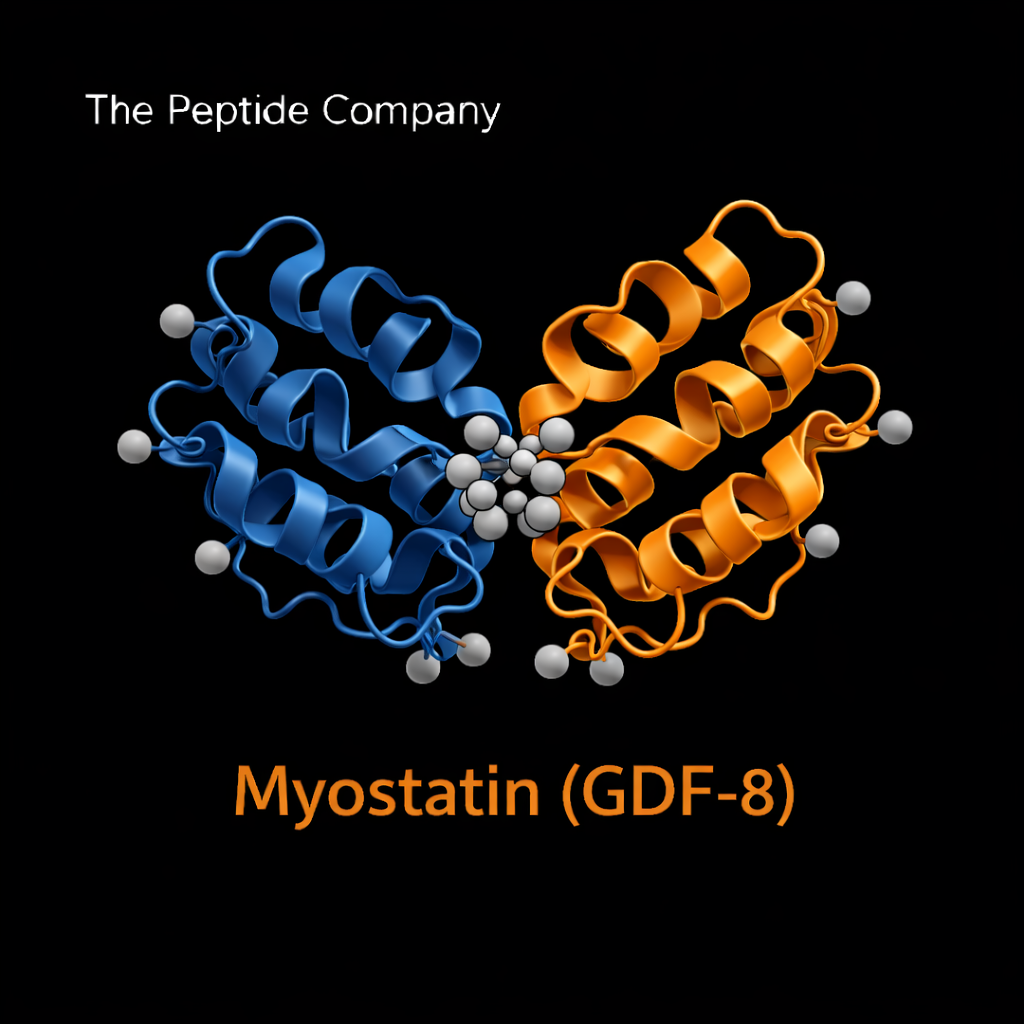
Molecular Structure and Processing
Myostatin is synthesized as a precursor protein consisting of a signal peptide, propeptide domain, and mature growth factor domain. Following proteolytic cleavage, the mature dimeric protein binds to activin type II receptors (ActRIIA and ActRIIB). Its activity is tightly regulated by binding proteins, including follistatin, decorin, and other extracellular modulators that influence bioavailability.
Receptor Binding and SMAD Signaling
Myostatin binds primarily to activin type II receptors, initiating recruitment of type I receptors and phosphorylation of SMAD2/3 transcription factors. These SMAD complexes translocate to the nucleus, where they regulate gene expression related to muscle protein synthesis and degradation. Through this pathway, myostatin suppresses myoblast proliferation and differentiation while modulating muscle fiber growth.
Role in Muscle Development and Homeostasis
During development, myostatin ensures controlled muscle formation by preventing excessive myogenesis. In adult tissue, it maintains muscle mass equilibrium by balancing anabolic and catabolic signaling. Experimental models lacking functional myostatin demonstrate increased muscle mass and fiber hypertrophy, highlighting its regulatory importance.
Interaction With Follistatin and Extracellular Regulators
Myostatin activity is modulated by extracellular binding proteins such as follistatin, which binds and neutralizes myostatin, reducing receptor activation. Decorin and other matrix-associated proteins also influence myostatin signaling by altering growth factor availability. These interactions create a regulatory network linking muscle biology with extracellular matrix signaling.
Myostatin and Fibrosis
Beyond skeletal muscle, myostatin has been implicated in fibrotic signaling pathways through its interaction with TGF-β family members. Elevated myostatin activity in certain experimental models correlates with increased extracellular matrix deposition and tissue remodeling. This connection places myostatin at the intersection of muscle regulation and fibrosis research.
Metabolic Cross-Talk
Myostatin signaling intersects with metabolic pathways, including insulin sensitivity and mitochondrial function. Studies suggest that alterations in myostatin expression influence systemic energy balance and adipose tissue dynamics. This cross-talk links muscle mass regulation with broader metabolic homeostasis.
Myostatin in Aging and Sarcopenia Research
Age-associated muscle decline (sarcopenia) has been linked to changes in myostatin signaling dynamics. Research examines whether modulation of myostatin activity may influence age-related muscle atrophy and functional capacity. These investigations contribute to broader understanding of anabolic resistance and muscle aging biology.
Comparison With IGF-1 and mTOR Signaling
Myostatin acts in functional opposition to anabolic pathways such as IGF-1 and mTOR. While IGF-1 promotes protein synthesis and muscle hypertrophy, myostatin restrains excessive growth, ensuring structural and metabolic balance. Understanding this regulatory tension provides insight into coordinated muscle adaptation.
Research Applications
Myostatin is studied extensively in models of muscle hypertrophy, muscle wasting, metabolic disorders, and fibrosis. Experimental approaches include genetic knockout systems, antibody-based inhibition, receptor modulation, and extracellular binding protein interactions. These models contribute to deeper understanding of muscle growth control mechanisms.
Limitations and Open Research Questions
Important questions remain regarding tissue-specific regulation, long-term signaling adaptations, and integration with endocrine networks. Further investigation is required to clarify how myostatin signaling coordinates with other TGF-β family members and systemic metabolic signals.
Summary
Myostatin (GDF-8) is a master regulator of skeletal muscle growth operating through TGF-β superfamily signaling and SMAD-mediated transcriptional control. By restraining excessive myogenesis and balancing anabolic pathways, it maintains structural and metabolic equilibrium within muscle tissue. Its study provides critical insight into muscle development, fibrosis signaling, and systemic metabolic integration.
Educational & Research Disclaimer
This document is provided for educational and scientific research purposes only. No medical, therapeutic, or usage claims are made. Myostatin and related signaling modulators are not approved for human use and are intended solely for controlled laboratory and academic investigation.
Myostatin (GDF-8) is a TGF-β superfamily protein that negatively regulates skeletal muscle growth. It controls myogenesis, muscle fiber size, and anabolic balance. Research models use myostatin to investigate muscle hypertrophy, sarcopenia, cachexia, fibrosis, and metabolic homeostasis.
Myostatin binds to activin type II receptors (primarily ActRIIB), triggering type I receptor recruitment, SMAD2/3 phosphorylation, and transcriptional regulation of genes governing muscle growth suppression. This pathway serves as a central node in TGF-β–related inhibitory signaling.
Myostatin coordinates muscle mass during embryonic development and maintains adult muscle equilibrium. Loss-of-function models show substantial increases in muscle fiber number and size, highlighting its essential role in growth limitation.
Elevated myostatin expression is associated with sarcopenia, cachexia, disuse atrophy, chronic illness, and aging. Research explores how modulating the myostatin pathway may influence muscle preservation in these contexts.
Beyond skeletal muscle, myostatin impacts systemic energy metabolism, adipose tissue dynamics, insulin sensitivity, and mitochondrial function. Studies suggest that suppressing myostatin signaling may shift metabolic balance toward greater energy expenditure and improved glucose handling.
Multiple investigational agents (ligand traps, antibodies, receptor blockers) have been evaluated, but here myostatin is presented strictly as a research-grade protein used for pathway studies. No therapeutic claims or usage implications apply.
Myostatin should be framed as a cytokine-like regulatory protein of the TGF-β superfamily, used experimentally to study inhibitory muscle signaling, hypertrophy control, SMAD pathways, and metabolic cross-talk. Content must avoid any implication of treatment or human administration.
PMID:
GHRP‑2 : Pituitary Axis Modulation, Ghrelin Receptor Activation, and Cellular Recovery Research
ACE‑031 : Myostatin Inhibition, Muscle Hypertrophy, and Regenerative Research