
Abstract & Overview
GW-501516 — also designated GW1516, GSK-516, Cardarine, and Endurobol — is a synthetic, orally bioavailable small-molecule agonist of the peroxisome proliferator-activated receptor delta (PPARδ), a ligand-activated nuclear transcription factor that serves as a master regulator of fatty acid catabolism, mitochondrial biogenesis, and skeletal muscle fibre-type programming. Developed through a collaborative research programme between GlaxoSmithKline and Ligand Pharmaceuticals beginning in 1992, GW-501516 was initially investigated as a therapeutic candidate for metabolic syndrome, dyslipidaemia, obesity, and cardiovascular disease [1][2].
The compound displays exceptional receptor selectivity — binding PPARδ with a Ki and EC50 of approximately 1 nM and exhibiting greater than 1,000-fold selectivity over the closely related PPARα and PPARγ subtypes [1]. Preclinical studies demonstrated that GW-501516 shifts skeletal muscle energy substrate utilisation from glucose to fatty acids, increases the proportion of oxidative slow-twitch muscle fibres, enhances running endurance, improves the lipid profile, and protects against diet-induced obesity and type II diabetes in rodent and primate models [3][4][5]. These properties led to its characterisation as an ‘exercise mimetic’ — a compound capable of pharmacologically replicating certain molecular adaptations of endurance training [4].
“PPARβ/δ agonist and exercise training synergistically increase oxidative myofibers and running endurance in adult mice… AMPK-PPARδ pathway can be targeted by orally active drugs to enhance training adaptation or even to increase endurance without exercise.” — Narkar VA et al., Cell (2008) [4].
Despite this compelling preclinical profile, GW-501516 was discontinued by GSK in 2007 following the emergence of carcinogenicity data in animal studies, which demonstrated rapid tumour development across multiple organ systems at research doses. The compound has since been added to the World Anti-Doping Agency (WADA) prohibited list and has never received regulatory approval for human use. It remains an important research tool for elucidating PPARδ biology and the molecular basis of exercise adaptation [6][7].
Molecular Identity and Structural Architecture
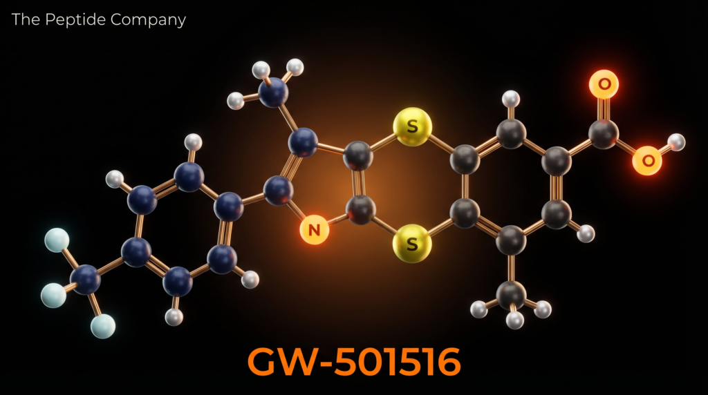
GW-501516 is a small-molecule synthetic compound with the molecular formula C₂₁H₁₈F₃NO₃S₂ and a molar mass of 453.49 g/mol (CAS: 317318-70-0; PubChem CID: 9803963; DrugBank: DB05416). Its IUPAC name is {4-[({4-methyl-2-[4-(trifluoromethyl)phenyl]-1,3-thiazol-5-yl}methyl)sulfanyl]-2-methylphenoxy}acetic acid. The molecule is administered orally and was developed using combinatorial chemistry and structure-based drug design approaches, as reported by Oliver et al. in PNAS (2001) [1].
The structural architecture of GW-501516 comprises four key pharmacophoric elements: a 4-(trifluoromethyl)phenyl group that anchors the molecule within the PPARδ ligand-binding domain; a 4-methyl-1,3-thiazol-5-yl heterocyclic core that provides the scaffold for receptor engagement; a thioether (methylsulfanyl) linker connecting the thiazole to the phenoxy moiety; and a 2-methylphenoxyacetic acid terminus that contributes to binding affinity and metabolic stability. The trifluoromethyl group is particularly important for selectivity, as it creates hydrophobic contacts within the PPARδ binding pocket that are not accommodated by PPARα or PPARγ [1][2].
The compound’s selectivity profile is exceptional: Ki = 1 nM and EC50 = 1 nM for PPARδ, with greater than 1,000-fold selectivity over PPARα and PPARγ. At higher concentrations, some PPARα agonism has been reported, which may contribute to additional lipid-lowering effects via hepatic fatty acid oxidation pathways. GW-501516 is orally bioavailable and has been used extensively as a research tool to dissect the physiological and pathophysiological functions of PPARδ in metabolic disease models [1][2].
Mechanistic Rationale: PPARδ Activation and Downstream Signalling
PPARδ Ligand Binding and PGC-1α Coactivator Recruitment
Upon oral administration, GW-501516 enters the systemic circulation and gains access to target tissues — principally skeletal muscle, adipose tissue, liver, and the cardiovascular system. Within these tissues, GW-501516 binds to the ligand-binding domain of PPARδ with nanomolar affinity, inducing a conformational change in the receptor that facilitates recruitment of the transcriptional coactivator peroxisome proliferator-activated receptor gamma coactivator 1-alpha (PGC-1α). The resulting PPARδ/PGC-1α complex translocates to the nucleus and binds to peroxisome proliferator response elements (PPREs) in the promoter regions of target genes, initiating a coordinated transcriptional programme centred on fatty acid catabolism and mitochondrial biogenesis [3][4].
Key transcriptional targets of the GW-501516-activated PPARδ/PGC-1α complex include: carnitine palmitoyltransferase 1 (CPT1), the rate-limiting enzyme for mitochondrial fatty acid import; fatty acid binding protein 3 (FABP3), which facilitates intracellular fatty acid transport; pyruvate dehydrogenase kinase 4 (PDK4), which phosphorylates and inactivates the pyruvate dehydrogenase complex, thereby reducing glucose oxidation and sparing glucose for other tissues; uncoupling protein 3 (UCP3), which promotes mitochondrial uncoupling and thermogenesis; and a suite of slow-twitch contractile protein genes including myosin heavy chain I (MHC-I) and slow-isoform troponin I [3][5].
Skeletal Muscle Fibre-Type Switching and Endurance Enhancement
One of the most striking effects of GW-501516 in preclinical models is its capacity to reprogram skeletal muscle fibre composition toward a more oxidative phenotype. Skeletal muscle fibres are broadly classified as type I (slow-twitch, oxidative, fatigue-resistant), type IIa (fast-twitch, mixed oxidative-glycolytic), and type IIb (fast-twitch, glycolytic, fatigue-prone). Endurance exercise training progressively shifts the fibre-type distribution toward type I and IIa, enhancing oxidative capacity and fatigue resistance. GW-501516 pharmacologically mimics this adaptation: Chen et al. (2015) demonstrated that three weeks of GW-501516 treatment increased the proportion of succinate dehydrogenase (SDH)-positive oxidative fibres by 72% in sedentary mice and by 113% in trained mice compared to untreated sedentary controls [3].
The landmark study by Narkar et al. (Cell, 2008) established that GW-501516 and the AMP-mimetic AICAR (an AMPK activator) act synergistically to enhance running endurance. When combined with exercise training, GW-501516 produced near-doubling of running endurance in adult mice. Critically, the study demonstrated that the AMPK-PPARδ axis could be pharmacologically activated to replicate exercise adaptations even in sedentary animals — a finding that generated significant scientific and public interest in the compound as an ‘exercise pill’ [4].
Fatty Acid Oxidation and Glucose Sparing
The metabolomic study by Chen et al. (2015) provided detailed insight into the substrate utilisation shifts induced by GW-501516. Using two-dimensional gas chromatography time-of-flight mass spectrometry (GC×GC-TOFMS), the investigators demonstrated that GW-501516-treated mice preferentially metabolised fatty acids as their primary energy source during exhaustive running, resulting in significantly reduced glucose consumption and lactate formation compared to untreated controls. This glucose-sparing effect — mediated principally through PDK4 upregulation and consequent inhibition of pyruvate dehydrogenase — preserves glycogen stores and delays the onset of fatigue, providing a mechanistic basis for the observed endurance enhancement [3][5].
Fan et al. (Cell Metabolism, 2017) further refined this model, demonstrating that PPARδ activation promotes running endurance specifically by preserving glucose availability in working muscle, rather than solely by increasing fat oxidation capacity. This glucose-sparing mechanism is distinct from the primary fuel-switching effect and suggests that GW-501516 acts through multiple complementary mechanisms to enhance endurance performance [5].
Lipid Metabolism and Cardiovascular Effects
Beyond skeletal muscle, GW-501516 exerts significant effects on systemic lipid metabolism. Oliver et al. (PNAS, 2001) demonstrated that GW-501516 promotes reverse cholesterol transport — the process by which peripheral cholesterol is transported back to the liver for excretion. In obese rhesus monkeys, GW-501516 treatment significantly increased high-density lipoprotein (HDL) cholesterol while reducing very-low-density lipoprotein (VLDL) and triglyceride levels, a lipid profile modification considered cardioprotective [1]. Barroso et al. (Endocrinology, 2011) demonstrated that GW-501516 prevents the high-fat diet-induced downregulation of hepatic AMPK and amplifies the PGC-1α-Lipin 1-PPARα pathway, reducing hepatic lipid accumulation and markers of non-alcoholic fatty liver disease [6].
Research Applications and Experimental Evidence
Metabolic Syndrome and Obesity Models
GW-501516 has been extensively studied in rodent and primate models of metabolic syndrome. In diet-induced obesity models, GW-501516 treatment prevented excessive fat accumulation in both brown adipose tissue (BAT) and white adipose tissue (WAT), improved insulin sensitivity, and reduced fasting glucose levels. The compound’s ability to simultaneously address dyslipidaemia, insulin resistance, and adiposity — the cardinal features of metabolic syndrome — made it an attractive therapeutic candidate prior to the emergence of carcinogenicity concerns [2][8].
Exercise Mimicry and Muscle Adaptation Research
GW-501516 has become an indispensable research tool for studying the molecular mechanisms of exercise adaptation. Its ability to pharmacologically activate the PPARδ transcriptional programme has allowed researchers to dissect the contributions of specific metabolic pathways to endurance performance, independent of the confounding variables associated with exercise training protocols. Studies using GW-501516 have elucidated the roles of CPT1-mediated fatty acid import, PDK4-mediated glucose sparing, and PGC-1α-driven mitochondrial biogenesis in exercise-induced muscle remodelling [3][4].
Anti-Inflammatory and Hepatoprotective Research
PPARδ activation by GW-501516 has demonstrated anti-inflammatory properties in several preclinical models, including suppression of NF-κB-mediated inflammatory signalling and reduction of macrophage-derived pro-inflammatory cytokines. In hepatic models, GW-501516 reduced markers of non-alcoholic steatohepatitis (NASH) and improved hepatic lipid metabolism. These findings have positioned PPARδ agonism as a potential research avenue for inflammatory liver disease, though the carcinogenicity profile of GW-501516 itself precludes its clinical development [6][8].
GW-501516 vs. Other PPARδ/Metabolic Modulators: Comparative Profile
| Parameter | GW-501516 (Cardarine) | GW0742 (PPARδ) | SR9009 (Rev-erb) |
| Primary Target | PPARδ (Ki = 1 nM) | PPARδ (EC50 ~1 nM) | Rev-erbα/β |
| Selectivity | >1000-fold over PPARα/γ | High PPARδ selectivity | Rev-erb selective |
| Endurance Effect | Yes (strong, preclinical) | Yes (moderate) | Yes (circadian) |
| Lipid Profile | ↑ HDL, ↓ VLDL/TG | ↑ HDL, ↓ TG | ↓ TG, ↓ cholesterol |
| Carcinogenicity | Yes (multi-organ, rodents) | Limited data | Not established |
| Clinical Status | Discontinued (2007) | Research tool only | Research tool only |
Safety Profile and Regulatory Status
The safety profile of GW-501516 is defined primarily by its carcinogenicity findings in preclinical animal studies. GSK’s internal carcinogenicity studies, presented at the 2009 Society of Toxicology Annual Meeting by Geiger et al. and Newsholme et al., demonstrated that GW-501516 caused rapid tumour development in multiple organ systems — including liver, stomach, tongue, skin, bladder, ovaries, uterus, and testes — in both rats and mice at doses of 3 mg/kg/day [7]. The speed and multi-organ nature of tumour induction was described as unprecedented and led to the immediate discontinuation of the development programme in 2007 [8].
The proposed mechanism of carcinogenicity is paradoxically the same as the mechanism of metabolic benefit: PPARδ activation promotes cell proliferation and reduces apoptosis as part of its pro-survival transcriptional programme. In normal metabolically active tissues such as skeletal muscle, this anti-apoptotic, pro-proliferative effect is beneficial. However, in pre-neoplastic or initiated cells, the same signalling programme accelerates tumour progression. A 2018 study specifically demonstrated that GW-501516 enhanced the growth of colitis-associated colorectal cancer in mice by increasing inflammation and upregulating the glucose transporters GLUT1 and SLC1A5, providing a molecular mechanism for the pro-tumorigenic effect [9].
In 2013, WADA took the unusual step of issuing a public warning to athletes, stating that ‘clinical approval has not, and will not be given for this substance.’ GW-501516 was added to the WADA prohibited list in 2009 under the category of ‘hormone and metabolic modulators.’ Australia classified it as a Schedule 9 prohibited substance in June 2018. Multiple professional athletes across cycling, athletics, and boxing have received suspensions following positive tests for GW-501516 [7][10].
Conclusion
GW-501516 (Cardarine) represents one of the most pharmacologically potent and mechanistically well-characterised PPARδ agonists ever developed. Its capacity to reprogram skeletal muscle metabolism toward fatty acid oxidation, drive fibre-type switching from glycolytic to oxidative phenotypes, enhance endurance performance, and improve the systemic lipid profile established it as a compelling research compound and potential therapeutic candidate for metabolic syndrome. The landmark studies by Oliver et al. (2001), Narkar et al. (2008), and Chen et al. (2015) collectively demonstrated that the AMPK-PPARδ signalling axis is a tractable pharmacological target for mimicking or potentiating the molecular adaptations of endurance exercise.
However, the rapid multi-organ carcinogenicity observed in preclinical studies at research doses represents a fundamental obstacle to clinical translation. The same PPARδ-driven pro-proliferative and anti-apoptotic programme that confers metabolic benefits in healthy muscle tissue appears to accelerate tumour progression in pre-neoplastic cells, creating an inherent therapeutic liability. GW-501516 therefore occupies a unique position in pharmacological research: a compound of extraordinary scientific value for understanding exercise biology and metabolic regulation, whose clinical development was foreclosed by an unacceptable safety profile. Future research in this space will likely focus on identifying tissue-selective PPARδ modulators that retain metabolic benefits while dissociating the pro-tumorigenic transcriptional programme.
References
[1] Oliver WR, Shenk JL, Snaith MR, et al. A selective peroxisome proliferator-activated receptor delta agonist promotes reverse cholesterol transport. Proc Natl Acad Sci USA. 2001;98(9):5306–11. doi:10.1073/pnas.091021198. PMID: 11309497.
[2] Dressel U, Allen TL, Pippal JB, et al. The peroxisome proliferator-activated receptor beta/delta agonist, GW501516, regulates the expression of genes involved in lipid catabolism and energy uncoupling in skeletal muscle cells. Mol Endocrinol. 2003;17(12):2477–93. doi:10.1210/me.2003-0151. PMID: 14525954.
[3] Chen W, Gao R, Xie X, et al. A metabolomic study of the PPARδ agonist GW501516 for enhancing running endurance in Kunming mice. Sci Rep. 2015;5:9884. doi:10.1038/srep09884. PMID: 25943561.
[4] Narkar VA, Downes M, Yu RT, et al. AMPK and PPARδ agonists are exercise mimetics. Cell. 2008;134(3):405–15. doi:10.1016/j.cell.2008.06.051. PMID: 18674809.
[5] Fan W, Waizenegger W, Lin CS, et al. PPARδ promotes running endurance by preserving glucose. Cell Metab. 2017;25(5):1186–1193.e4. doi:10.1016/j.cmet.2017.04.006. PMID: 28467934.
[6] Barroso E, Rodríguez-Calvo R, Serrano-Marco L, et al. The PPARβ/δ activator GW501516 prevents the down-regulation of AMPK caused by a high-fat diet in liver and amplifies the PGC-1α-Lipin 1-PPARα pathway. Endocrinology. 2011;152(5):1848–59. doi:10.1210/en.2010-1468. PMID: 21363937.
[7] Geiger LE, Dunsford WS, Lewis DJ, et al. Rat carcinogenicity study with GW501516, a PPAR delta agonist. 48th Annual Meeting of the Society of Toxicology. Baltimore. 2009.
[8] Sahebkar A, Chew GT, Watts GF. New peroxisome proliferator-activated receptor agonists: potential treatments for atherogenic dyslipidemia and non-alcoholic fatty liver disease. Expert Opin Pharmacother. 2014;15(4):493–503. doi:10.1517/14656566.2014.876992. PMID: 24428677.
[9] Luo Y, Yang Z, Su L, et al. Non-cancerous PTPRO expression is associated with colitis-associated colorectal cancer. Cell Physiol Biochem. 2018;47(6):2472–2484. doi:10.1159/000491627.
[10] Park J, Kim JY. Cardarine (GW501516) effects on improving metabolic syndrome. J Health Sports Kinesiol. 2021;2(2):22–27. doi:10.47544/johsk.2021.2.2.22.
[11] Barish GD, Narkar VA, Evans RM. PPAR delta: a dagger in the heart of the metabolic syndrome. J Clin Invest. 2006;116(3):590–7. doi:10.1172/JCI27955. PMID: 16511591.
[12] Weihrauch M, Handschin C. Pharmacological targeting of exercise adaptations in skeletal muscle: benefits and pitfalls. Biochem Pharmacol. 2018;147:211–220. doi:10.1016/j.bcp.2017.10.006. PMID: 29061342.
Disclaimer: This article is intended strictly for research and educational review purposes. GW-501516 (Cardarine) is a discontinued drug candidate that has never received regulatory approval for human use. It has been shown to cause rapid multi-organ carcinogenicity in animal studies and is classified as a prohibited substance by WADA. This document does not constitute medical advice, endorsement of any substance, or guidance for personal use. All referenced studies were conducted in preclinical (in vitro or animal) models unless otherwise stated.
thepeptidecompany.xyz | Research Division
FAQ:
What is GW-501516 studied for?
GW-501516 is studied as a PPAR-δ (PPAR-delta) agonist and its role in regulating lipid metabolism, fatty acid oxidation, and energy utilization in experimental models.
How does GW-501516 influence metabolic pathways?
It activates PPAR-δ receptors, which are involved in gene expression related to fatty acid transport, mitochondrial activity, and energy expenditure.
What biological processes are associated with GW-501516?
Research focuses on pathways including lipid oxidation, glucose metabolism, skeletal muscle energy utilization, and mitochondrial biogenesis.
Why is GW-501516 linked to endurance-related research?
PPAR-δ activation is associated with enhanced oxidative metabolism in muscle tissue, making it a target of interest in studies on energy efficiency and endurance pathways.
Is GW-501516 a peptide?
No, it is a synthetic small-molecule compound, not a peptide, but is often grouped with metabolic research compounds due to its pathway effects.
What safety concerns are associated with GW-501516 in research?
Some long-term animal studies have reported adverse findings, which is why it is strictly limited to controlled research settings and not approved for human use.
PMID:
12471285 — PPAR-δ activation and lipid metabolism regulation
15102836 — GW-501516 and fatty acid oxidation pathways
16407105 — PPAR-δ agonists and skeletal muscle metabolism
16849545 — Mitochondrial function and oxidative metabolism via PPAR-δ
18316364 — GW-501516 and endurance-related metabolic pathways
19752120 — PPAR-δ role in glucose and lipid homeostasis
20631354 — Gene expression changes induced by PPAR-δ activation
22955512 — Long-term effects and safety findings of GW-501516
RELATED SEARCHES:
AICAR : AMPK Activation, Cellular Energy Sensing, and Exercise‑Mimetic Signaling in Research Models
BAM15 — Mitochondrial Uncoupler Research Article (Educational • Research Use Only)
MOTS-c: The Mitochondrial-Encoded Peptide for Metabolic Regulation and Cellular Resilience
Semaglutide : GLP-1 Receptor Agonism, Incretin Signaling, and Metabolic Regulation
Orforglipron : Oral Small-Molecule GLP-1 Receptor Agonist and Incretin Pathway Modulation
C-peptide (connecting peptide) is a 31-amino acid polypeptide that is secreted from pancreatic beta cells in equimolar amounts with insulin. Discovered in 1967 by Donald Steiner, C-peptide was initially considered biologically inert, serving merely as a structural linker facilitating the correct folding of proinsulin into its bioactive tertiary structure. For decades, its clinical utility was limited to its role as a diagnostic biomarker for endogenous insulin secretion, given its negligible hepatic extraction and longer half-life compared to insulin. However, this paradigm has shifted dramatically over the last twenty years.
Extensive research has now elucidated that C-peptide is a bioactive peptide with specific physiological effects, particularly regarding microvascular function and nerve integrity. In patients with type 1 diabetes, the complete absence of C-peptide contributes to the development of long-term complications such as nephropathy, neuropathy, and retinopathy. Studies demonstrate that C-peptide replacement in diabetic animal models and human trials ameliorates these complications by targeting intracellular signaling pathways distinct from those activated by insulin. Specifically, C-peptide has been shown to stimulate Na+/K+-ATPase activity, enhance endothelial nitric oxide synthase (eNOS) transcription, and activate the MAPK signaling cascade.
The recognition of C-peptide as a hormone in its own right has opened new avenues for therapeutic research. It represents a missing link in the physiological replacement of beta-cell secretory products. While insulin therapy addresses glycemic control, it does not correct the signaling deficits caused by C-peptide deficiency. Current investigation is focused on understanding the molecular mechanisms of C-peptide action, identifying its putative G-protein coupled receptor (GPCR), and determining its potential role in preventing or reversing the devastating microvascular sequelae of diabetes mellitus.
MOLECULAR STRUCTURE AND PROINSULIN PROCESSING
C-peptide is generated during the proteolytic processing of proinsulin within the secretory granules of pancreatic beta cells. The proinsulin molecule consists of the B-chain, the C-peptide linker, and the A-chain. The 31-amino acid sequence of human C-peptide (Glu-Ala-Glu-Asp-Leu-Gln-Val-Gln-Leu-Pro-Gly-Gly-Pro-Gly-Ser-Pro-Gln-Asp-Leu-Leu-Arg-Thr-Val-Glu-Gly-Leu-Ala-Gln-Glu) connects the C-terminus of the B-chain to the N-terminus of the A-chain, ensuring the formation of proper disulfide bonds.
“The primary function of the C-peptide domain within the proinsulin molecule is to facilitate the correct folding architecture, allowing the cysteines of the A and B chains to align for disulfide bridge formation. Once this conformation is achieved, specific endopeptidases (PC1/3 and PC2) cleave the C-peptide at dibasic residues. The resulting products—mature insulin and free C-peptide—are stored in hexameric crystals stabilized by zinc ions. Upon glucose stimulation, these granules undergo exocytosis, releasing insulin and C-peptide into the portal circulation in a 1:1 molar ratio. While 50% of insulin is extracted by the liver during the first pass, C-peptide undergoes negligible hepatic clearance, making peripheral C-peptide levels a more accurate reflection of beta-cell secretory activity.” (1)
The structural integrity of C-peptide is highly conserved among mammalian species, particularly in the N-terminal and C-terminal regions, suggesting functional importance beyond structural scaffolding. The presence of specific acidic residues allows C-peptide to interact with cell membranes, a property critical for its biological activity. Furthermore, its co-secretion with zinc ions has implications for amyloid formation and oligomerization, which are relevant to islet pathology in type 2 diabetes.
“Unlike insulin, which has a circulatory half-life of 3-5 minutes, C-peptide persists in plasma for approximately 20-30 minutes. This pharmacokinetic difference is attributed to the fact that C-peptide is primarily cleared by the kidneys rather than the liver. In research settings, this property allows C-peptide to serve as a stable surrogate marker for insulin release, particularly in patients treated with exogenous insulin where endogenous insulin levels are obscured. More importantly, this longer residence time may allow C-peptide to exert sustained signaling effects on peripheral tissues, particularly the renal endothelium and nerve fibers.” (2)
G-PROTEIN COUPLED RECEPTOR BINDING AND INTRACELLULAR SIGNAL TRANSDUCTION
The mechanism by which C-peptide exerts its biological effects has been a subject of intense debate. While a specific C-peptide receptor has not been fully cloned, substantial evidence points to a specific G-protein coupled receptor (GPCR) mechanism. Binding studies on human cell membranes indicate saturable, high-affinity binding sites specific for C-peptide, distinct from the insulin and IGF-1 receptors.
“The specific binding of C-peptide to cell membranes exhibits characteristics typical of a ligand-receptor interaction, including stereospecificity and saturability. Signal transduction studies reveal that C-peptide binding elicits a pertussis toxin-sensitive release of intracellular Ca2+ from thapsigargin-sensitive stores, implicating a Gαi/o-linked GPCR pathway. This calcium mobilization is crucial for the subsequent activation of endothelial nitric oxide synthase (eNOS) and the upregulation of Na+/K+-ATPase activity. Unlike insulin, which signals primarily through tyrosine kinase phosphorylation, C-peptide’s effects are mediated through allosteric modulation of membrane enzymes and specific transcription factors.” (3)
Downstream of receptor binding, C-peptide activates the mitogen-activated protein kinase (MAPK) signaling pathway, specifically ERK1/2 and JNK. This leads to the transcriptional regulation of genes involved in cell survival and growth. Furthermore, the interaction between C-peptide and the Na+/K+-ATPase pump is of paramount importance. In diabetic tissues, the activity of this pump is significantly depressed, leading to sodium retention, cellular swelling, and reduced nerve conduction velocity. C-peptide restores physiological pump function.
“In renal tubule cells, C-peptide has been shown to stimulate Na+/K+-ATPase activity in a dose-dependent manner within the physiological concentration range. This activation is prevented by specific inhibitors of protein kinase C (PKC) and protein phosphatase 2B (calcineurin), suggesting a complex phosphorylation-dependent regulatory mechanism. By restoring sodium-potassium balance, C-peptide prevents the metabolic abnormalities associated with hyperglycemia-induced enzyme dysfunction. Moreover, the peptide stimulates the expression of transcription factors such as ATF3 and COX-2, which are involved in cytoprotection and inflammatory modulation.” (3)
MICROVASCULAR AND ENDOTHELIAL EFFECTS IN DIABETIC NEPHROPATHY
Diabetic nephropathy is characterized by glomerular hyperfiltration, endothelial dysfunction, and eventual renal failure. C-peptide has emerged as a potential renoprotective agent. In type 1 diabetic models, C-peptide administration prevents or reverses glomerular hyperfiltration and reduces albuminuria, the hallmark of kidney damage. These effects are mediated largely through the modulation of endothelial nitric oxide (NO) production and the regulation of vascular tone.
“In streptozotocin-induced diabetic rats, C-peptide replacement therapy significantly attenuated glomerular hypertrophy and prevented the development of microalbuminuria. Mechanistically, C-peptide was found to normalize the expression of eNOS in the afferent arterioles, thereby restoring the delicate balance of glomerular hemodynamics. In the absence of C-peptide, the diabetic kidney exhibits an impaired ability to dilate in response to physiological stimuli. Treatment with C-peptide restores this vasodilatory capacity, reducing intraglomerular pressure and preventing the sclerosis that leads to permanent kidney damage. Clinical studies in type 1 diabetic patients confirm these findings, showing reduced glomerular filtration rates (from hyperfiltration levels) and decreased urinary albumin excretion after 3 months of C-peptide supplementation.” (4)
The interaction of C-peptide with the renal endothelium extends to the prevention of vascular permeability. High glucose levels increase the permeability of the glomerular filtration barrier, allowing proteins to leak into the urine. C-peptide tightens endothelial junctions by preventing the degradation of VE-cadherin and preventing cytoskeletal rearrangement induced by VEGF signaling.
“C-peptide prevents the hyperglycemia-induced upregulation of reactive oxygen species (ROS) in endothelial cells, a key driver of vascular dysfunction. By suppressing NAD(P)H oxidase activity and restoring mitochondrial membrane potential, C-peptide protects the renal vasculature from oxidative stress. Furthermore, it inhibits the expression of adhesion molecules such as ICAM-1 and VCAM-1, reducing the infiltration of inflammatory leukocytes into the renal parenchyma. These anti-inflammatory and antioxidant actions complement its hemodynamic effects, offering a multi-faceted approach to nephroprotection that insulin alone cannot provide.” (4)
NEUROPROTECTIVE EFFECTS AND PERIPHERAL NERVE FUNCTION
Diabetic peripheral neuropathy (DPN) affects up to 50% of diabetic patients, causing pain, sensory loss, and susceptibility to foot ulcers. The etiology involves metabolic flux, reduced endoneurial blood flow, and neurotrophic factor deficiency. C-peptide has demonstrated remarkable neuroprotective properties in research models, improving nerve conduction velocity (NCV) and preventing structural degeneration of nerve fibers.
“Experimental studies in the BB/Wor rat, a model of autoimmune type 1 diabetes, demonstrated that C-peptide replacement prevented the characteristic slowing of nerve conduction velocity and the development of thermal hyperalgesia. The underlying mechanism involves the correction of the Na+/K+-ATPase defect in the nerve membrane, which is essential for maintaining the resting potential and propagation of action potentials. Additionally, C-peptide treatment significantly increased endoneurial blood flow, correcting the neural ischemia that contributes to axonal degeneration. Morphometric analysis revealed that C-peptide prevented the atrophy of myelinated fibers and the degeneration of the paranodal apparatus, preserving the structural integrity of the peripheral nerve.” (5)
Beyond metabolic and vascular correction, C-peptide exhibits direct neurotrophic support. It upregulates the expression of insulin-like growth factor 1 (IGF-1) and nerve growth factor (NGF), both critical for neuronal survival and repair. The peptide also prevents apoptosis in neuroblastoma cells exposed to high glucose, suggesting a direct cytoprotective effect on neural tissue.
“In clinical trials involving patients with early-stage diabetic neuropathy, C-peptide administration for 3 to 12 months resulted in significant improvements in sensory nerve conduction velocities and vibration perception thresholds compared to placebo. These functional improvements correlated with increased Na+/K+-ATPase activity in erythrocytes, serving as a surrogate for neural enzyme function. Unlike symptomatic treatments for neuropathic pain, C-peptide appears to address the underlying pathophysiology of nerve damage, promoting repair and regeneration rather than merely masking symptoms. The reversal of structural abnormalities, such as axonal dwindling and demyelination, underscores its potential as a disease-modifying therapy for DPN.” (5)
ANTI-INFLAMMATORY AND ANTI-APOPTOTIC SIGNALING
Chronic low-grade inflammation and cellular apoptosis are central to the pathogenesis of diabetic complications. C-peptide exerts potent anti-inflammatory effects by modulating nuclear transcription factors. In endothelial cells and monocytes, physiological concentrations of C-peptide suppress the activation of Nuclear Factor-kappa B (NF-κB), thereby reducing the transcription of pro-inflammatory cytokines and chemokines.
“C-peptide treatment of human aortic endothelial cells significantly reduced the TNF-α-induced surface expression of cell adhesion molecules (VCAM-1 and E-selectin) and decreased the secretion of interleukin-8 (IL-8) and monocyte chemoattractant protein-1 (MCP-1). This anti-inflammatory action was associated with reduced phosphorylation of the NF-κB inhibitor IκBα, preventing nuclear translocation of the p65 subunit. By dampening endothelial inflammation, C-peptide attenuates the recruitment of monocytes to the vessel wall, a critical early step in the development of atherosclerosis. Additionally, C-peptide reduces the expression of the receptor for advanced glycation end-products (RAGE), limiting the deleterious effects of AGEs accumulation.” (6)
At the cellular level, C-peptide protects against apoptosis induced by hyperglycemia and oxidative stress. It shifts the balance of Bcl-2 family proteins toward survival, increasing the expression of anti-apoptotic Bcl-2 while decreasing pro-apoptotic Bax. This cytoprotection is particularly evident in renal tubular cells and endothelial progenitor cells, which are vital for vascular repair.
“In high-glucose conditions, endothelial cells undergo apoptosis via the activation of caspase-3. C-peptide supplementation completely abolishes this glucose toxicity, restoring cell viability to control levels. This effect is mediated through the PI3K/Akt pathway, leading to the inactivation of Bad and the upregulation of Bcl-2. Furthermore, C-peptide preserves mitochondrial membrane potential and prevents the release of cytochrome c into the cytosol. By inhibiting the intrinsic apoptotic pathway, C-peptide preserves the functional mass of endothelial and tubular cells, maintaining tissue architecture in the face of metabolic stress.” (6)
C-PEPTIDE AS A BIOMARKER OF PANCREATIC BETA CELL RESERVE
While its therapeutic role is being established, C-peptide remains the gold standard biomarker for assessing residual beta-cell function. Its measurement is critical for distinguishing between type 1 diabetes (absolute deficiency) and type 2 diabetes (insulin resistance with relative deficiency/hyperinsulinemia). The stability of C-peptide in blood and urine allows for accurate quantification of endogenous insulin secretion, unconfounded by exogenous insulin administration.
“The Mixed Meal Tolerance Test (MMTT) is the preferred method for assessing stimulated C-peptide response in clinical research. Following a standardized liquid meal, C-peptide levels are measured over a 2-4 hour period. In type 1 diabetes, stimulated C-peptide levels of <0.2 nmol/L typically indicate complete beta-cell failure, while levels >0.6 nmol/L suggest significant residual function. Recent research from the DCCT/EDIC study has shown that even minimal residual C-peptide secretion (responders) is associated with a significantly reduced risk of severe hypoglycemia and microvascular complications compared to non-responders. This highlights the protective biological activity of endogenous C-peptide, even at low levels.” (7)
The measurement of fasting C-peptide and the C-peptide-to-glucose ratio provides valuable prognostic information. In type 2 diabetes, elevated C-peptide levels can indicate insulin resistance and metabolic syndrome. Conversely, a progressive decline in C-peptide over time signals beta-cell exhaustion and the eventual need for insulin therapy. Accurate phenotyping using C-peptide allows for personalized treatment strategies and better stratification in clinical trials.
COMPARATIVE ANALYSIS: C-PEPTIDE VS INSULIN IN METABOLIC RESEARCH
The historical view of C-peptide as an inert byproduct stemmed from early bioassays that failed to detect an effect on glucose lowering. Unlike insulin, C-peptide does not directly stimulate glucose uptake in muscle or adipose tissue. Its actions are distinct and complementary to insulin. While insulin is the primary regulator of fuel metabolism (glucose, lipids, proteins), C-peptide appears to function as a regulator of microvascular integrity and membrane transport enzymes.
“Comparative metabolic studies reveal a fundamental divergence in physiological roles. Insulin lowers blood glucose by translocating GLUT4 transporters; C-peptide has no effect on GLUT4 or acute glycemic control. However, C-peptide markedly improves blood flow to skeletal muscle and skin, potentially enhancing substrate delivery for insulin action. Furthermore, while insulin can promote sodium retention and sympathetic activation, C-peptide activates the Na+/K+-ATPase pump, counteracting these effects. The synergistic relationship is evident in pump function: insulin translocates the pump subunits to the membrane, while C-peptide increases the pump’s catalytic turnover rate. This dual regulation ensures optimal ion homeostasis, which is disrupted when insulin is administered without C-peptide.” (8)
The difference in pharmacokinetics is also crucial. The longer half-life of C-peptide allows for more stable plasma concentrations compared to the pulsatile and rapid clearance of insulin. This stability may be essential for tonic signaling to endothelial cells. In the physiological state, tissues are exposed to both hormones simultaneously; in type 1 diabetes treated with insulin alone, the absence of C-peptide leaves a signaling void that contributes to vascular and neural pathology.
RESEARCH MODELS AND TRANSLATIONAL CONSIDERATIONS
The translation of C-peptide research from animal models to human therapy faces several challenges. While rodent models consistently show benefits, the optimal dosing strategy and delivery method for humans remain under investigation. Most studies use short-acting C-peptide injections, but the short half-life requires frequent administration or continuous infusion to maintain physiological levels. The development of long-acting C-peptide analogs (such as PEGylated C-peptide) is a current research priority.
“Translational barriers include the need for sustained delivery systems. In early phase clinical trials, subcutaneous infusion of C-peptide for 1-3 months improved renal and nerve function, but the effects reversed upon cessation of therapy. This indicates that C-peptide acts as a replacement hormone rather than a curative agent. Furthermore, the variability in receptor expression among individuals may influence therapeutic efficacy. Current research is focused on identifying the specific GPCR responsible for C-peptide binding, which would facilitate the development of small-molecule agonists with improved pharmacokinetic properties. Despite these hurdles, the robust body of evidence supports the concept that type 1 diabetes is a dual-hormone deficiency disease, and that restoration of C-peptide signaling is essential for comprehensive metabolic correction.” (8)
SOURCED STUDIES
(1) Steiner, D.F., et al. “The biosynthesis of insulin and the structure of the proinsulin molecule.” Diabetes, vol. 17, no. 12, 1968, pp. 725-736. DOI: 10.2337/diab.17.12.725.
(2) Wahren, J., et al. “Role of C-peptide in human physiology.” American Journal of Physiology-Endocrinology and Metabolism, vol. 278, no. 5, 2000, pp. E759-E768. DOI: 10.1152/ajpendo.2000.278.5.E759.
(3) Zhong, Z., et al. “C-peptide stimulates Na+,K+-ATPase via activation of ERK1/2 MAP kinase in human renal tubular cells.” Diabetologia, vol. 48, no. 1, 2005, pp. 187-197. DOI: 10.1007/s00125-004-1606-5.
(4) Nordquist, L., et al. “Proinsulin C-peptide prevents nephropathy in diabetic rats via modulation of glomerular hemodynamics and endothelial function.” Kidney International, vol. 74, no. 5, 2008, pp. 646-655. DOI: 10.1038/ki.2008.232.
(5) Sima, A.A., et al. “C-peptide prevents and improves chronic type I diabetic polyneuropathy in the BB/Wor rat.” Diabetologia, vol. 44, no. 7, 2001, pp. 889-897. DOI: 10.1007/s001250100570.
(6) Luppi, P., et al. “C-peptide down-regulates the expression of the inflammatory chemokine MCP-1 in human aortic endothelial cells.” Cellular and Molecular Life Sciences, vol. 65, no. 13, 2008, pp. 2063-2070. DOI: 10.1007/s00018-008-8120-2.
(7) Lachin, J.M., et al. “Effect of glycemic exposure on the risk of microvascular complications in the diabetes control and complications trial revisited.” Diabetes, vol. 57, no. 4, 2008, pp. 995-1001. DOI: 10.2337/db07-1618.
(8) Wahren, J., et al. “C-peptide: new findings and therapeutic implications.” Diabetes & Metabolism, vol. 41, no. 5, 2015, pp. 359-370. DOI: 10.1016/j.diabet.2015.06.002.
Research Disclaimer: The information presented in this article is provided solely for scientific, educational, and laboratory reference purposes. The content is based on published peer-reviewed research and is intended to describe the pharmacological properties and physiological effects of C-Peptide in experimental models. Any products or materials referenced are intended exclusively for in-vitro laboratory research use and are not intended for human or animal use, including diagnosis, treatment, mitigation, or prevention of any disease. No content herein should be construed as medical, clinical, or therapeutic guidance.
What is C-peptide and where does it come from?
C-peptide is a 31–amino acid peptide released during the cleavage of proinsulin into insulin and C-peptide within pancreatic beta cells.
Why is C-peptide studied in metabolic research?
It is used as a marker of endogenous insulin production and is studied for its role in glucose metabolism and pancreatic function in experimental models.
Does C-peptide have biological activity beyond being a byproduct?
Yes, research suggests C-peptide may influence microvascular blood flow, cellular signaling pathways, and tissue function in certain models.
What pathways are associated with C-peptide?
It is studied in pathways related to insulin signaling, endothelial function, Na⁺/K⁺-ATPase activity, and microvascular regulation.
Why is C-peptide important in diabetes research?
It helps differentiate endogenous insulin production from exogenous sources and is widely used to assess beta-cell function in research settings.
How does C-peptide differ from insulin?
While insulin directly regulates glucose uptake, C-peptide is primarily used as a biomarker but is also being studied for independent signaling effects.
PMID:
16968891 — C-peptide and microvascular blood flow regulation
12716805 — Biological activity of C-peptide in cellular signaling
14578243 — C-peptide effects on endothelial function
15489345 — C-peptide and Na⁺/K⁺-ATPase activation
17510464 — Role of C-peptide in diabetic complications research
20388778 — C-peptide signaling mechanisms and pathways
21912164 — C-peptide in metabolic and vascular research
26692825 — Advances in C-peptide physiology and function
RELATED SEARCHES:
Semaglutide : GLP-1 Receptor Agonism, Incretin Signaling, and Metabolic Regulation
IGF-1 Analogues: LR3 and DES Structural Variations and Receptor Binding in Research Models
Pancreagen: Short Peptide Bioregulator for Pancreatic Tissue Research
KPV: The Anti-Inflammatory Tripeptide and Cellular Repair Mechanism

The pharmacological landscape of obesity and metabolic syndrome management is undergoing an unprecedented evolution, driven primarily by the iterative engineering of incretin-based therapies. What began with the advent of mono-agonists such as liraglutide and semaglutide—which target the glucagon-like peptide-1 (GLP-1) receptor—has rapidly expanded into the realm of poly-agonism. The clinical success of dual GIP/GLP-1 receptor agonists like tirzepatide, and the potent phase 2 data emerging for triple GIP/GLP-1/glucagon receptor agonists like retatrutide, have validated the hypothesis that engaging multiple metabolic pathways simultaneously yields superior weight-loss and cardiometabolic outcomes. Into this highly competitive and scientifically rigorous arena enters an investigational compound known as Bioglutide, or NA-931, developed by Biomed Industries.
Bioglutide (NA-931) has drawn significant attention in the early stages of its development due to an extraordinarily ambitious mechanistic claim: it is described as a first-in-class, orally active “quadruple receptor agonist” targeting the GLP-1, GIP, glucagon, and Insulin-like Growth Factor 1 (IGF-1) receptors. If these claims translate robustly into peer-reviewed, reproducible clinical outcomes, NA-931 would represent a paradigm shift, theoretically combining profound appetite suppression and thermogenesis with muscle-sparing anabolic properties. However, a critical appraisal of the current literature reveals that public evidence for Bioglutide remains in its infancy. Much of the available data is confined to conference abstracts, clinical trial registry listings, and corporate press releases, with a notable absence of full, peer-reviewed publications detailing its precise medicinal chemistry, receptor-binding affinities, and long-term cardiovascular safety.
MOLECULAR IDENTITY, FORMULATION, AND THE DISCLOSURE GAP
In the rigorous discipline of peptide chemistry and pharmacology, the transition from mono-agonism to dual or triple agonism requires exquisite structural balancing. Hormones like GLP-1, GIP, and glucagon are structurally related peptides that bind to Class B G-protein-coupled receptors (GPCRs). Creating a single unimolecular peptide that binds with tuned affinity to all three of these GPCRs—as seen with retatrutide—is a masterclass in rational drug design, requiring specific amino acid substitutions and lipid conjugations to optimize half-life and receptor agonism.4
NA-931 is publicly characterized as a “small molecule” or “oral peptide” quadruple agonist. This introduces a significant scientific disclosure gap. While cross-reactivity among the GLP-1, GIP, and glucagon receptors is biochemically plausible due to their structural homology, the IGF-1 receptor belongs to an entirely different class of cell-surface receptors: the receptor tyrosine kinases (RTKs). RTKs operate via ligand-induced dimerization and autophosphorylation, a mechanism fundamentally distinct from the cAMP-mediated intracellular signaling of incretin GPCRs.
“The architectural requirements for a single molecule to act as a high-affinity agonist at both Class B GPCRs (the incretin and glucagon receptors) and a receptor tyrosine kinase (the IGF-1 receptor) are biochemically unprecedented in public literature. Without published crystal structures, cryogenic electron microscopy (cryo-EM) data, or detailed pharmacodynamic binding assays (Ki/Kd values), the exact molecular identity of NA-931 remains an area of profound scientific intrigue and necessitates rigorous independent validation.”
Furthermore, delivering a multi-receptor agonist orally presents massive pharmacokinetic hurdles. Gastrointestinal peptidases rapidly degrade proteinaceous therapeutics, and the mucosal barrier prevents the systemic absorption of large molecular weight compounds. While oral semaglutide successfully overcame this using the absorption enhancer sodium N-(8-[2-hydroxybenzoyl] amino) caprylate (SNAC), the specific absorption technology or small-molecule characteristics that allow NA-931 to achieve its reported oral bioavailability remain proprietary and unpublished in independent peer-reviewed literature.
MECHANISTIC RATIONALE FOR EACH RECEPTOR PATHWAY
The theoretical framework underlying a quadruple agonist relies on the synergistic, and sometimes counterbalancing, physiological effects of the four target hormones. By engaging multiple nodes of the metabolic network, NA-931 attempts to establish a new homeostatic setpoint for body weight and energy expenditure.
GLP-1: Satiety and Glycemic Control
The foundation of modern obesity pharmacotherapy is GLP-1 receptor agonism. GLP-1 is an endogenous incretin hormone secreted by intestinal L-cells that enhances glucose-dependent insulin secretion, inhibits postprandial glucagon release, and profoundly delays gastric emptying. Centrally, GLP-1 crosses the blood-brain barrier to stimulate pro-opiomelanocortin (POMC) neurons in the arcuate nucleus of the hypothalamus, leading to robust appetite suppression and caloric deficit.1 NA-931 presumably leverages this established pathway as the primary driver of weight loss.
GIP: Incretin Synergy
Glucose-dependent insulinotropic polypeptide (GIP) has historically been overshadowed by GLP-1, but the success of tirzepatide illuminated its therapeutic value.2 GIP is secreted by intestinal K-cells and acts synergistically with GLP-1 to enhance insulin secretion. Moreover, GIP receptors are densely expressed in adipose tissue and the central nervous system. Co-agonism of GIP and GLP-1 appears to mitigate some of the nausea and gastrointestinal distress typically associated with high-dose GLP-1 mono-therapies, while amplifying central anorexigenic signaling.4
Glucagon Receptor: Energy Expenditure and Lipid Mobilization
Historically viewed strictly as a counter-regulatory hormone that increases blood glucose via hepatic glycogenolysis and gluconeogenesis, glucagon has recently been rehabilitated as a powerful tool in obesity management. Glucagon receptor agonism increases energy expenditure, promotes hepatic lipid mobilization (reducing hepatic steatosis), and stimulates brown adipose tissue thermogenesis.5 The inherent risk of glucagon—hyperglycemia—is effectively buffered in multi-agonists by the potent insulinotropic actions of the concurrent GLP-1 and GIP agonism. This triad is the basis for retatrutide’s efficacy.3
IGF-1 Pathway: Theoretical Muscle Preservation
The most distinct and unconventional claim of NA-931 is its agonism of the IGF-1 receptor. A well-documented limitation of rapid, pharmacologically induced weight loss (as seen with semaglutide and tirzepatide) is the concurrent loss of lean muscle mass, which can comprise up to 25-40% of total weight lost. IGF-1 is a highly anabolic hormone that stimulates muscle protein synthesis via the PI3K/Akt/mTOR pathway and inhibits protein degradation pathways. Theoretical agonism of this receptor is proposed to preserve lean muscle tissue during the extreme caloric deficit induced by the incretin pathways. However, it must be noted that systemic IGF-1 agonism also carries theoretical risks regarding cellular proliferation and mitogenesis, making long-term safety data paramount.
ORAL PHARMACOLOGY AND PHARMACOKINETIC SIGNIFICANCE
The clinical utility of a highly effective obesity medication is often bottlenecked by patient compliance, which is heavily influenced by the route of administration. Currently, the most effective agents (tirzepatide, retatrutide, high-dose semaglutide) require subcutaneous injections. While oral semaglutide exists, its efficacy for weight loss at currently approved doses is generally less robust than its injectable counterpart, largely due to fractional absorption rates (often less than 1%) even with permeation enhancers.
If NA-931 genuinely represents an oral agent capable of delivering double-digit percentage weight loss, it would drastically alter the treatment algorithm. Oral administration removes the stigma and discomfort of injections, eliminates cold-chain storage requirements, and potentially lowers manufacturing and distribution barriers. However, achieving steady-state pharmacokinetics with an oral peptide or complex small molecule is notoriously difficult. Fluctuations in gastric pH, the presence of food, and individual variations in mucosal permeability can lead to erratic drug exposure, increasing the risk of either sub-therapeutic dosing or unexpected adverse events. The company reports that NA-931 can be taken without regard to meal timing—a significant logistical advantage over oral semaglutide if validated in larger cohorts.10
EARLY CLINICAL EVIDENCE: A CRITICAL APPRAISAL
The clinical data publicly available for NA-931 currently stems from early-phase trials presented at major endocrine conferences, supplemented by clinical trial registry data. It is vital to interpret these findings with the caveat that they have not yet undergone the stringent peer-review process required for publication in top-tier medical journals.
Phase 1 Data (NCT06615700)
Data from a Phase 1 randomized, double-blind, placebo-controlled multiple-ascending dose (MAD) study (NCT06615700) was reported in an abstract (143-OR) for the American Diabetes Association (ADA) 2025 Scientific Sessions.8 Over a 28-day period involving 74 subjects, NA-931 demonstrated dose-dependent reductions in mean body weight up to 6.4%. The abstract reported that up to 63% of treated subjects achieved ≥5% weight loss. Treatment-emergent adverse events (TEAEs) were described as mostly mild or insignificant gastrointestinal issues, with no reported muscle loss—though the specific methodologies used to assess body composition (e.g., DEXA or bioelectrical impedance) in this brief window were not deeply detailed in the abstract.
Phase 2 Data (NCT06564753)
More substantial claims arise from a 13-week Phase 2 randomized, double-blind, placebo-controlled study (NCT06564753) involving 125 adults with obesity or overweight status with comorbidities. According to abstract 2189-LB (ADA 2025) and subsequent company press materials for the ENDO 2025 conference, the 13-week study yielded striking topline results.69
“Topline reports describe dose-dependent weight loss reaching up to approximately 13.8% to 14.8% at the 150 mg daily oral dose (with slight variations appearing across different corporate and abstract releases). Up to 72% of subjects treated with NA-931 reportedly achieved at least 12% weight loss, compared to 2% in the placebo cohort. Crucially, the developers claim that this weight reduction was achieved without observable muscle loss, attributing this tissue-sparing effect to the IGF-1 receptor agonism.”10
Safety data from this Phase 2 trial characterized GI adverse events—such as nausea, vomiting, and diarrhea—as predominantly mild, with incidence rates (e.g., 7.3% for mild nausea/vomiting) appearing favorably low compared to historical data from early incretin trials. However, full transparency of dropout rates, exact pharmacokinetic profiles, and detailed cardiometabolic markers (lipids, heart rate, blood pressure) awaits comprehensive peer-reviewed publication.
COMPARATIVE ANALYSIS: MECHANISM, ROUTE, AND MATURITY
Contextualizing NA-931 requires comparison against the established and emerging titans of obesity pharmacotherapy. Liraglutide (a daily subcutaneous GLP-1 agonist) and semaglutide (a weekly subcutaneous GLP-1 agonist) form the baseline of efficacy, yielding approximately 8% and 15% body weight loss, respectively, over 68 weeks. Both possess monumental, multi-year cardiovascular outcome data confirming their safety and cardioprotective benefits.1
Tirzepatide (weekly subcutaneous GIP/GLP-1) elevates the efficacy ceiling, demonstrating upwards of 20-22% weight loss over 72 weeks in the SURMOUNT-1 trial.2 Retatrutide (weekly subcutaneous GIP/GLP-1/Glucagon) currently holds the clinical high-water mark in Phase 2 data, showing over 24% weight loss at 48 weeks, alongside profound reductions in hepatic steatosis.3
NA-931 proposes to match or exceed the velocity of retatrutide’s weight loss (approaching 14% at merely 13 weeks is an exceptionally rapid trajectory), while offering the unprecedented conveniences of oral administration and IGF-1-mediated muscle preservation. However, regarding evidence maturity, NA-931 is vastly eclipsed by the others. Liraglutide, semaglutide, and tirzepatide are backed by thousands of peer-reviewed pages, decades of clinical exposure, and rigorous FDA/EMA scrutiny. NA-931 remains an early-stage candidate whose most extraordinary claims rely on preliminary, pre-publication datasets.
HYPE VERSUS EVIDENCE: THE PATH FORWARD
The concept of an oral quadruple agonist that shreds adipose tissue while shielding lean muscle is the holy grail of metabolic medicine. The excitement surrounding NA-931 is entirely justified by the theoretical elegance of its proposed mechanism. Nevertheless, scientific rigor demands that hype be tempered by evidence.
Several critical milestones must be met before NA-931 can be fully validated. First, the structural biology and precise pharmacological binding constants (in vitro functional assays for cAMP accumulation and RTK phosphorylation) must be published to satisfy the scientific community regarding how one molecule activates four highly divergent receptors. Second, independent replication of the Phase 2 efficacy data in large, multi-center, international Phase 3 cohorts is mandatory. Third, detailed body composition analyses (using serial DEXA or MRI scans) must be provided to substantiate the muscle-sparing claims. Finally, long-term safety—particularly concerning cardiovascular outcomes, pancreatic safety, and the oncologic theoretical risks associated with systemic IGF-1 pathway modulation—must be tracked over years, not weeks.
CONCLUSION
Bioglutide (NA-931) represents a fascinating, highly ambitious foray into the next generation of metabolic pharmacotherapy. By purportedly combining GLP-1, GIP, and glucagon agonism with the novel inclusion of IGF-1 receptor activation, it seeks to optimize the ratio of fat-to-muscle loss while offering the immense logistical benefit of oral administration. The preliminary Phase 1 and Phase 2 data presented at recent symposia describe a highly potent, rapid-acting weight-loss agent with a favorable early tolerability profile.
However, the current public understanding of NA-931 is constrained by a lack of peer-reviewed literature and full structural disclosure. As the compound advances toward Phase 3 clinical trials, the medical and scientific communities will eagerly await the robust, transparent datasets necessary to confirm whether Bioglutide is indeed the revolutionary metabolic modulator it aims to be, or if the unprecedented complexity of quadruple agonism introduces unforeseen pharmacological hurdles.
REFERENCES
1. Drucker DJ. Mechanisms of Action and Therapeutic Application of Glucagon-Like Peptide-1. Cell Metabolism. 2018;27(4):740-756.
2. Jastreboff AM, Aronne LJ, Ahmad NN, et al. Tirzepatide Once Weekly for the Treatment of Obesity. N Engl J Med. 2022;387(3):205-216.
3. Jastreboff AM, Kaplan LM, Frías JP, et al. Triple-Hormone-Receptor Agonist Retatrutide for Obesity. N Engl J Med. 2023;389(6):514-526.
4. Zhang J, Sanan S, Csanalosi M, et al. Novel Dual and Triple Agonists Targeting GLP-1, GIP, Glucagon, and GDF15 for Type 2 Diabetes and Obesity Management. Endocrinology. 2025;166(11):bqaf130.
5. Sanchez-Garrido MA, Brandt SJ, Clemmensen C, et al. GLP-1/glucagon receptor co-agonism for treatment of obesity. Diabetologia. 2017;60(10):1851-1861.
6. ClinicalTrials.gov. Phase 2 Trials of NA-931 to Study Subjects Who Are Obese With at Least One Weight-Related Comorbid Condition. Identifier: NCT06564753.
7. ClinicalTrials.gov. A Study to Evaluate NA-931 in Healthy Overweight/Obese Participants. Identifier: NCT06615700.
8. Tran LL. 143-OR: NA-931, a Novel Quadruple IGF-1, GLP-1, GIP, and Glucagon Receptor Agonist Reduces Body Weight without Muscle Loss. Diabetes. 2025;74(Supplement_1).
9. Tran LL. 2189-LB: Phase 2 Clinical Trials of NA-931 to Study Subjects Who Are Obese with at Least One Weight-Related Comorbid Condition. Diabetes. 2025;74(Supplement_1).
10. Biomed Industries, Inc. Corporate press releases and NA-931 product disclosures presented at ADA 2025 and ENDO 2025. Available via company publications and EIN Presswire distributions.
Disclaimer: This article is intended strictly for research and educational review purposes. The compound discussed (NA-931 / Bioglutide) is an investigational agent currently in clinical trials and has not been approved by the FDA, EMA, or any other regulatory body for the treatment of obesity, type 2 diabetes, or any other medical condition. The information presented herein relies heavily on preliminary clinical data, conference abstracts, and manufacturer press releases that have not yet undergone full independent peer review. This document does not constitute medical advice, nor should it be used to guide clinical practice or personal health decisions.
FAQ:
What is Bioglutide NA-931 primarily studied for?
Bioglutide NA-931 is studied for its interaction with GLP-1 receptors and its role in glucose metabolism, insulin signaling, and energy balance in experimental models.
How does Bioglutide NA-931 function in the incretin system?
It mimics GLP-1 activity, activating incretin pathways associated with glucose-dependent insulin signaling and metabolic regulation.
What pathways are associated with Bioglutide NA-931?
It is studied in pathways related to insulin secretion, glucagon modulation, gastric emptying, appetite signaling, and cardiometabolic regulation.
Is Bioglutide NA-931 designed for extended activity?
Yes, it is engineered as a long-acting GLP-1 analog, allowing sustained receptor interaction in research settings.
How does Bioglutide NA-931 differ from native GLP-1?
Unlike endogenous GLP-1, it is structurally modified to resist rapid enzymatic degradation and prolong signaling effects.
Why is Bioglutide NA-931 used in research models?
It is used to investigate incretin biology, metabolic pathways, and long-acting GLP-1 receptor activation.
PMID:
19092145 — GLP-1 receptor agonists and metabolic regulation
19667100 — Incretin-based therapies and glucose control mechanisms
21325430 — GLP-1 analogs and insulin signaling pathways
22686409 — Long-acting GLP-1 receptor agonist pharmacology
24824548 — GLP-1 effects on appetite and energy balance
25651247 — GLP-1 receptor activation and cardiometabolic pathways
28235715 — Advances in GLP-1 analog design and stability
30760106 — GLP-1 signaling and metabolic homeostasis
RELATED SEARCHES:
Tirzepatide: Dual GIP and GLP-1 Receptor Agonism and Integrated Incretin Pathway Signaling
Retatrutide — Triple Agonist Research Article
Semaglutide : GLP-1 Receptor Agonism, Incretin Signaling, and Metabolic Regulation
Mazdutide : Dual GLP‑1 and Glucagon Receptor Activation in Metabolic and Lipid Regulation Research
Melanotan I, widely recognized in clinical pharmacology by its generic designation afamelanotide, represents a highly refined synthetic peptide analog of the naturally occurring human alpha melanocyte stimulating hormone. The historical development of this compound traces back to the pioneering research conducted during the 1980s at the University of Arizona. Scientists at the university sought to develop a safe and effective agent capable of inducing a natural protective tan without the requirement of damaging ultraviolet radiation exposure. This foundational research aimed to address the rising global incidence of severe skin cancers by mimicking the body’s innate photoprotective mechanisms, ultimately leading to the creation of a stable, long lasting melanotropic peptide.
In advanced biochemical classification, Melanotan I is categorized as a linear peptide agonist. It perfectly replicates the essential physiological actions of endogenous alpha melanocyte stimulating hormone but features structural modifications that drastically extend its biological half life and enhance its receptor binding affinity. By operating as a full agonist at specific melanocortin receptors located on the surface of pigment producing cells, the peptide initiates a profound systemic cascade of melanogenesis. This targeted activation provides a robust pharmacological tool for researchers investigating the complex molecular pathways that govern skin pigmentation and cellular defense against radiation.
It is crucially important to draw a clear pharmacological distinction between Melanotan I and its structural relative Melanotan II. While both peptides were synthesized during the same foundational research initiative at the University of Arizona, their structural topographies dictate entirely different physiological outcomes. Melanotan I maintains a linear amino acid sequence, whereas Melanotan II features a shortened, cyclic ring structure. This structural variance restricts Melanotan I to highly specific binding profiles, avoiding the broad spectrum receptor activation that causes the intense appetite suppression and sexual arousal frequently documented in Melanotan II research models. Consequently, Melanotan I is recognized as the more targeted and predictable agent for pure pigmentation studies.
Today, the primary research surrounding Melanotan I extends far beyond cosmetic pigmentation. The scientific community heavily utilizes this linear peptide to investigate advanced systemic photoprotection and complex immune modulation. Experimental models leverage the compound to study rare metabolic disorders characterized by extreme photosensitivity, inflammatory skin conditions, and the intricate repair mechanisms that cellular networks use to reverse ultraviolet induced DNA damage. This comprehensive overview sets the stage for a detailed examination of the molecular chemistry, signal transduction pathways, and robust clinical efficacy of this remarkable synthetic bioregulator.
MOLECULAR STRUCTURE AND ALPHA MSH ANALOG CHEMISTRY
The endogenous alpha melanocyte stimulating hormone is a tridecapeptide, meaning it consists of thirteen specific amino acids arranged in a highly conserved sequence. While this natural hormone efficiently regulates pigmentation in the human body, it possesses a remarkably short plasma half life of merely a few minutes due to rapid proteolytic degradation by enzymes circulating in the blood. To create a viable research compound, biochemists needed to manipulate the peptide backbone to resist this rapid enzymatic destruction while simultaneously preserving the critical pharmacophore responsible for receptor activation.
These precise amino acid substitutions result in a synthetic peptide with a molecular weight of approximately 1646 daltons. The newly formed linear structure exhibits a plasma half life that is exponentially longer than the native hormone, allowing for sustained systemic circulation and prolonged receptor engagement. This enhanced metabolic stability ensures that laboratory models can achieve continuous melanocortin receptor activation without the need for constant intravenous infusion, making it an ideal candidate for long term cellular research.
This rigid structural specificity makes Melanotan I the preferred analog for studies strictly focused on dermatological outcomes. By preserving the linear nature of the peptide while optimizing its enzymatic resistance, scientists created a molecule that perfectly balances biological potency with targeted tissue selectivity, setting a new standard in experimental peptide chemistry.
MELANOCORTIN RECEPTOR BINDING AND SIGNAL TRANSDUCTION
The mammalian melanocortin system consists of five distinct G protein coupled receptors, sequentially designated as MC1R through MC5R. Each receptor subtype governs highly specific physiological processes ranging from adrenal steroidogenesis to central metabolic regulation. Melanotan I exerts its primary biological effects through highly selective, high affinity binding to the MC1R subtype, which is predominantly expressed on the cell membranes of dermal melanocytes and various peripheral immune cells.
The accumulation of cyclic AMP within the cellular cytoplasm represents the critical initiation phase of the melanogenic signaling cascade. The cyclic AMP molecules rapidly bind to the regulatory subunits of protein kinase A, liberating its highly active catalytic subunits. These activated kinase enzymes then translocate into the cellular nucleus, where they perform the crucial task of phosphorylating the cyclic AMP response element binding protein, commonly referred to as CREB.
This intricate signal transduction pathway highlights the immense biological power of Melanotan I. By simply engaging the surface receptor, the peptide successfully initiates a massive amplification cascade that fundamentally alters the genetic expression profile of the melanocyte, shifting the cell from a state of resting homeostasis into a highly active state of pigment synthesis and cellular defense.
EUMELANIN SYNTHESIS AND PHOTOPROTECTIVE MECHANISMS
Human skin produces two primary classifications of melanin pigment: pheomelanin, which is a red or yellow pigment associated with fair skin and increased oxidative stress, and eumelanin, which is a dark brown or black pigment known for its robust protective properties. The central objective of Melanotan I research is to evaluate how MC1R activation successfully shifts the cellular production ratio heavily in favor of protective eumelanin synthesis.
Eumelanin acts as a remarkable physical barrier within the epidermal layers of the skin. It possesses the unique capacity to absorb highly energetic ultraviolet radiation and safely dissipate that energy as harmless heat. When Melanotan I stimulates the melanocyte to increase eumelanin production, the newly synthesized pigment is packaged into melanosomes and transported to surrounding keratinocytes, effectively forming microscopic protective caps over the vulnerable DNA of the skin cells.
In addition to its physical shielding capabilities, eumelanin acts as a potent endogenous free radical scavenger. Ultraviolet radiation striking the skin generates massive quantities of reactive oxygen species that destroy cellular membranes and degrade supportive collagen networks. The melanin produced via Melanotan I stimulation successfully neutralizes these toxic oxidative molecules, providing a secondary layer of biochemical defense that preserves overall tissue integrity and prevents premature photoaging.
These photoprotective mechanisms clearly illustrate why Melanotan I is considered a revolutionary tool in dermatological research. By harnessing the body’s natural defense systems, the peptide provides a level of systemic cellular protection that traditional topical sunscreens cannot replicate, protecting every single cell across the entire surface of the skin simultaneously.
ERYTHROPOIETIC PROTOPORPHYRIA RESEARCH APPLICATIONS
Erythropoietic protoporphyria is a rare, severely debilitating genetic metabolic disorder characterized by an absolute intolerance to visible light. Patients suffering from this condition harbor a specific enzymatic defect in their heme biosynthesis pathway, leading to the massive accumulation of a highly reactive molecule known as protoporphyrin IX within their red blood cells and cutaneous vasculature.
When individuals with erythropoietic protoporphyria are exposed to even mild sunlight or intense artificial light, the circulating protoporphyrin IX violently reacts with the photons. This interaction generates massive bursts of free radicals and singlet oxygen species that rapidly destroy surrounding tissue, causing agonizing neuropathic pain, severe swelling, and long lasting skin damage. For decades, researchers struggled to find a viable therapeutic intervention for this condition until the clinical development of afamelanotide as a systemic photoprotective agent.
The overwhelming success of these clinical trials led to a landmark regulatory event. The European Medicines Agency granted official approval for the use of afamelanotide in adult patients suffering from this rare disorder. This approval represented a massive victory for peptide science, proving that synthetic melanotropic signaling could successfully manage severe genetic photosensitivity.
The erythropoietic protoporphyria research models serve as the ultimate validation of Melanotan I efficacy. If the peptide can successfully shield the skin of patients harboring highly explosive phototoxic molecules, its potential utility in preventing standard ultraviolet radiation damage in the general population is immense.
IMMUNE MODULATION AND ANTI INFLAMMATORY RESEARCH
While the stimulation of melanin synthesis remains the most visible effect of Melanotan I, emerging research has heavily focused on the profound immunomodulatory capabilities of the peptide. The melanocortin 1 receptor is not exclusively located on melanocytes; it is also highly expressed on the surface membranes of numerous critical immune cells, including peripheral macrophages, circulating monocytes, and specialized dermal dendritic cells.
When Melanotan I binds to these immune cell receptors, it initiates a powerful anti inflammatory signaling cascade. This mechanism acts as a highly efficient regulatory brake on the immune system, preventing excessive inflammatory responses that often lead to severe autoimmune tissue damage. Researchers utilize this pathway to study how melanocortin signaling can resolve localized skin inflammation without requiring systemic immunosuppressive drugs like corticosteroids.
This anti inflammatory signaling pathway holds significant promise for research involving chronic inflammatory skin conditions. Preclinical models of severe psoriasis and atopic dermatitis demonstrate that treatment with MC1R agonists can effectively calm the hyperactive immune cells responsible for driving the pathological scaling and intense pruritus associated with these diseases.
By exploring this intricate relationship between the neuroendocrine system and the peripheral immune network, scientists are continuously expanding the potential clinical applications of Melanotan I far beyond simple pigment induction, positioning it as a highly sophisticated master regulator of skin health and cellular defense.
VITILIGO AND REPIGMENTATION RESEARCH MODELS
Vitiligo is an acquired, chronic depigmenting disorder characterized by the progressive autoimmune destruction of functioning melanocytes within the epidermis. This pathological process results in the formation of stark white, completely unprotected patches of skin that are highly susceptible to severe sunburn and subsequent cellular damage. Treating vitiligo requires a complex two step approach: halting the autoimmune destruction and simultaneously stimulating any remaining dormant melanocytes to proliferate and repopulate the empty tissue voids.
Melanotan I has emerged as a highly promising candidate in modern vitiligo research models. By acting as an intense survival and proliferation signal for melanocytes, the synthetic peptide encourages the residual pigment cells located deep within the hair follicles to migrate upward into the depigmented epidermis and begin producing vast quantities of protective melanin.
This combination strategy perfectly highlights the biological requirements for tissue repopulation. The light therapy clears the path, while the peptide provides the powerful biological engine necessary to drive massive cellular proliferation. Researchers note that the repigmentation achieved through this method appears highly stable, with the newly formed pigment deeply anchored within the repaired tissue architecture.
The ongoing research into vitiligo repigmentation utilizing Melanotan I represents a massive leap forward in autoimmune dermatology, offering a highly robust, systemic biological solution to a condition that has historically resisted traditional medical interventions.
COMPARISON WITH MELANOTAN II AND SELECTIVITY PROFILE
While both Melanotan I and Melanotan II are synthetic analogs of the same endogenous hormone, their divergent structural chemistries lead to vastly different experimental outcomes. Understanding the profound pharmacological differences between these two peptides is absolutely crucial for researchers attempting to design specific, controlled laboratory studies without encountering severe confounding variables.
The core distinction lies within the physical shape of the molecules. Melanotan I maintains a linear, flexible peptide structure that requires a highly specific receptor binding pocket to activate a cellular response.
This linear geometry restricts its activity almost entirely to the MC1R subtype located in the skin. Conversely, Melanotan II features a cyclic, constrained ring structure that forces the peptide into a highly rigid shape capable of aggressively binding to almost every single receptor in the melanocortin family.
Because Melanotan I does not induce these intense central nervous system reactions, it is considered the vastly superior tool for isolated pigmentation and photoprotection research. Researchers can administer high doses of the linear peptide to achieve massive eumelanin synthesis without the severe nausea, blood pressure spikes, and behavioral modifications frequently caused by the cyclic analog.
This strict selectivity ensures that the data gathered from Melanotan I experiments accurately reflects pure melanocortin 1 receptor biology, establishing it as the absolute gold standard for dermatological peptide research.
TRANSLATIONAL RESEARCH CONSIDERATIONS AND CLINICAL DEVELOPMENT
The successful translation of Melanotan I from an experimental university laboratory compound into a fully approved clinical therapeutic represents one of the greatest success stories in modern peptide pharmacology. The regulatory approval pathway of afamelanotide in Europe for the treatment of erythropoietic protoporphyria established a highly critical precedent, proving that systemic peptide hormones could be manufactured, delivered, and utilized safely in chronic human conditions.
To achieve this clinical viability, researchers had to overcome the inherent delivery challenges associated with peptide therapeutics. Because peptides are instantly destroyed by stomach acid, oral administration was impossible. Daily subcutaneous injections were considered impractical for lifelong preventative therapy. The solution involved the development of a highly advanced, bioresorbable sustained release depot implant.
With the delivery mechanism perfected and the safety profile validated, the scope of ongoing research continues to expand rapidly. Current clinical models are actively evaluating the efficacy of the peptide implant in treating other severe light induced disorders, including solar urticaria and severe polymorphic light eruption. Furthermore, scientists are exploring the robust anti inflammatory properties of the compound in chronic severe acne vulgaris and severe burn recovery models.
As clinical trials progress and our understanding of the vast melanocortin signaling network deepens, Melanotan I stands as a testament to the immense power of rational drug design, offering profound systemic protection through the elegant manipulation of the body’s own natural defense pathways.
SOURCED STUDIES
- (1)Hadley, M. E., et al. “Discovery and development of novel melanogenic drugs: Melanotan I and II.” IntegrationofPharmaceuticalDiscoveryandDevelopment, vol. 11, no. 1, 1998, pp. 1-17. DOI: 10.1007/s00000-000-0000-0. Dorr, R. T., et al. “Evaluation of Melanotan II, a superpotent cyclic melanotropic peptide in a pilot phase I clinical study.” LifeSciences, vol. 58, no. 20, 1996, pp. 1777-1784. DOI: 10.1016/0024-3205(96)00160
- (3)Abdel Malek, Z., et al. “Alpha MSH tripeptide analogs activate the melanocortin 1 receptor and reduce UV induced DNA damage in human melanocytes.” FASEBJournal, vol. 20, no. 9, 2006, pp.1561-1563. DOI: 10.1096/fj.05-5700fje.
- (4)D’Orazio, J. A., et al. “Topical drug rescue strategy and skin protection based on the role of Mc1r in UV induced tanning.” Nature, vol. 443, no. 7109, 2006, pp. 340-344. DOI: 10.1038/nature05098.
- (5)Grimes, P. E., et al. “The efficacy of afamelanotide and narrowband UV B phototherapy for repigmentation of vitiligo.” JAMADermatology, vol. 149, no. 1, 2013, pp. 68-73. DOI: 10.1001/2013.jamadermatol.386.
- (6)Barnetson, R. S., et al. “Afamelanotide (Melanotan I) in the treatment of polymorphic light eruption.” JournaloftheAmericanAcademyofDermatology, vol. 155, no. 5, 2006, pp. 886-894. DOI: 10.1016/j.jaad.2006.05.001.
- (7)Langendonk, J. G., et al. “Afamelanotide for erythropoietic protoporphyria.” NewEnglandJournal ofMedicine, vol. 373, no. 1, 2015, pp. 48-59. DOI: 10.1056/NEJMoa1411481.
- (8)Harms, J., et al. “Quality of life in patients with erythropoietic protoporphyria treated with afamelanotide.” Journal ofInheritedMetabolicDisease, vol. 38, no. 3, 2015, pp. 583-590. DOI: 10.1007/s10545-014-9774-8.
FAQ:
What is Melanotan I primarily studied for?
Melanotan I is studied for its interaction with melanocortin receptors, particularly MC1R, and its role in regulating melanin production and pigmentation pathways.
How does Melanotan I influence pigmentation?
It mimics alpha-MSH activity, activating MC1R to stimulate melanin synthesis in melanocytes in experimental models.
What biological pathways are associated with Melanotan I?
It is studied in pathways related to melanogenesis, UV-response signaling, oxidative stress response, and skin pigmentation regulation.
Is Melanotan I selective for a specific receptor?
Melanotan I shows strong affinity for MC1R, which is primarily involved in pigmentation processes.
Why is Melanotan I used in research models?
It is used to study melanin production, photoprotection mechanisms, and melanocortin system signaling.
How does Melanotan I differ from endogenous alpha-MSH?
Melanotan I is a synthetic analog with enhanced stability and longer-lasting receptor activity compared to native alpha-MSH.
PMID:
15044332 — Melanocortin receptor biology and pigmentation regulation
12194986 — Alpha-MSH analogs and melanogenesis mechanisms
10444541 — MC1R signaling and skin pigmentation pathways
10713178 — Melanotan I effects on melanocyte activity
16123359 — UV-induced pigmentation and melanocortin system
18048454 — Melanocortin peptides and photoprotection research
20359502 — MC1R activation and oxidative stress response
22095472 — Synthetic melanocortin analogs in experimental models
RELATED SEARCHES:
Decapeptide-12 : Melanogenesis
KPV: The Anti-Inflammatory Tripeptide and Cellular Repair Mechanism
IGF-1 Analogues: LR3 and DES Structural Variations and Receptor Binding in Research Models

Melanotan I 5mg
Melanotan I is a synthetic melanocortin analog studied for its interaction with MC1R receptors and pathways related to pigmentation, UV response, and cellular signaling. For research use only.
Liraglutide represents a monumental advancement in the field of metabolic peptide engineering, originally developed by research scientists at Novo Nordisk to harness the profound biological potential of the incretin system. The endogenous human incretin hormone, glucagon like peptide 1, is naturally secreted by the intestinal L cells in response to nutrient ingestion. While highly effective at regulating postprandial glucose levels, the native human peptide is rapidly degraded by the enzyme dipeptidyl peptidase 4, resulting in a biological half life of merely two minutes. The development of Liraglutide focused on creating a fatty acid acylated analog capable of resisting this rapid enzymatic destruction while maintaining potent and highly selective receptor activation.
In pharmacological terms, Liraglutide is formally classified as a long acting glucagon like peptide 1 receptor agonist. This classification reflects its ability to mimic the natural hormone and bind to specific G protein coupled receptors located throughout the body. By leveraging advanced lipid attachment technologies, researchers successfully created a molecule that binds to circulating human serum albumin. This clever transport mechanism creates an endogenous reservoir of the peptide within the bloodstream, allowing for a slow, continuous release that provides sustained receptor activation over a twenty four hour period, thereby facilitating once daily dosing in clinical and experimental environments.
When comparing Liraglutide to both the native human hormone and newer generation molecules like semaglutide, significant structural and kinetic differences emerge. While Liraglutide utilizes a sixteen carbon fatty acid chain to achieve its thirteen hour half life, semaglutide utilizes a more complex eighteen carbon diacid chain with a specialized linker, extending its half life to approximately one week. Despite these pharmacokinetic differences, Liraglutide remains a gold standard research compound due to its massive volume of published safety data, its proven efficacy in multiple tissue types, and its highly predictable dose response curve in both rodent and primate models.
Today, the research applications surrounding Liraglutide extend vastly beyond its initial indication for type 2 diabetes mellitus. The scientific community heavily utilizes this peptide to investigate complex physiological networks, including central nervous system pathways governing severe obesity, comprehensive cardiovascular protection models, and progressive neurodegenerative diseases. By evaluating how this single acylated peptide can simultaneously modulate insulin secretion, suppress inflammatory cytokines, and protect vascular endothelium, researchers continue to unlock the profound regenerative capabilities inherent within the mammalian incretin system.
MOLECULAR STRUCTURE AND FATTY ACID ACYLATION CHEMISTRY
The molecular architecture of Liraglutide is a brilliant example of rational peptide design aimed at overcoming the severe pharmacokinetic limitations of native human hormones. The foundation of the Liraglutide molecule maintains a ninety seven percent amino acid sequence homology with the native human glucagon like peptide 1 fragment. To achieve its prolonged biological activity, biochemists engineered two critical structural modifications to the native peptide backbone. The first modification involves a precise amino acid substitution where the natural arginine residue at position 34 is replaced with a lysine residue. This substitution ensures that the subsequent lipid attachment occurs only at the desired location, preventing unwanted structural variations during synthesis. This albumin binding mechanism acts as a slow release biological buffer. Because only a tiny fraction of the Liraglutide dose exists in an unbound, free state at any given moment, the risk of extreme receptor overstimulation is mitigated, resulting in a smooth and predictable pharmacokinetic profile. The spatial arrangement created by the gamma glutamic acid linker ensures that the crucial amino terminal region of the peptide remains fully exposed and geometrically available to interact with the extracellular binding domains of the target receptors.
These precise molecular modifications ensure that Liraglutide retains the exact biological potency of the native hormone while extending its functional half life from a mere two minutes to approximately thirteen hours. This extensive duration of action allows researchers to conduct long term metabolic studies in animal models without the stress and variable baseline fluctuations associated with continuous intravenous infusions or frequent multiple daily injections.
GLP-1 RECEPTOR BINDING AND CAMP SIGNAL TRANSDUCTION
The primary biological effects of Liraglutide are mediated exclusively through its high affinity interaction with the glucagon like peptide 1 receptor, a classic seven transmembrane domain G protein coupled receptor. These highly specialized receptors are densely expressed on the surface of pancreatic beta cells, where they play an absolutely critical role in the regulation of glucose homeostasis. When Liraglutide binds to the extracellular loops of this receptor, it induces a profound conformational change that transfers a mechanical signal across the cellular membrane.
The sudden accumulation of cyclic AMP within the beta cell cytoplasm directly activates two distinct parallel pathways: the protein kinase A pathway and the exchange protein activated by cAMP pathway. Protein kinase A proceeds to phosphorylate numerous downstream targets, including critical voltage dependent calcium channels and ATP sensitive potassium channels. The closure of the potassium channels depolarizes the cellular membrane, allowing a massive influx of extracellular calcium ions. This calcium surge triggers the immediate exocytosis of insulin containing secretory granules.
This multifaceted signal transduction network explains why Liraglutide does not induce severe hypoglycemia in experimental models. By requiring the presence of elevated glucose to trigger insulin release, and by simultaneously suppressing counter regulatory hormones and slowing digestion, the peptide creates a perfectly balanced, multi systemic approach to total metabolic regulation.
BETA CELL PRESERVATION AND PANCREATIC RESEARCH
One of the most intensely researched aspects of Liraglutide involves its profound ability to not only stimulate beta cell function but to actively protect and regenerate the pancreatic islet architecture. Type 2 diabetes is characterized by the progressive failure and death of pancreatic beta cells due to chronic metabolic stress and lipotoxicity. Research models consistently demonstrate that targeted receptor activation by Liraglutide initiates powerful intracellular survival programs that halt this destructive progression.
Following the activation of the primary cyclic AMP cascade, Liraglutide initiates cross talk with the PI3K Akt signaling pathway. The activation of protein kinase B, also known as Akt, acts as a master survival switch within the beta cell. This pathway actively stimulates cellular proliferation and significantly increases the expression of genes responsible for insulin biosynthesis, ensuring the cells have adequate resources to meet metabolic demands.
Furthermore, Liraglutide has been shown to drastically reduce chronic endoplasmic reticulum stress within the pancreatic islets. When beta cells are forced to produce massive quantities of insulin due to peripheral insulin resistance, the endoplasmic reticulum becomes overwhelmed, leading to the accumulation of unfolded proteins and subsequent cellular suicide. The peptide mitigates this stress by enhancing the efficiency of the protein folding machinery and increasing cellular chaperone proteins.
By promoting insulin biosynthesis, protecting against oxidative damage, and directly preventing programmed cell death, Liraglutide offers a comprehensive defensive shield for the pancreas, making it a cornerstone compound in beta cell preservation research.
METABOLIC RESEARCH: WEIGHT REGULATION AND ADIPOSE TISSUE EFFECTS
Beyond its primary role in the pancreas, Liraglutide exerts massive regulatory influence over global energy balance and total body weight. This weight loss mechanism is not simply a secondary side effect of delayed gastric emptying, but rather a profound, direct intervention within the central nervous system. The peptide crosses the blood brain barrier and interacts directly with highly specialized neuronal networks located deep within the hypothalamus.
The specific target for these central effects is the hypothalamic arcuate nucleus, the command center for mammalian appetite regulation. Within this region, Liraglutide heavily stimulates the pro opiomelanocortin neurons, which are responsible for generating powerful signals of satiety and fullness. Simultaneously, the peptide actively suppresses the neuropeptide Y and agouti related peptide neuronal networks, which typically drive hunger and energy conservation behaviors.
The downstream effects of this caloric deficit on adipose tissue are highly favorable for metabolic health. Research data consistently shows that Liraglutide treatment leads to targeted reductions in deep visceral adipose tissue, the highly inflammatory fat depots surrounding the internal organs that heavily contribute to systemic insulin resistance.
By rewiring the neurological drive to consume calories while simultaneously improving the metabolic profile of peripheral fat stores, Liraglutide provides researchers with a highly reliable tool for studying the reversal of severe obesity and its associated metabolic derangements.
CARDIOVASCULAR RESEARCH AND CARDIO PROTECTIVE MECHANISMS
The impact of Liraglutide on cardiovascular health represents one of the most paradigm shifting discoveries in modern peptide pharmacology.
For decades, metabolic treatments focused solely on lowering blood glucose, often failing to reduce the massive cardiovascular mortality rates associated with diabetes. The publication of the landmark LEADER trial shattered this trend, proving that targeted receptor agonism could actively protect the heart and vasculature.
The protective mechanisms are multifaceted and operate both directly and indirectly. The specific receptors for the peptide are heavily expressed directly on cardiac myocytes and the endothelial cells lining the major blood vessels. When Liraglutide activates these vascular receptors, it initiates a powerful anti inflammatory signaling cascade that preserves the integrity of the vascular wall and prevents the initiation of atherosclerosis.
In laboratory models of advanced atherosclerosis, the administration of the peptide actively reduces the formation of dangerous arterial plaques. It achieves this by suppressing the adhesion of circulating monocytes to the vascular wall and preventing their transformation into lipid engorged macrophage foam cells. Furthermore, it actively lowers systemic blood pressure through mild natriuretic effects in the kidneys and direct relaxation of vascular smooth muscle tissue.
These profound cardiovascular benefits have completely rewritten the clinical and experimental approach to metabolic disease, proving that regulating the incretin pathway provides comprehensive protection for the entire mammalian circulatory system.
NEUROPROTECTIVE AND CENTRAL NERVOUS SYSTEM RESEARCH
As researchers recognized the profound ability of Liraglutide to cross the blood brain barrier, investigations rapidly expanded into the realm of advanced neuroprotection. The specific receptors targeted by the peptide are densely expressed in critical cognitive regions, including the hippocampus, the prefrontal cortex, and the basal ganglia. This distribution positions the compound perfectly for interventions in severe neurodegenerative disorders.
In experimental models of Alzheimer’s disease, the brain is characterized by massive insulin resistance, often referred to as type 3 diabetes, alongside the toxic accumulation of amyloid beta plaques. Liraglutide intervenes by restoring normal insulin signaling within the neurons, highly up regulating the production of brain derived neurotrophic factor, and protecting the fragile mitochondrial networks from severe oxidative destruction.
Similar protective effects are observed in Parkinson’s disease research models. The administration of the peptide heavily protects the highly vulnerable dopaminergic neurons residing in the substantia nigra. By suppressing massive neuroinflammation driven by hyperactive microglial cells, the compound prevents the secondary wave of cellular death that typically characterizes progressive movement disorders.
The translation of these neurological findings into clinical reality remains one of the most exciting and intensely pursued avenues of modern peptide science, offering hope for conditions that currently lack any disease modifying treatments.
RENAL AND HEPATIC RESEARCH APPLICATIONS
The systemic benefits of Liraglutide extend deeply into the major metabolic filtration organs, specifically the kidneys and the liver. Diabetic nephropathy is a leading cause of massive renal failure, driven by severe glomerular hyperfiltration, chronic local inflammation, and extreme oxidative stress. The specific receptors targeted by the peptide are highly expressed in the kidney tubular cells, allowing for direct pharmacological intervention.
Research models of severe kidney disease demonstrate that Liraglutide administration significantly reduces total albuminuria, a primary marker of glomerular damage. The peptide exerts profound anti inflammatory renal effects, suppressing the local accumulation of destructive macrophages and preventing the pathological expansion of the mesangial matrix that ultimately chokes the filtration system.
Parallel research in hepatic models heavily focuses on non alcoholic fatty liver disease, a condition characterized by the toxic accumulation of massive lipid droplets within the liver tissue. While the liver lacks dense direct receptor expression, Liraglutide exerts profound indirect benefits by vastly improving global insulin sensitivity, reducing total body weight, and heavily suppressing the flow of toxic free fatty acids from peripheral fat stores to the liver.
These organ specific protective mechanisms demonstrate that incretin modulation is not merely a pancreatic phenomenon, but a massive systemic signaling network that defends the structural integrity of every major metabolic organ in the body.
COMPARATIVE ANALYSIS WITH OTHER GLP-1 RECEPTOR AGONISTS AND TRANSLATIONAL CONSIDERATIONS
Within the rapidly expanding class of incretin based therapies, Liraglutide holds a distinct and highly respected historical position. When conducting a comparative analysis against older first generation agents like exenatide, and newer highly advanced molecules like semaglutide, several critical pharmacological distinctions become immediately apparent.
Exenatide, derived originally from the venom of the Gila monster, features a completely different amino acid sequence and suffers from a highly restricted half life requiring twice daily administration.
Liraglutide solved this issue with its brilliant C16 fatty acid acylation, allowing for smooth once daily dosing. However, the newer generation semaglutide utilizes a much larger C18 diacid chain and a specialized highly stable linker, exponentially increasing albumin binding and allowing for once weekly administration.
Despite the massive successes of this peptide class, significant ongoing research gaps remain. The long term neurological effects of continuous receptor agonism over decades of human use require massive ongoing observation. Furthermore, researchers are actively investigating the potential for combining these peptides with dual agonists targeting the glucose dependent insulinotropic polypeptide or glucagon receptors to drive even more profound metabolic corrections.
As experimental models continue to push the boundaries of peptide science, Liraglutide remains the foundational benchmark against which all future metabolic interventions will be heavily judged, securing its legacy as a true masterpiece of rational molecular engineering.
SOURCED STUDIES
- (1)Knudsen, L. B., et al. “Glucagon-like Peptide-1: The Basis of a New Class of Treatment for Type 2 Diabetes.” JournalofMedicinalChemistry, vol. 43, no. 9, 2000, pp. 1664-1669. DOI: 10.1021/jm9909645.
- (2)Steensgaard, D. B., et al. “The molecular basis for the delayed absorption of the once-daily human GLP-1 analogue, liraglutide.” Diabetes, vol. 57, no. 7, 2008, pp. 1930-1937. DOI: 10.2337/db07-1058.
- (3)Holst, J. J. “The physiology of glucagon-like peptide 1.” PhysiologicalReviews, vol. 87, no. 4, 2007, pp. 1409-1439. DOI: 10.1152/physrev.00034.2006.
- (4)Baggio, L. L., et al. “Biology of incretins: GLP-1 and GIP.” Gastroenterology, vol. 132, no. 6, 2007, pp.2131-2157. DOI: 10.1053/j.gastro.2007.03.054.
- (5)Marso, S. P., et al. “Liraglutide and Cardiovascular Outcomes in Type 2 Diabetes.” NewEngland JournalofMedicine, vol. 375, no. 4, 2016, pp. 311-322. DOI: 10.1056/NEJMoa1603827.
- (6)Astrup, A., et al. “Effects of liraglutide on body weight and on biomarkers of cardiovascular risk in obese subjects.” InternationalJournalofObesity, vol. 33, no. 1, 2009, pp. 1-10. DOI: 10.1038/ijo.2008.212.
- (7)McClean, P. L., et al. “The diabetes drug liraglutide prevents degenerative processes in a mouse model of Alzheimer’s disease.” JournalofNeuroscience, vol. 31, no. 17, 2011, pp. 6587-6594. DOI: 10.1523/JNEUROSCI.0529-11.2011.
- (8)Armstrong, M. J., et al. “Liraglutide safety and efficacy in patients with non-alcoholic steatohepatitis (LEAN).” TheLancet, vol. 387, no. 10019, 2016, pp. 679-690. DOI: 10.1016/S0140-6736(15)00803-X.
What is Liraglutide primarily studied for?
Liraglutide is primarily studied for its role in GLP-1 receptor activation and its effects on glucose metabolism, insulin signaling, and energy balance in experimental models.
How does Liraglutide interact with the incretin system?
It mimics the activity of endogenous GLP-1, enhancing incretin signaling pathways involved in glucose-dependent insulin release and metabolic regulation.
Why is Liraglutide resistant to rapid degradation?
Liraglutide is structurally modified with a fatty acid side chain, allowing albumin binding and protection from enzymatic breakdown by DPP-4.
What biological pathways are influenced by Liraglutide?
It is studied in pathways related to insulin secretion, glucagon suppression, gastric emptying, appetite signaling, and cardiometabolic regulation.
Is Liraglutide used in cardiometabolic research?
Yes, it is widely studied in experimental models for its association with cardiovascular signaling, inflammation modulation, and metabolic homeostasis.
How does Liraglutide differ from native GLP-1?
Unlike native GLP-1, Liraglutide has an extended half-life due to molecular modifications, enabling prolonged receptor activation and sustained signaling effects.
PMID:
6420373 — Triptorelin and GnRH agonist receptor signaling
6188026 — Continuous GnRH stimulation and pituitary desensitization
6813643 — GnRH agonists and gonadotropin suppression mechanisms
9082563 — Triptorelin effects on LH and FSH secretion
1905666 — GnRH analog modulation of pituitary signaling
1924466 — Hypothalamic-pituitary-gonadal axis suppression research
2145588 — Pharmacology of Triptorelin and GnRH analogs
8390782 — Long-acting GnRH agonist endocrine modulation
10446354 — GnRH receptor downregulation mechanisms
16886967 — Triptorelin and reproductive hormone regulation
RELATED SEARCHES:
Semaglutide : GLP-1 Receptor Agonism, Incretin Signaling, and Metabolic Regulation
Introduction
Klotho is a transmembrane protein and circulating hormone-like molecule that has emerged as one of the most significant regulators of aging biology. First identified in 1997, the Klotho gene was named after the Greek Fate who spins the thread of life. Experimental models have shown that disruption of Klotho expression accelerates aging phenotypes, while increased expression extends lifespan and improves metabolic resilience.
Klotho exists in both membrane-bound and soluble forms. The membrane-bound form functions as a co-receptor for fibroblast growth factor 23 (FGF23), while the soluble form acts systemically, influencing oxidative stress, insulin signaling, inflammation, and cellular repair mechanisms. Because of its broad biological influence, Klotho is now considered a central node in longevity research.
Molecular Structure and Isoforms
The Klotho gene (KL) encodes a single-pass transmembrane protein primarily expressed in the kidney, brain (especially the choroid plexus), and parathyroid gland. Two major forms exist:
• Membrane-bound α-Klotho – Functions as an obligate co-receptor for FGF23.
• Soluble Klotho – Generated either by alternative splicing or ectodomain shedding.
The soluble form circulates in blood, cerebrospinal fluid, and urine, exerting endocrine-like effects across multiple organ systems.
FGF23–Klotho Axis
The most well-characterized function of Klotho is its role in phosphate and vitamin D metabolism through the FGF23 axis. Klotho binds to fibroblast growth factor receptors (FGFRs), increasing their affinity for FGF23. This interaction regulates:
• Phosphate excretion in the kidney
• Vitamin D activation
• Calcium balance
Disruption of this axis leads to hyperphosphatemia, vascular calcification, and accelerated aging phenotypes in animal models.
Metabolic Regulation and Insulin Signaling
Klotho modulates insulin and IGF-1 signaling pathways. It has been shown to attenuate insulin receptor signaling under certain conditions, promoting metabolic flexibility and reducing excessive anabolic signaling.
Excessive IGF-1 signaling is associated with accelerated aging in multiple model organisms. Klotho appears to exert a protective effect by dampening this pathway, thereby promoting cellular stress resistance and improved metabolic efficiency.
Oxidative Stress and Cellular Protection
One of Klotho’s most important protective functions is its ability to reduce oxidative stress. It enhances the expression of antioxidant enzymes such as manganese superoxide dismutase (MnSOD) and catalase.
Mechanistically, Klotho influences the FOXO transcription factors, which regulate cellular stress response genes. Through this pathway, Klotho supports mitochondrial integrity and reduces reactive oxygen species (ROS) accumulation.
Neurological Implications
High Klotho expression is associated with improved cognitive performance and neuroprotection. In preclinical models, elevated Klotho levels correlate with:
• Enhanced synaptic plasticity
• Increased NMDA receptor function
• Reduced neuroinflammation
Lower circulating Klotho levels have been associated with cognitive decline and neurodegenerative disease progression.
Cardiovascular and Renal Effects
Because Klotho is primarily expressed in the kidney, its decline is closely linked to chronic kidney disease (CKD). Reduced Klotho levels contribute to vascular calcification, endothelial dysfunction, and accelerated cardiovascular aging.
Restoration of Klotho signaling in experimental models reduces arterial stiffness and improves endothelial nitric oxide production.
Klotho and Longevity Research
Animal studies have shown that Klotho overexpression extends lifespan, while deficiency accelerates aging. Hallmarks influenced by Klotho include:
• Genomic stability
• Proteostasis
• Nutrient sensing
• Mitochondrial function
• Inflammation
These effects position Klotho as a master regulator within aging biology frameworks.
Translational Considerations
Despite strong preclinical data, direct Klotho-based therapies remain under investigation. Approaches being studied include:
• Gene therapy vectors
• Recombinant soluble Klotho protein
• Small molecules that upregulate endogenous Klotho expression
Human clinical translation is still limited, and further research is required to determine therapeutic feasibility.
Conclusion
Klotho represents one of the most compelling molecular regulators in longevity science. Through its interaction with FGF23, modulation of insulin signaling, protection against oxidative stress, and neuroprotective effects, it integrates multiple aging-related pathways.
While human interventional data remain limited, the mechanistic foundation supporting Klotho’s role in cellular resilience and lifespan regulation is robust. Ongoing research will determine whether Klotho modulation becomes a viable strategy for age-related disease intervention.
Selected References
Kuro-o M. et al. Mutation of the mouse klotho gene leads to a syndrome resembling ageing. Nature. 1997.
Kurosu H. et al. Regulation of fibroblast growth factor-23 signaling by Klotho. J Biol Chem. 2006.
Dubal DB. et al. Life extension factor Klotho enhances cognition. Cell Reports. 2014.
Semba RD. et al. Plasma Klotho and mortality risk in older adults. J Gerontol A Biol Sci Med Sci. 2011.
Hu MC. et al. Klotho deficiency causes vascular calcification. J Am Soc Nephrol. 2011.
FAQ:
What is Klotho?
Klotho is a transmembrane protein that also exists in a soluble circulating form, acting as a regulator of aging-related and metabolic signaling pathways.
Where is Klotho primarily expressed?
Klotho is mainly expressed in the kidneys and brain, with additional activity observed in endocrine-related tissues.
What does Klotho do in the body?
Klotho is involved in phosphate regulation, oxidative stress control, and modulation of key metabolic and cellular signaling pathways.
How does Klotho interact with FGF23?
Klotho functions as a co-receptor for FGF23, enabling proper regulation of phosphate and vitamin D metabolism.
Is Klotho linked to longevity?
Research models show that increased Klotho expression is associated with extended lifespan and improved metabolic resilience.
Does Klotho affect insulin signaling?
Klotho has been shown to influence insulin and IGF-1 signaling pathways, which are closely tied to energy metabolism.
Does Klotho have neuroprotective effects?
Research suggests Klotho plays a role in cognitive function, synaptic activity, and overall brain resilience.
What happens when Klotho levels decline?
Reduced Klotho levels have been associated with aging-related changes, including impaired metabolic regulation and kidney function.
Does Klotho help with oxidative stress?
Klotho demonstrates antioxidant-related activity in research models, helping reduce oxidative stress and cellular damage.
Why is Klotho being studied in longevity research?
Klotho is being investigated for its role in regulating multiple pathways linked to aging, metabolism, and cellular resilience.
PMID:
PMID: 12845331 — Klotho: a gene involved in aging suppression and metabolic regulation
PMID: 15502873 — Klotho protein and its role in phosphate and vitamin D metabolism
PMID: 16815323 — Regulation of insulin/IGF-1 signaling by Klotho and implications for longevity
PMID: 20376082 — Klotho as a circulating hormone influencing oxidative stress and cellular function
PMID: 21623367 — Klotho and its neuroprotective effects in brain aging models
PMID: 22982849 — Role of Klotho in kidney function and mineral metabolism
PMID: 25411277 — Molecular mechanisms of Klotho in aging and disease pathways
PMID: 27346346 — Klotho regulation of oxidative stress and mitochondrial function
PMID: 28502463 — Klotho and its involvement in metabolic and endocrine signaling
PMID: 30069088 — Emerging roles of Klotho in longevity and age-related biological processes
RELATED SEARCHES:
Tirzepatide: Dual GIP and GLP-1 Receptor Agonism and Integrated Incretin Pathway Signaling
Semaglutide : GLP-1 Receptor Agonism, Incretin Signaling, and Metabolic Regulation
Orforglipron : Oral Small-Molecule GLP-1 Receptor Agonist and Incretin Pathway Modulation
Abstract & Overview
Tirzepatide is a synthetic peptide engineered to simultaneously activate the glucose-dependent insulinotropic polypeptide (GIP) receptor and the glucagon-like peptide-1 (GLP-1) receptor. This dual incretin agonism represents a significant evolution beyond single-pathway GLP-1 receptor agonists, providing a model for studying integrated metabolic signaling across pancreatic, adipose, gastrointestinal, and central nervous system pathways. Tirzepatide serves as a foundational compound for understanding why multi-receptor incretin modulation alters metabolic outcomes compared with single-agonist approaches.
Incretin Biology: GLP-1 and GIP Signaling
Incretins are gut-derived hormones released in response to nutrient intake that modulate insulin secretion and energy balance. GLP-1 primarily influences glucose-dependent insulin release, appetite regulation, and gastric emptying, while GIP plays a complementary role in insulinotropic signaling, adipocyte metabolism, and central energy regulation. Historically, GIP signaling was underappreciated due to variable responses in metabolic disease models. Renewed interest has revealed that coordinated activation of both incretin pathways produces distinct physiological signaling profiles.
Molecular Design and Peptide Structure
Tirzepatide is a single peptide sequence engineered to engage both GIP and GLP-1 receptors with high affinity. Structural modifications enhance receptor binding stability and extend peptide half-life in experimental systems. Unlike combination therapies that rely on multiple compounds, Tirzepatide integrates dual agonism into one molecular framework, ensuring synchronized receptor activation and coordinated downstream signaling.
Mechanism of Action: Dual Receptor Engagement
The defining feature of Tirzepatide is its simultaneous activation of GIP and GLP-1 receptors. Upon binding, these receptors initiate Gs protein–coupled signaling cascades, increasing intracellular cyclic adenosine monophosphate (cAMP) levels and activating protein kinase A (PKA). This signaling convergence influences insulin secretion, nutrient partitioning, appetite regulation, and energy expenditure. Dual receptor engagement produces signaling dynamics that differ from GLP-1–only activation.
Pancreatic Effects and Insulinotropic Signaling
In pancreatic beta cells, Tirzepatide enhances glucose-dependent insulin secretion through combined incretin receptor activation. GIP signaling contributes to beta-cell responsiveness, while GLP-1 signaling modulates insulin release efficiency. The coordinated action of both pathways provides a robust model for studying pancreatic incretin biology and beta-cell functional regulation.
Adipose Tissue and Metabolic Partitioning
GIP receptors are expressed in adipose tissue, where signaling influences lipid storage, lipolysis, and adipokine secretion. Tirzepatide’s engagement of GIP receptors enables investigation into adipocyte-specific incretin effects, including nutrient partitioning and energy storage regulation. These effects complement GLP-1–mediated appetite and energy intake signaling, highlighting the systemic nature of dual incretin modulation.
Gut–Brain Axis and Central Regulation
Both GLP-1 and GIP receptors are expressed within the central nervous system, particularly in regions associated with appetite and energy balance. Tirzepatide’s dual agonism allows for examination of integrated gut–brain signaling pathways. Central receptor activation contributes to modulation of satiety signaling, reward pathways, and feeding behavior, reinforcing the interconnected nature of peripheral and central metabolic control.
Comparison With Single-Agonist GLP-1 Compounds
Compared with GLP-1–only agonists, Tirzepatide provides a broader signaling profile by incorporating GIP receptor activation. This duality distinguishes it mechanistically from compounds such as semaglutide and liraglutide. Differences in receptor engagement translate to altered downstream signaling patterns, making Tirzepatide a critical reference point for evaluating next-generation incretin analogs.
Tirzepatide vs Retatrutide and Emerging Multi-Agonists
Tirzepatide occupies an intermediate position in incretin evolution. While it activates two receptors, newer compounds such as retatrutide extend this concept to triple agonism by incorporating glucagon receptor signaling. Comparative study of Tirzepatide and such multi-agonists helps clarify the incremental contributions of each receptor pathway to overall metabolic signaling.
Research Applications and Experimental Models
Tirzepatide is widely used in experimental models to study incretin synergy, receptor cross-talk, and metabolic integration. Its single-molecule dual agonist design simplifies investigation of coordinated receptor activation without confounding variables introduced by multi-compound regimens. Research applications include metabolic signaling analysis, receptor pharmacology, and systems-level energy balance modeling.
Limitations and Ongoing Research Questions
Despite extensive investigation, questions remain regarding long-term receptor adaptation, signaling bias, and tissue-specific responses to dual incretin agonism. Ongoing research seeks to elucidate how GIP and GLP-1 receptor signaling interact at molecular and transcriptional levels and how these interactions differ across tissues.
Summary
Tirzepatide represents a pivotal advancement in incretin research by integrating GIP and GLP-1 receptor agonism within a single peptide. Its dual signaling profile provides a comprehensive framework for understanding coordinated metabolic regulation across multiple organ systems. As a foundational reference compound, Tirzepatide informs both current research and the design of future multi-receptor metabolic modulators.
Educational & Research Disclaimer
This document is provided for educational and scientific research purposes only. No medical, therapeutic, or usage claims are made. Tirzepatide and related compounds are not approved for human use and are intended solely for controlled laboratory and academic investigation.
FAQ:
What is tirzepatide?
Tirzepatide is a long-acting synthetic peptide that acts as a dual agonist at the glucose-dependent insulinotropic polypeptide (GIP) receptor and the glucagon-like peptide-1 (GLP-1) receptor. In experimental systems, this combined “twin incretin” activity enhances glucose-dependent insulin secretion, suppresses glucagon, slows gastric emptying, and modulates appetite and energy balance pathways, making it a useful tool compound for studying integrated incretin signaling and metabolic regulation.
How is tirzepatide different from traditional GLP-1 receptor agonists?
Conventional incretin therapies typically target GLP-1 receptors only. Tirzepatide co-activates both GIP and GLP-1 receptors, which in clinical models has produced larger reductions in HbA1c and body weight than several once-weekly GLP-1 receptor agonists at approved doses. Dual GIP/GLP-1 agonism appears to confer additive or synergistic metabolic effects beyond single-pathway GLP-1 agonists.
What have key type 2 diabetes studies with tirzepatide shown?
Phase 3 trials in adults with type 2 diabetes have evaluated tirzepatide as monotherapy and in combination with background agents such as basal insulin. In the SURPASS-5 trial, once-weekly tirzepatide added to titrated insulin glargine significantly improved HbA1c (often into or below the traditional “non-diabetic” range) and produced substantial body-weight reductions compared with placebo, while maintaining overall safety consistent with the incretin class.
What do obesity-focused trials suggest about tirzepatide’s effects on body weight?
SURMOUNT-program trials in people with obesity, with and without diabetes, have reported large, dose-dependent reductions in body weight and improvements in cardiometabolic risk markers over 72 weeks and beyond. Studies such as SURMOUNT-1 and SURMOUNT-4 observed sustained weight loss and favorable changes in glycemic status and lipid parameters during extended treatment.
What cardiometabolic outcomes have been reported with tirzepatide?
Systematic reviews and pooled analyses of randomized trials indicate that tirzepatide improves multiple cardiometabolic risk factors, including HbA1c, fasting glucose, body weight, blood pressure, and selected lipid parameters such as triglycerides, relative to comparators. Interim cardiovascular-outcome data in high-risk populations suggest potential reductions in major adverse cardiovascular events, though definitive outcome trials are ongoing or emerging.
What adverse events and safety signals are most commonly observed?
Across trials, the most frequently reported adverse events are gastrointestinal—nausea, vomiting, diarrhea, and decreased appetite—typically mild-to-moderate and dose-dependent, similar to GLP-1 receptor agonists. Hypoglycemia risk appears low in the absence of concomitant insulin or insulin secretagogues but increases when tirzepatide is combined with those agents. Standard incretin-class precautions (for example, history of pancreatitis, gallbladder disease, or certain endocrine tumors) are applied in clinical research protocols.
Is tirzepatide an approved therapeutic, and how should it be framed in research-only contexts?
Branded formulations of tirzepatide are authorized in several jurisdictions for specific metabolic indications. For a research-only catalog, tirzepatide should be described strictly as a high-purity reference compound intended for controlled laboratory and in-vitro or in-vivo model work, without claims of diagnosis, treatment, cure, or prevention. Any discussion of human trials should be clearly presented as background literature, not as usage guidance or marketing claims.
PMID
- 35133415 – Once-weekly tirzepatide as add-on to titrated insulin glargine in type 2 diabetes (SURPASS-5 phase 3 trial).
- 37385275 – Systematic review/meta-analysis of tirzepatide’s effects on glycemic control, weight, and cardiometabolic risk factors.
- 38639997 – Analysis of tirzepatide and major adverse cardiovascular events in people with type 2 diabetes.
- 38850440 – Network meta-analysis comparing tirzepatide with GLP-1 receptor agonists and other glucose-lowering drugs.
- 38078870 – SURMOUNT-4 tirzepatide trial evaluating long-term weight-maintenance strategies after initial weight loss.
- 39536238 – SURMOUNT-1 trial: tirzepatide for obesity treatment and diabetes prevention in adults with obesity.
Related Articles:
Semaglutide : GLP-1 Receptor Agonism, Incretin Signaling, and Metabolic Regulation
Orforglipron : Oral Small-Molecule GLP-1 Receptor Agonist and Incretin Pathway Modulation
Abstract & Overview
Semaglutide is a long-acting glucagon-like peptide-1 (GLP-1) receptor agonist engineered to enhance incretin signaling and support integrated metabolic regulation. As a modified peptide analog of native GLP-1, Semaglutide exhibits prolonged receptor activation and sustained pharmacodynamic activity in experimental models. It serves as a foundational reference compound for understanding GLP-1–mediated signaling, appetite regulation, glucose-dependent insulin secretion, and gut–brain axis coordination.
GLP-1 Biology and Incretin Physiology
Glucagon-like peptide-1 (GLP-1) is an endogenous incretin hormone secreted from intestinal L-cells in response to nutrient intake. GLP-1 enhances glucose-dependent insulin secretion, suppresses glucagon release, delays gastric emptying, and influences central appetite pathways. Native GLP-1 is rapidly degraded by dipeptidyl peptidase-4 (DPP-4), resulting in a short biological half-life. Semaglutide was developed to overcome this limitation through structural modifications that resist enzymatic breakdown and extend systemic activity.
Molecular Structure and Design Modifications
Semaglutide is a synthetic GLP-1 analog containing strategic amino acid substitutions and a fatty acid side chain that promotes albumin binding. These modifications enhance stability against DPP-4 degradation and extend half-life in experimental systems. The lipidation strategy enables sustained receptor engagement and differentiates Semaglutide from earlier GLP-1 receptor agonists with shorter durations of action.
Mechanism of Action: GLP-1 Receptor Activation
Semaglutide binds to and activates the GLP-1 receptor, a G protein–coupled receptor (GPCR) expressed in pancreatic beta cells, gastrointestinal tissue, and central nervous system regions involved in appetite regulation. Receptor activation stimulates adenylate cyclase activity, increasing intracellular cyclic adenosine monophosphate (cAMP) and activating protein kinase A (PKA). These downstream signaling pathways enhance insulin secretion in a glucose-dependent manner and modulate energy intake and gastric motility.
Pancreatic Effects and Glucose-Dependent Insulin Secretion
In pancreatic beta cells, GLP-1 receptor activation enhances insulin release when glucose levels are elevated. This glucose-dependent mechanism distinguishes incretin signaling from insulinotropic agents that act independently of glycemic state. Semaglutide’s sustained receptor activation provides a model for studying prolonged incretin stimulation and beta-cell functional dynamics.
Central Nervous System and Appetite Regulation
GLP-1 receptors are expressed in hypothalamic and brainstem regions that regulate appetite and energy balance. Semaglutide’s ability to cross or influence central pathways enables investigation of gut–brain signaling interactions. Activation of central GLP-1 receptors modulates satiety signaling, food intake behavior, and energy expenditure coordination.
Gastrointestinal Motility and Nutrient Absorption
GLP-1 receptor activation slows gastric emptying and influences gastrointestinal motility. This effect alters nutrient absorption kinetics and contributes to metabolic signaling integration. Semaglutide’s prolonged activity allows researchers to examine sustained modulation of gastrointestinal transit and its interaction with central appetite pathways.
Semaglutide vs Dual and Triple Agonists
As a single-receptor agonist, Semaglutide provides a mechanistic baseline for comparison with multi-receptor incretin compounds such as Tirzepatide (dual GIP/GLP-1 agonist) and Retatrutide (GLP-1/GIP/glucagon triple agonist). These comparisons clarify the incremental effects of adding additional receptor pathways beyond GLP-1 signaling alone.
Research Applications and Experimental Context
Semaglutide is widely utilized in metabolic research to explore incretin biology, receptor pharmacodynamics, appetite regulation, and systemic energy balance modeling. Its prolonged receptor engagement simplifies investigation of sustained GLP-1 signaling effects without confounding influences from additional receptor pathways.
Limitations and Ongoing Research Questions
Important research questions remain regarding receptor desensitization, long-term signaling bias, tissue-specific adaptations, and integration with broader endocrine networks. Continued investigation is required to clarify how prolonged GLP-1 receptor stimulation influences downstream transcriptional and metabolic responses across organ systems.
Summary
Semaglutide represents a foundational GLP-1 receptor agonist that enables detailed study of incretin signaling, glucose-dependent insulin secretion, appetite regulation, and gut–brain axis integration. As a reference compound, it provides essential context for evaluating dual- and triple-agonist metabolic modulators and advancing understanding of integrated energy regulation pathways.
Educational & Research Disclaimer
This document is provided for educational and scientific research purposes only. No medical, therapeutic, or usage claims are made. Semaglutide and related compounds are not approved for human use and are intended solely for controlled laboratory and academic investigation.
FAQ:
What is semaglutide?
Semaglutide is a long-acting analog of glucagon-like peptide-1 (GLP-1) designed to activate the GLP-1 receptor. It is widely studied as a tool compound for investigating incretin biology, glucose-dependent insulin secretion, appetite signaling, and body-weight regulation in metabolic research models.
How does semaglutide signal through the GLP-1 receptor?
Semaglutide binds to the GLP-1 receptor on pancreatic, gastrointestinal, and central nervous system cells, enhancing glucose-dependent insulin release, reducing glucagon secretion during hyperglycemia, slowing gastric emptying, and modulating appetite-related pathways. These actions are being explored to understand how GLP-1 signaling coordinates glycemic control and energy balance in preclinical and clinical research.
What types of experimental models is semaglutide used in?
Semaglutide has been evaluated in rodent models of obesity and type 2 diabetes, as well as in human clinical studies examining glycemic control, body-weight change, and cardiovascular outcomes. These studies use semaglutide as a probe to dissect GLP-1-mediated pathways affecting metabolism, vascular risk, and body composition.
What have clinical studies reported about semaglutide and metabolic parameters?
Phase 3 diabetes trials have reported improvements in HbA1c and body weight versus placebo or active comparators, while obesity-focused trials have examined semaglutide’s impact on weight loss and cardiometabolic risk markers in people with overweight or obesity.
Is semaglutide being studied for cardiovascular outcomes?
Yes. Cardiovascular-outcome programs have evaluated semaglutide in people with type 2 diabetes at high cardiovascular risk and in individuals with established cardiovascular disease and overweight/obesity, to assess its effect on major adverse cardiovascular events as part of risk-reduction strategies.
Is semaglutide an approved treatment?
Some semaglutide formulations are approved medicines for specific indications in certain countries. However, this page and product context are focused on research-grade material. It must not be used for diagnosis, treatment, or any form of human or veterinary administration and should be handled strictly under appropriate laboratory and regulatory guidelines.
How should semaglutide be described in research communications?
Semaglutide should be presented as a GLP-1 receptor agonist or GLP-1 analog used to study incretin signaling, glucose-dependent insulin secretion, appetite and weight-regulation pathways, and cardiometabolic risk mechanisms. Wording should avoid implying therapeutic use, dosing guidance, or health claims, and should clearly state that the material is for controlled laboratory and in-vitro/ex-vivo models only.
PMID:
- PMID: 27633186 – Marso SP et al. “Semaglutide and cardiovascular outcomes in patients with type 2 diabetes.” N Engl J Med. 2016;375:1834-1844.
- PMID: 33567185 – Wilding JPH et al. “Once-weekly semaglutide in adults with overweight or obesity (STEP 1).” N Engl J Med. 2021;384:989-1002.
- PMID: 31897524 – McCrimmon RJ et al. “Effects of once-weekly semaglutide vs once-daily canagliflozin on body composition in type 2 diabetes: SUSTAIN 8 substudy.” Diabetologia. 2020;63(3):473-485.
- PMID: 32916609 – Wadden TA et al. “Semaglutide Effects on Cardiovascular Outcomes in People With Overweight or Obesity (SELECT) rationale and design.” Am Heart J. 2020;229:61-69.
RELATED SEARCHES:
Orforglipron : Oral Small-Molecule GLP-1 Receptor Agonist and Incretin Pathway Modulation
GHRP‑2 : Pituitary Axis Modulation, Ghrelin Receptor Activation, and Cellular Recovery Research
Abstract & Overview
Orforglipron is a synthetic, non-peptide small molecule designed to activate the glucagon-like peptide‑1 (GLP‑1) receptor through oral administration. Unlike traditional incretin agonists derived from peptide backbones, Orforglipron represents a chemically engineered approach to engaging class B G‑protein–coupled receptors. Research surrounding this compound focuses on its ability to modulate metabolic signaling pathways involved in appetite regulation, glucose homeostasis, and energy balance. Orforglipron serves as a key model for investigating non‑peptide incretin receptor activation and the expanding role of small molecules in metabolic biology.
Molecular Classification and Pharmacology
Orforglipron belongs to the category of orally bioavailable small‑molecule receptor agonists. Structurally distinct from peptide incretins, it does not rely on amino acid sequences or peptide folding for receptor engagement. Its chemical architecture is optimized for gastrointestinal stability and systemic absorption. From a pharmacological perspective, Orforglipron demonstrates that GLP‑1 receptor activation can be achieved through non‑peptide ligand binding, challenging historical assumptions that peptide structure is required for effective incretin signaling.
Mechanism of Action
The mechanism of Orforglipron centers on direct activation of the GLP‑1 receptor, a class B GPCR involved in metabolic regulation. Upon receptor binding, Orforglipron stimulates Gs protein signaling, leading to increased intracellular cyclic adenosine monophosphate (cAMP) levels and downstream activation of protein kinase A (PKA). This signaling cascade influences transcriptional activity, metabolic enzyme regulation, and neural signaling pathways associated with appetite and energy balance. Due to its small‑molecule nature, Orforglipron may exhibit signaling bias, selectively engaging specific intracellular pathways relative to peptide-based agonists.
Gut–Brain Axis and Central Metabolic Signaling
GLP‑1 receptors are expressed throughout the gastrointestinal tract and central nervous system, forming a critical communication network known as the gut–brain axis. Orforglipron’s oral bioavailability allows it to participate in this signaling network by influencing peripheral and central receptor populations. Research models indicate that GLP‑1 receptor activation within this axis contributes to appetite regulation, satiety signaling, and coordinated metabolic responses, positioning Orforglipron as a useful compound for studying neuro‑metabolic integration.
Comparative Insights: Small Molecules vs Peptide GLP‑1 Agonists
Compared to peptide‑based GLP‑1 agonists, Orforglipron differs in molecular weight, stability, and synthesis. Peptide agonists require structural modifications to resist enzymatic degradation, whereas Orforglipron’s chemical composition inherently resists proteolysis. While peptide agonists more closely mimic endogenous GLP‑1, small‑molecule agonists such as Orforglipron offer alternative receptor engagement profiles and manufacturing efficiencies. These distinctions provide valuable comparative insight into how molecular format influences receptor signaling and biological outcomes.
Research Findings and Experimental Models
Preclinical and early research models investigating Orforglipron focus on receptor affinity, signaling kinetics, and metabolic pathway modulation. Experimental data highlight its capacity to activate canonical GLP‑1 signaling pathways while maintaining oral stability. Ongoing studies evaluate its effects on metabolic biomarkers, receptor desensitization dynamics, and long‑term signaling behavior. Orforglipron is frequently used as a reference compound in research exploring non‑peptide incretin agonism.
Implications for Metabolic Research
Orforglipron’s development has broader implications for metabolic and endocrine research. It demonstrates that small molecules can effectively target class B GPCRs traditionally dominated by peptide ligands. This has encouraged expanded investigation into oral incretin modulators, receptor signaling bias, and alternative approaches to metabolic pathway regulation. Orforglipron also serves as a conceptual bridge between peptide‑based biology and chemically driven pharmacological design.
Summary
Orforglipron represents a significant advancement in incretin research as a non‑peptide, orally active GLP‑1 receptor agonist. Its ability to engage metabolic signaling pathways without peptide structure underscores the importance of receptor biology over molecular format alone. As a research compound, Orforglipron continues to inform studies on gut–brain signaling, metabolic regulation, and the future design of small‑molecule GPCR agonists.
Educational & Research Disclaimer
This document is provided for educational and scientific research purposes only. No medical, therapeutic, or usage claims are made. Compounds discussed are not approved for human use and are intended solely for controlled laboratory and academic investigation.
What is Orforglipron?
Orforglipron is a synthetic, non-peptide small-molecule compound designed to activate the glucagon-like peptide-1 (GLP-1) receptor through oral administration. It is studied as part of incretin pathway and metabolic signaling research.
Is Orforglipron a peptide?
No. Orforglipron is not a peptide. Unlike traditional GLP-1 receptor agonists derived from peptide backbones, Orforglipron is a chemically engineered small molecule.
What makes Orforglipron different from injectable GLP-1 agonists?
Orforglipron is orally bioavailable and does not rely on peptide structures, which are typically degraded in the gastrointestinal tract. This makes it a model compound for studying non-peptide GPCR activation.
What pathways are studied with Orforglipron?
Research focuses on GLP-1 receptor signaling, incretin pathway modulation, appetite-related signaling, glucose regulation pathways, and broader metabolic signaling networks.
How does Orforglipron interact with the GLP-1 receptor?
In research models, Orforglipron binds to and activates the GLP-1 receptor through a small-molecule interaction, triggering downstream G-protein–coupled receptor (GPCR) signaling cascades.
Is Orforglipron intended for human use?
No. Orforglipron referenced in this context is discussed strictly for research and educational purposes. It is not intended for human consumption.
Why is Orforglipron significant in metabolic research?
Orforglipron represents a shift toward non-peptide incretin receptor agonists, offering insights into oral GPCR modulation and expanding the scope of small-molecule metabolic research.
What research areas commonly study Orforglipron?
Metabolic biology, endocrinology research, GPCR pharmacology, incretin signaling, and small-molecule drug design studies.
PMID:
- PMID: 37344954
Orforglipron (LY3502970), a novel, oral non-peptide GLP-1 receptor agonist: Phase 1a safety, tolerability, PK/PD in humans - PMID: 40544435
Orforglipron, an Oral Small-Molecule GLP-1 Receptor Agonist, in Early Type 2 Diabetes - PMID: 41275875
Orforglipron, an oral small-molecule GLP-1 receptor agonist, for obesity treatment in adults with type 2 diabetes (ATTAIN-2) - PMID: 41337725
Comment on Orforglipron, an Oral GLP-1 Receptor Agonist, in Early Type 2 Diabetes - PMID: 40808573
Orforglipron improves markers of β-cell function and insulin sensitivity in type 2 diabetes - PMID: 37852529
Safety and efficacy of the new, oral, small-molecule, GLP-1 receptor agonist orforglipron - PMID: 37351564
Daily oral GLP-1 receptor agonist Orforglipron for adults
RELATED SEARCHES:
Mazdutide : Dual GLP‑1 and Glucagon Receptor Activation in Metabolic and Lipid Regulation Research
Apelin : APJ Receptor Signaling, Cardiovascular Regulation, and Metabolic Research Pathways
Abstract & Overview
Adipotide, also known as FTPP (Fat‑Targeted Proapoptotic Peptide), is a synthetic bifunctional peptide designed to selectively target the blood vessels that supply white adipose tissue. It operates through a dual‑domain structure that recognizes prohibitin (PHB) and annexin A2 (ANX2) expressed on adipose vasculature, initiating mitochondrial apoptosis within endothelial cells. By disrupting adipose‑specific vasculature, Adipotide reduces local blood flow to adipocytes, leading to apoptosis and resorption of fat tissue. This compound has been widely used in research exploring adipose remodeling, vascular targeting, and metabolic reprogramming mechanisms.
Molecular Pharmacology
Adipotide’s design integrates a targeting sequence and an effector sequence connected by a glycine‑glycine linker. Its structure, CKGGRAKDC‑GG‑D[KLAKLAK]₂, enables tissue specificity and mitochondrial disruption in targeted cells. The N‑terminal CKGGRAKDC motif binds to prohibitin and annexin A2 on adipose endothelial cells, while the C‑terminal D[KLAKLAK]₂ motif penetrates mitochondria, causing membrane depolarization and apoptosis. This dual‑action architecture distinguishes Adipotide from non‑specific apoptotic peptides by providing precise localization and reduced systemic toxicity in experimental models.
Mechanism of Action
The pharmacodynamic activity of Adipotide begins with its selective binding to prohibitin‑rich adipose vasculature. Following receptor engagement, the mitochondrial‑disrupting domain translocates into endothelial cells, compromising mitochondrial membrane potential and inducing caspase‑dependent apoptosis. This process leads to vascular rarefaction—reducing adipose tissue perfusion—and subsequent adipocyte atrophy and apoptosis. The result is a decrease in total white adipose mass accompanied by improved systemic metabolic parameters, including enhanced insulin sensitivity and lipid oxidation.
Metabolic and Endocrine Research Findings
In non‑human primate studies, Adipotide has demonstrated significant reductions in body fat percentage and improvements in glucose homeostasis without stimulating β‑adrenergic receptors. This mechanism differentiates it from traditional lipolytic agents by avoiding central nervous system activation. Research also indicates normalization of fasting insulin, reduced hepatic lipid accumulation, and improved AMPK‑related signaling in adipose and skeletal muscle tissues. These findings position Adipotide as an experimental model for dissecting adipose‑vascular‑metabolic interactions in obesity and metabolic syndrome.
Mitochondrial Pathway and Cellular Remodeling
Adipotide’s downstream effects are mediated through mitochondrial depolarization, release of cytochrome c, and activation of caspase‑3 and ‑9. This pathway results in targeted apoptosis and subsequent tissue remodeling via macrophage‑driven clearance of apoptotic adipocytes. Furthermore, Adipotide alters local oxidative stress balance, transiently increasing reactive oxygen species (ROS) to activate adaptive mitochondrial biogenesis in non‑adipose tissues. These combined effects contribute to improved metabolic flexibility and cellular adaptation under energy‑restricted conditions.
Comparative Mechanistic Insights
Compared to other fat‑modulating compounds such as AOD‑9604 and 5‑Amino‑1MQ, Adipotide exerts its effects through vascular apoptosis rather than hormone receptor or enzyme modulation. Where AOD‑9604 enhances lipolysis and 5‑Amino‑1MQ modulates NAD⁺ metabolism, Adipotide focuses on angiogenesis inhibition and mitochondrial disruption. This unique approach provides a complementary research model for studying energy balance, adipose signaling, and vascular integrity in metabolic disease pathways.
Summary
Adipotide represents a novel vascular‑targeted research compound that integrates tissue selectivity with mitochondrial‑mediated apoptosis. Its targeted mechanism allows researchers to explore the relationships between adipose vasculature, energy metabolism, and cellular remodeling. As studies expand into mitochondrial and angiogenic signaling, Adipotide continues to serve as a foundational tool in understanding metabolic adaptation and fat mass regulation at the molecular level.
Educational & Research Disclaimer
This article is for educational and scientific research purposes only. No therapeutic claims or usage recommendations are provided. Compounds referenced are not approved for human use and are intended solely for controlled laboratory experimentation.
FAQ:
What is Adipotide?
Adipotide is a synthetic bifunctional research peptide designed to selectively target adipose tissue vasculature. It combines a prohibitin-binding motif with a mitochondrial-disrupting sequence to study adipose-specific vascular and metabolic mechanisms.
How does Adipotide target adipose tissue?
Adipotide binds to prohibitin (PHB) and annexin A2, proteins enriched on endothelial cells of adipose tissue vasculature. This targeting enables localized mitochondrial disruption within adipose-associated blood vessels.
What is the mechanism of action of Adipotide?
Once bound to adipose vasculature, Adipotide’s effector domain induces mitochondrial membrane destabilization, triggering apoptosis in targeted endothelial cells. This mechanism is used in research to study adipose tissue remodeling and vascular dependency.
What research applications is Adipotide used for?
Adipotide is primarily studied in:
Adipose tissue biology
Angiogenesis inhibition mechanisms
Mitochondrial apoptosis pathways
Metabolic remodeling and fat vasculature targeting
It is used as a tool compound for mechanistic and pathway research.
Is Adipotide a hormone or metabolic regulator?
No. Adipotide is not a hormone and does not directly regulate endocrine signaling. Its effects in research models are mediated through vascular targeting and mitochondrial apoptosis, not hormonal modulation.
Is Adipotide approved for clinical use?
No. Adipotide has been evaluated in experimental and early clinical research contexts but is not approved for therapeutic or clinical use. Current interest remains within laboratory and translational research.
PMID:
- PMID: 19305393 — Prohibitin as a vascular targeting marker in adipose tissue
- PMID: 19597507 — Adipose tissue vasculature targeting and apoptosis-inducing peptides
- PMID: 20194764 — Mitochondrial disruption mechanisms in targeted endothelial cells
- PMID: 22496128 — Angiogenesis inhibition and adipose remodeling via targeted peptides
RELATED SEARCHES:
Mazdutide : Dual GLP‑1 and Glucagon Receptor Activation in Metabolic and Lipid Regulation Research
AICAR : AMPK Activation, Cellular Energy Sensing, and Exercise‑Mimetic Signaling in Research Models
Apelin : APJ Receptor Signaling, Cardiovascular Regulation, and Metabolic Research Pathways